
Main menu

  • Home
  • Content
    • Current
    • Ahead of print
    • Past Issues
    • JNM Supplement
    • SNMMI Annual Meeting Abstracts
    • Continuing Education
    • JNM Podcasts
  • Subscriptions
    • Subscribers
    • Institutional and Non-member
    • Rates
    • Journal Claims
    • Corporate & Special Sales
  • Authors
    • Submit to JNM
    • Information for Authors
    • Assignment of Copyright
    • AQARA requirements
  • Info
    • Reviewers
    • Permissions
    • Advertisers
  • About
    • About Us
    • Editorial Board
    • Contact Information
  • More
    • Alerts
    • Feedback
    • Help
    • SNMMI Journals
  • SNMMI
    • JNM
    • JNMT
    • SNMMI Journals
    • SNMMI

User menu

  • Subscribe
  • My alerts
  • Log in
  • My Cart

Search

  • Advanced search
Journal of Nuclear Medicine
  • SNMMI
    • JNM
    • JNMT
    • SNMMI Journals
    • SNMMI
  • Subscribe
  • My alerts
  • Log in
  • My Cart
Journal of Nuclear Medicine

Advanced Search

  • Home
  • Content
    • Current
    • Ahead of print
    • Past Issues
    • JNM Supplement
    • SNMMI Annual Meeting Abstracts
    • Continuing Education
    • JNM Podcasts
  • Subscriptions
    • Subscribers
    • Institutional and Non-member
    • Rates
    • Journal Claims
    • Corporate & Special Sales
  • Authors
    • Submit to JNM
    • Information for Authors
    • Assignment of Copyright
    • AQARA requirements
  • Info
    • Reviewers
    • Permissions
    • Advertisers
  • About
    • About Us
    • Editorial Board
    • Contact Information
  • More
    • Alerts
    • Feedback
    • Help
    • SNMMI Journals
  • View or Listen to JNM Podcast
  • Visit JNM on Facebook
  • Join JNM on LinkedIn
  • Follow JNM on Twitter
  • Subscribe to our RSS feeds
OtherBASIC SCIENCE INVESTIGATIONS

Imaging of HIF-1-Active Tumor Hypoxia Using a Protein Effectively Delivered to and Specifically Stabilized in HIF-1-Active Tumor Cells

Takashi Kudo, Masashi Ueda, Yuji Kuge, Takahiro Mukai, Shotaro Tanaka, Maki Masutani, Yasushi Kiyono, Shinae Kizaka-Kondoh, Masahiro Hiraoka and Hideo Saji
Journal of Nuclear Medicine June 2009, 50 (6) 942-949; DOI: https://doi.org/10.2967/jnumed.108.061119
Takashi Kudo
  • Find this author on Google Scholar
  • Find this author on PubMed
  • Search for this author on this site
Masashi Ueda
  • Find this author on Google Scholar
  • Find this author on PubMed
  • Search for this author on this site
Yuji Kuge
  • Find this author on Google Scholar
  • Find this author on PubMed
  • Search for this author on this site
Takahiro Mukai
  • Find this author on Google Scholar
  • Find this author on PubMed
  • Search for this author on this site
Shotaro Tanaka
  • Find this author on Google Scholar
  • Find this author on PubMed
  • Search for this author on this site
Maki Masutani
  • Find this author on Google Scholar
  • Find this author on PubMed
  • Search for this author on this site
Yasushi Kiyono
  • Find this author on Google Scholar
  • Find this author on PubMed
  • Search for this author on this site
Shinae Kizaka-Kondoh
  • Find this author on Google Scholar
  • Find this author on PubMed
  • Search for this author on this site
Masahiro Hiraoka
  • Find this author on Google Scholar
  • Find this author on PubMed
  • Search for this author on this site
Hideo Saji
  • Find this author on Google Scholar
  • Find this author on PubMed
  • Search for this author on this site
  • Article
  • Figures & Data
  • Supplemental
  • Info & Metrics
  • PDF
Loading

Article Figures & Data

Figures

  • Tables
  • Additional Files
  • FIGURE 1. 
    • Download figure
    • Open in new tab
    • Download powerpoint
    FIGURE 1. 

    Concept of hypoxia imaging using 123I-IPOS. PTD enables 123I-IPOS to be delivered to normoxic and hypoxic tissue. In normoxic tissue, POS is degraded in manner similar to HIF-1α degradation, and 123I-IBB is cleared. In contrast, in HIF-1-active tissues, POS escapes degradation and radioactivity is retained within cell. Thus, POS enables specific imaging of HIF-1-active hypoxic region.

  • FIGURE 2. 
    • Download figure
    • Open in new tab
    • Download powerpoint
    FIGURE 2. 

    Analysis of fusion protein expression with sodium dodecylsulfate-polyacrylamide gel electrophoresis (lane M, molecular weight marker; lane 1, Escherichia coli lysate before IPTG induction; lane 2, E. coli lysate after IPTG induction; lane 3, protein solution after purification). New band was expressed in lane 2 at about 60 kDa, corresponding to GST-fused POS. After purification, only 1 band was detected in lane 3 at 34 kDa. IPTG = isopropyl-β-d-thiogalactopyranoside.

  • FIGURE 3. 
    • Download figure
    • Open in new tab
    • Download powerpoint
    FIGURE 3. 

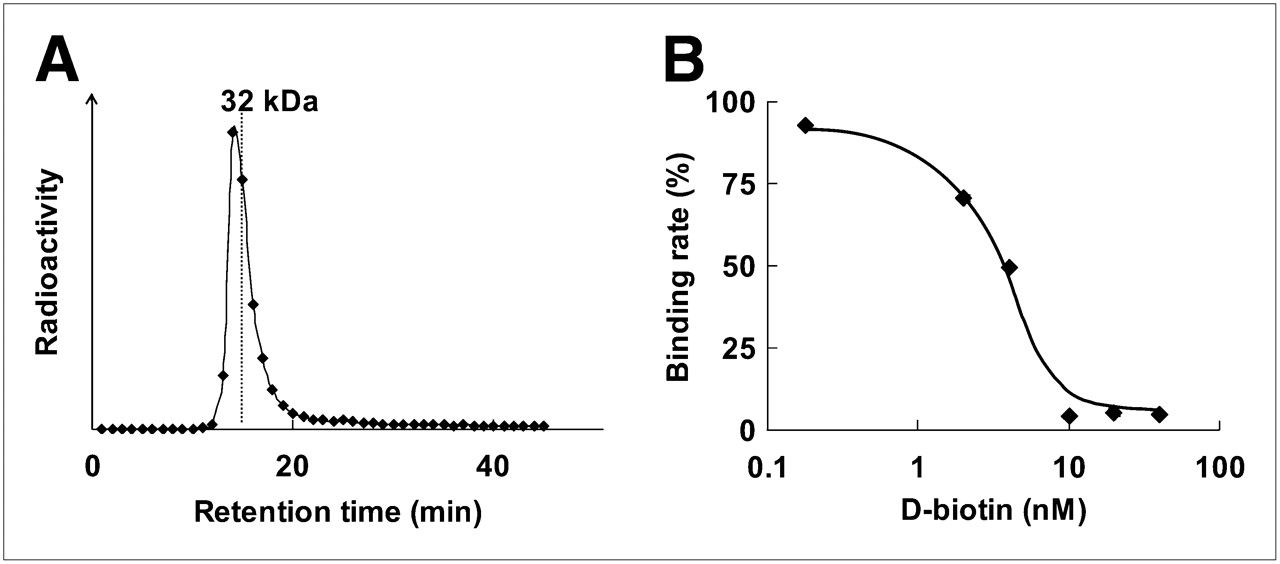
    Binding of 125I-IBB to POS. (A) Size-exclusion HPLC chromatogram after incubation of 125I-IBB with POS. Symbols represent radioactivity of each eluate collected every 1 min. (B) Concentration-dependent inhibition of 125I-IBB binding to POS by D-biotin. Symbols and bars represent mean and SD.
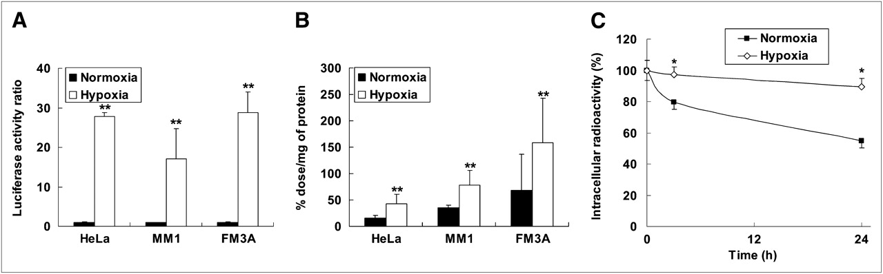
  • FIGURE 4. 
    • Download figure
    • Open in new tab
    • Download powerpoint
    FIGURE 4. 

    (A) HIF-1-dependent luciferase activity in HeLa, MM1, and FM3A cells. Data were normalized by protein concentration of cells. Results are shown as ratio of activity after hypoxia to activity after normoxia. Columns and bars represent mean and SD (n = 3–6 **P < 0.01 vs. normoxia). (B) Intracellular radioactivity in HeLa, MM1, and FM3A cells after 24-h incubation under either normoxic or hypoxic conditions. Radioactivity was normalized by protein concentration of cells. Columns and bars represent mean and SD (n = 3–6 **P < 0.01 vs. normoxia). (C) Degradation of intracellularly accumulated IPOS after reoxygenation. HeLa cells were incubated with 125I-IPOS for 24 h under hypoxic conditions. Then, medium was replaced with fresh medium, and cells were subjected to further incubation under normoxic or hypoxic conditions. Results are shown as percentage of radioactivity at start of second incubation. Symbols and bars represent mean and SD (n = 3–5; *P < 0.05 vs. normoxia).
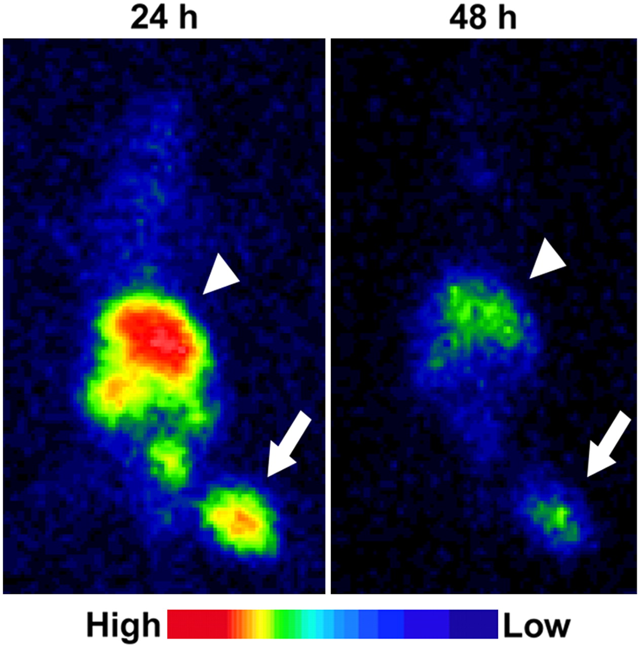
  • FIGURE 5. 
    • Download figure
    • Open in new tab
    • Download powerpoint
    FIGURE 5. 

    Typical planar images of FM3A-implamted mice at 24 or 48 h after injection of 123I-IPOS. Tumors were clearly visualized in both images (arrow). Arrowheads indicate liver.

  • FIGURE 6. 
    • Download figure
    • Open in new tab
    • Download powerpoint
    FIGURE 6. 

    Correlation between accumulation of 125I-IPOS and HIF-1 activity within same tumor. Ordinate represents accumulated radioactivity (%ID), and abscissa represents HIF-1-dependent luciferase activity. Correlation coefficient (R) was 0.71, indicating highly significant correlation (P < 0.05).
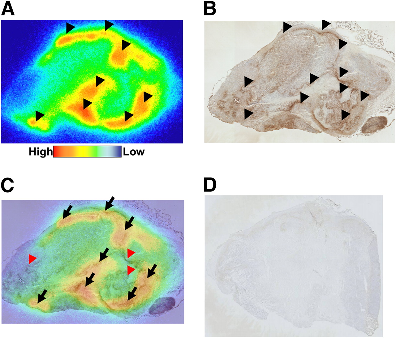
  • FIGURE 7. 
    • Download figure
    • Open in new tab
    • Download powerpoint
    FIGURE 7. 

    Comparison between intratumoral distribution of 125I-IPOS and pimonidazole-positive hypoxic region. Typical autoradiogram (A) and pimonidazole immunohistochemical staining (B) in identical section are shown. Merged image (C) is also presented. Black arrowheads indicate area of 125I-IPOS accumulation in A and pimonidazole-positive area in B. Black arrows show areas in which both signals are positive, and red arrowheads indicate pimonidazole-positive areas without accumulation of 125I-IPOS. No signal was observed in pimonidazole-untreated tumor (D).

Tables

  • Figures
  • Additional Files
    • View popup
    TABLE 1

    Biodistribution of 125I-IPOS in FM3A-Implanted Mice

    Time after injection (h)
    Organ162448
    Blood15.55 ± 1.063.14 ± 0.540.18 ± 0.120.01 ± 0.01
    Liver32.97 ± 1.0313.80 ± 0.870.97 ± 0.440.30 ± 0.06
    Spleen17.29 ± 2.356.07 ± 0.731.21 ± 0.530.19 ± 0.05
    Kidney5.51 ± 0.362.79 ± 0.470.74 ± 0.600.09 ± 0.02
    Stomach2.25 ± 0.570.62 ± 0.320.49 ± 0.310.04 ± 0.02
    Neck2.39 ± 0.060.80 ± 0.080.09 ± 0.040.00 ± 0.01
    Intestine7.96 ± 0.099.82 ± 1.610.91 ± 0.130.24 ± 0.08
    Tumor1.47 ± 1.021.49 ± 0.380.28 ± 0.080.07 ± 0.05
    Muscle0.94 ± 0.650.28 ± 0.050.07 ± 0.060.00 ± 0.01
    Tumor/blood0.10 ± 0.070.49 ± 0.181.79 ± 0.596.72 ± 6.61
    • Organ uptake values are expressed as %ID/g of tissue except for tumor/blood. Values are mean ± SD; n = 3–5.

    • View popup
    TABLE 2

    Effect of POS Concentration on Biodistribution of 125I-IPOS in FM3A-Implanted Mice at 24 Hours After Injection

    Protein concentration (μg)
    Organ0.050.5530
    Blood0.12 ± 0.040.16 ± 0.100.16 ± 0.010.27 ± 0.07
    Liver1.92 ± 0.241.27 ± 0.535.53 ± 1.0513.61 ± 1.90
    Spleen1.07 ± 0.301.15 ± 0.412.33 ± 0.257.87 ± 2.20
    Kidney0.35 ± 0.200.59 ± 0.511.03 ± 0.104.32 ± 1.87
    Stomach0.28 ± 0.140.55 ± 0.420.52 ± 0.200.86 ± 0.27
    Neck0.05 ± 0.040.09 ± 0.040.11 ± 0.020.23 ± 0.08
    Intestine4.57 ± 0.821.95 ± 2.032.17 ± 0.242.18 ± 0.28
    Tumor0.23 ± 0.080.25 ± 0.070.49 ± 0.091.37 ± 0.33
    Muscle0.05 ± 0.030.05 ± 0.050.07 ± 0.010.10 ± 0.03
    Tumor/blood2.04 ± 0.611.61 ± 0.103.00 ± 0.685.14 ± 0.34
    • Organ uptake values are expressed as %ID/g of tissue except for tumor/blood. Values are mean ± SD; n = 5.

Additional Files

  • Figures
  • Tables
  • Supplemental Data

    Files in this Data Supplement:

    • Supplemental Figure
PreviousNext
Back to top

In this issue

Journal of Nuclear Medicine: 50 (6)
Journal of Nuclear Medicine
Vol. 50, Issue 6
June 2009
  • Table of Contents
  • Table of Contents (PDF)
  • About the Cover
  • Index by author
Print
Download PDF
Article Alerts
Sign In to Email Alerts with your Email Address
Email Article

Thank you for your interest in spreading the word on Journal of Nuclear Medicine.

NOTE: We only request your email address so that the person you are recommending the page to knows that you wanted them to see it, and that it is not junk mail. We do not capture any email address.

Enter multiple addresses on separate lines or separate them with commas.
Imaging of HIF-1-Active Tumor Hypoxia Using a Protein Effectively Delivered to and Specifically Stabilized in HIF-1-Active Tumor Cells
(Your Name) has sent you a message from Journal of Nuclear Medicine
(Your Name) thought you would like to see the Journal of Nuclear Medicine web site.
Citation Tools
Imaging of HIF-1-Active Tumor Hypoxia Using a Protein Effectively Delivered to and Specifically Stabilized in HIF-1-Active Tumor Cells
Takashi Kudo, Masashi Ueda, Yuji Kuge, Takahiro Mukai, Shotaro Tanaka, Maki Masutani, Yasushi Kiyono, Shinae Kizaka-Kondoh, Masahiro Hiraoka, Hideo Saji
Journal of Nuclear Medicine Jun 2009, 50 (6) 942-949; DOI: 10.2967/jnumed.108.061119

Citation Manager Formats

  • BibTeX
  • Bookends
  • EasyBib
  • EndNote (tagged)
  • EndNote 8 (xml)
  • Medlars
  • Mendeley
  • Papers
  • RefWorks Tagged
  • Ref Manager
  • RIS
  • Zotero
Share
Imaging of HIF-1-Active Tumor Hypoxia Using a Protein Effectively Delivered to and Specifically Stabilized in HIF-1-Active Tumor Cells
Takashi Kudo, Masashi Ueda, Yuji Kuge, Takahiro Mukai, Shotaro Tanaka, Maki Masutani, Yasushi Kiyono, Shinae Kizaka-Kondoh, Masahiro Hiraoka, Hideo Saji
Journal of Nuclear Medicine Jun 2009, 50 (6) 942-949; DOI: 10.2967/jnumed.108.061119
Twitter logo Facebook logo LinkedIn logo Mendeley logo
  • Tweet Widget
  • Facebook Like
  • Google Plus One
Bookmark this article

Jump to section

  • Article
    • Abstract
    • MATERIALS AND METHODS
    • RESULTS
    • DISCUSSION
    • CONCLUSION
    • Acknowledgments
    • Footnotes
    • References
  • Figures & Data
  • Supplemental
  • Info & Metrics
  • PDF

Related Articles

  • This Month in JNM
  • PubMed
  • Google Scholar

Cited By...

  • 18F-Fluoromisonidazole PET Uptake Is Correlated with Hypoxia-Inducible Factor-1{alpha} Expression in Oral Squamous Cell Carcinoma
  • Google Scholar

More in this TOC Section

  • Design and Fabrication of Kidney Phantoms for Internal Radiation Dosimetry Using 3D Printing Technology
  • Synthesis and Biologic Evaluation of Novel 18F-Labeled Probes Targeting Prostate-Specific Membrane Antigen for PET of Prostate Cancer
  • Tumor-Specific Binding of Radiolabeled PEGylated GIRLRG Peptide: A Novel Agent for Targeting Cancers
Show more Basic Science Investigations

Similar Articles

SNMMI

© 2025 SNMMI

Powered by HighWire