


Article Figures & Data
Figures
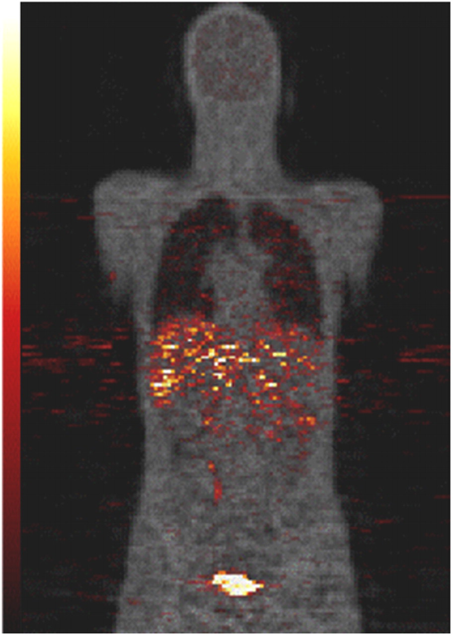
- FIGURE 1.
11C-NPA coronal PET image (hot metal scale) overlaid on attenuation image reconstructed from transmission data (gray scale) for male subject. PET data have been summed over first 4 scans and the image roughly represents activity distribution averaged over first 20 min after injection.
- FIGURE 2.
Activity concentration, normalized by injected dose, in various organs as function of time.


Tables
Subject no. Sex Height (cm) Weight (kg) Age (y) 1 F 161 69 22 2 F 168 66 38 3 F 170 73 39 4 M 180 82 20 5 M 175 64 24 6 M 180 80 21 Organ Residence time (s) Liver 439.0 Duodenum 252.3 Urinary bladder 106.0 Kidney 97.6 Lung 89.9 Brain 77.2 Cortical bone 46.4 Gallbladder 33.3 Upper large intestine 30.4 Red marrow 25.0 Lower large intestine 17.9 Heart 13.5 Dose from… Target organ β Photon Total SD Adrenals 5.12 × 10⁻⁴ 3.29 × 10⁻³ 3.80 × 10⁻³ 5.50 × 10⁻⁴ Brain 3.65 × 10⁻³ 1.83 × 10⁻³ 5.48 × 10⁻³ 4.89 × 10⁻⁴ Breasts 5.12 × 10⁻⁴ 9.68 × 10⁻⁴ 1.48 × 10⁻³ 2.57 × 10⁻⁴ Gallbladder wall 1.99 × 10⁻² 8.26 × 10⁻³ 2.81 × 10⁻² 1.08 × 10⁻² LLI wall 4.46 × 10⁻³ 3.16 × 10⁻³ 7.62 × 10⁻³ 1.03 × 10⁻³ Small intestine 1.99 × 10⁻² 4.97 × 10⁻³ 2.48 × 10⁻² 3.98 × 10⁻³ Stomach wall 5.12 × 10⁻⁴ 2.17 × 10⁻³ 2.68 × 10⁻³ 3.78 × 10⁻⁴ ULI wall 4.76 × 10⁻³ 5.45 × 10⁻³ 1.02 × 10⁻² 2.14 × 10⁻³ Heart wall 1.47 × 10⁻³ 2.20 × 10⁻³ 3.67 × 10⁻³ 4.35 × 10⁻⁴ Kidneys 2.09 × 10⁻² 6.34 × 10⁻³ 2.72 × 10⁻² 5.72 × 10⁻³ Liver 1.68 × 10⁻² 8.27 × 10⁻³ 2.50 × 10⁻² 4.44 × 10⁻³ Lungs 6.24 × 10⁻³ 2.30 × 10⁻³ 8.54 × 10⁻³ 2.07 × 10⁻³ Muscle 5.12 × 10⁻⁴ 1.43 × 10⁻³ 1.94 × 10⁻³ 2.70 × 10⁻⁴ Ovaries 5.88 × 10⁻⁴ 3.84 × 10⁻³ 4.43 × 10⁻³ 2.10 × 10⁻⁴ Pancreas 5.12 × 10⁻⁴ 3.09 × 10⁻³ 3.60 × 10⁻³ 4.87 × 10⁻⁴ Red marrow 9.33 × 10⁻⁴ 1.84 × 10⁻³ 2.78 × 10⁻³ 6.87 × 10⁻⁴ Osteogenic cells 2.39 × 10⁻³ 1.44 × 10⁻³ 3.83 × 10⁻³ 9.23 × 10⁻⁴ Skin 5.12 × 10⁻⁴ 7.75 × 10⁻⁴ 1.29 × 10⁻³ 2.16 × 10⁻⁴ Spleen 5.12 × 10⁻⁴ 1.93 × 10⁻³ 2.44 × 10⁻³ 3.77 × 10⁻⁴ Testes 4.35 × 10⁻⁴ 9.07 × 10⁻⁴ 1.34 × 10⁻³ 1.45 × 10⁻⁴ Thymus 5.12 × 10⁻⁴ 1.17 × 10⁻³ 1.68 × 10⁻³ 3.03 × 10⁻⁴ Thyroid 5.12 × 10⁻⁴ 7.66 × 10⁻⁴ 1.28 × 10⁻³ 2.29 × 10⁻⁴ Urinary bladder wall 1.82 × 10⁻² 6.71 × 10⁻³ 2.49 × 10⁻² 6.28 × 10⁻³ Uterus 5.88 × 10⁻⁴ 3.85 × 10⁻³ 4.44 × 10⁻³ 2.06 × 10⁻⁴ Total body 1.49 × 10⁻³ 1.68 × 10⁻³ 3.17 × 10⁻³ 4.01 × 10⁻⁴ LLI wall = lower large intestine wall; ULI = upper large intestine wall.
The organ receiving the highest dose was the gallbladder wall. Final column lists standard deviation of total dose across subjects.
Supplemental Data
Files in this Data Supplement:


