


Article Figures & Data
Figures
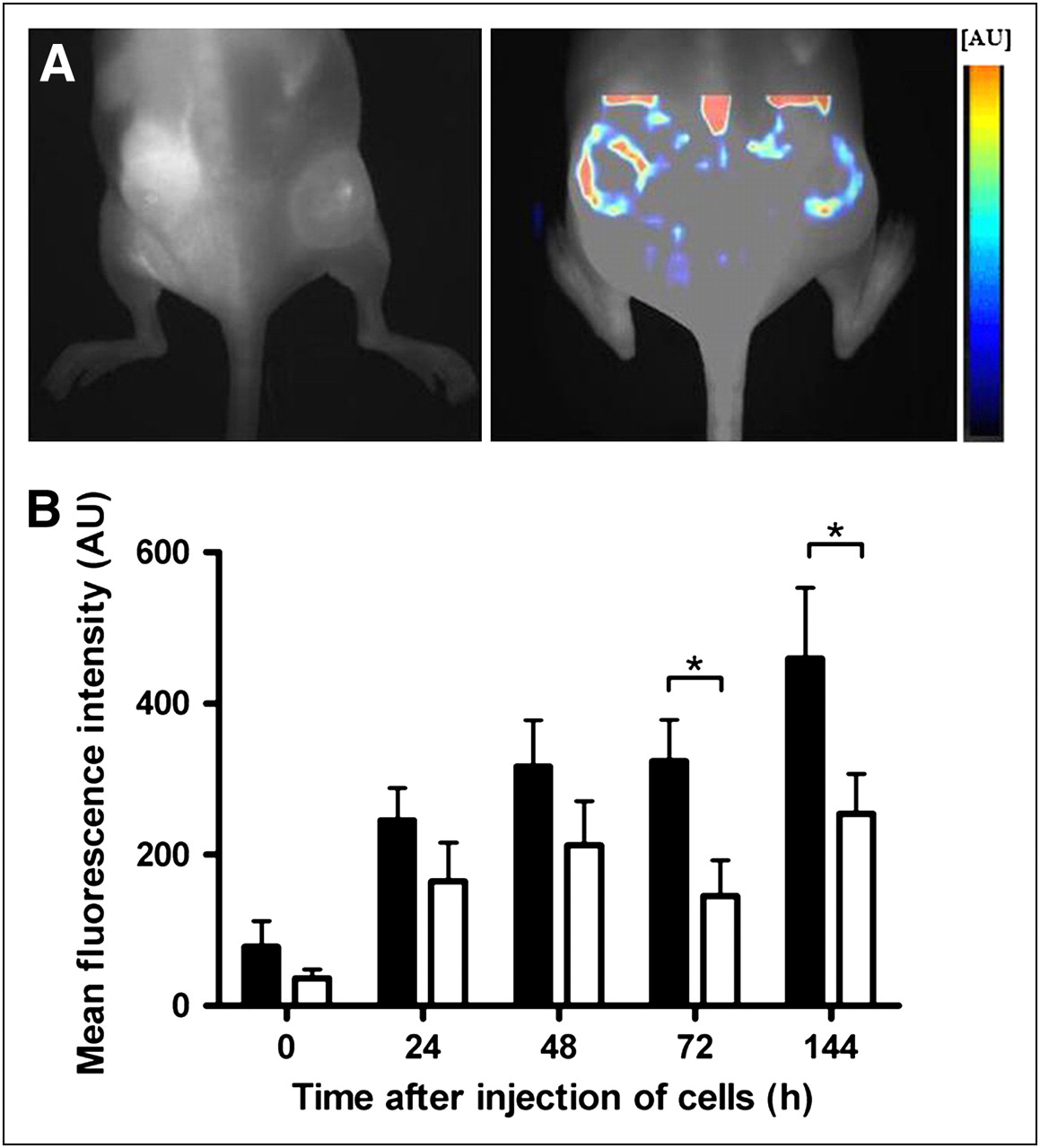
- FIGURE 1.
Contrast enhancement of biogel pellets after injection of labeled Mϕs. (A) Fluorescence reflectance images were repeatedly acquired up to 144 h after injection of labeled Mϕs (5 × 10⁶). (B) Note stronger enhancement of lipopolysaccharide (LPS) pellets (right flank, ▪), compared with control pellets without lipopolysaccharide (left flank, □). Lipopolysaccharide pellets showed increasing conspicuity over first 72 h, with slight drop of signal intensity at 144 h. Means ± SEMs are shown (n = 5). *P < 0.05, Student t test. AU = arbitrary units.
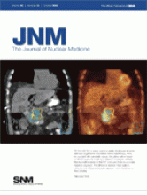
- FIGURE 2.
FMT of mice showed distribution of Mϕs inside pellets. (A) FMT was performed 72 h after injection of labeled Mϕs (5 × 10⁶) to resolve 3-dimensional distribution of Mϕs in target tissue (left, NIRF image; right, color-encoded FMT). FMT confirmed FRI results and showed that Mϕs distributed mainly in periphery of pellets. (B) Fluorescence quantification by FMT showed approximately 2.7 times higher signal in lipopolysaccharide-containing pellets (▪) than in controls (□). Means ± SEMs are shown (n = 10). *P < 0.05, Student t test. AU = arbitrary units.
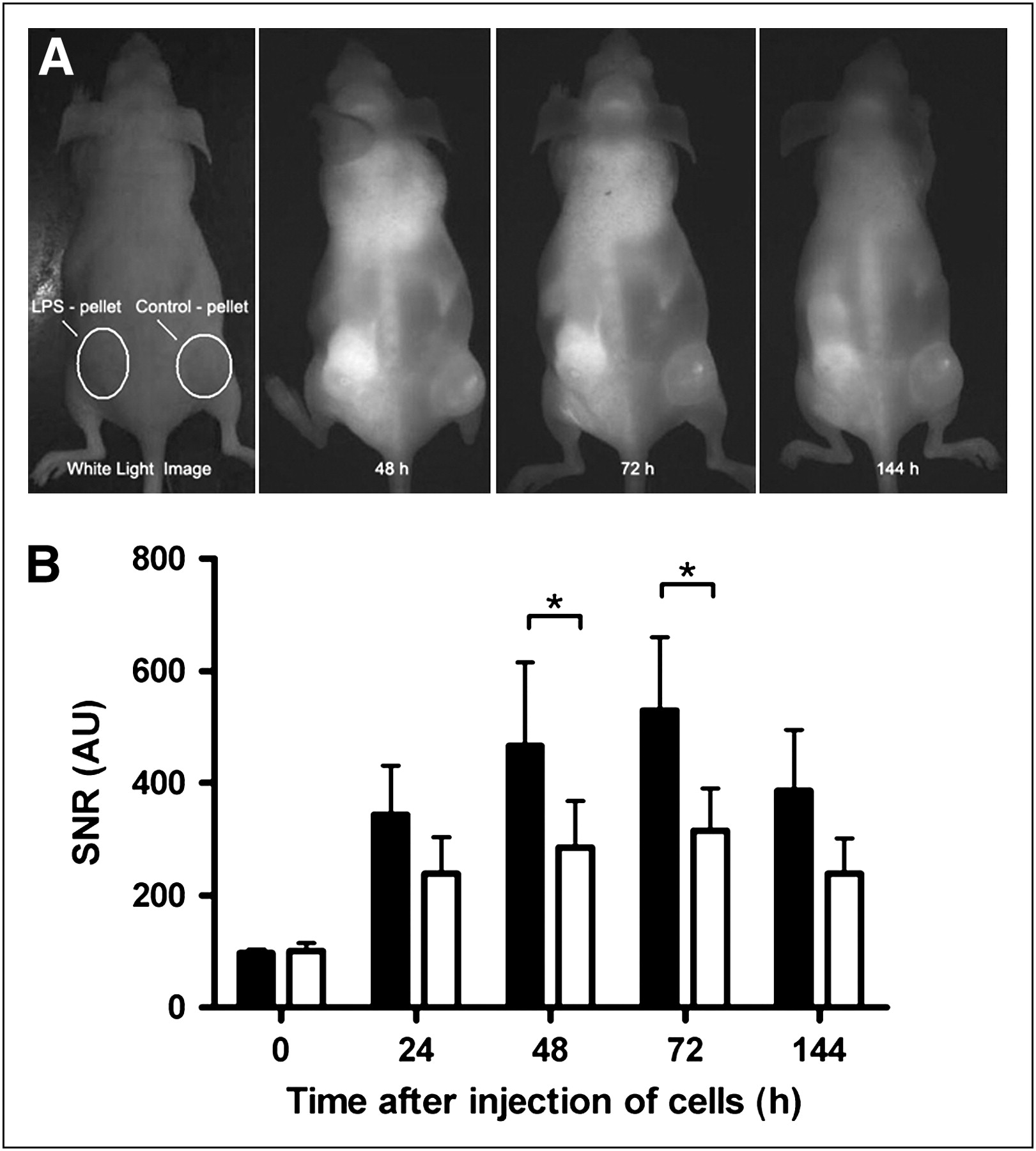
- FIGURE 3.
(A) Signal intensity of pellets after injection of different numbers of labeled Mϕs. (B) Numbers of DiR-labeled Mϕs recovered from pellets. Lipopolysaccharide-containing biogel pellets were implanted into mice, and different amounts of DiR-labeled Mϕs were injected. FMT and cell recovery were analyzed 72 h after cell injection. FMT showed clear increase of signal intensity dependant on number of injected cells (A). There was also clear dependency between amount of injected cells and numbers of DiR-positive Mϕs recovered from pellets (B). Means ± SEMs are shown (n = 5). *P < 0.05. **P < 0.01, ANOVA. AU = arbitrary units.
- FIGURE 4.
Migration of DiR-stained Mϕs in vivo. Mice were injected with 5 × 10⁶ DiR-stained Mϕs. Biogel pellets were recovered 2 d after injection of Mϕs, and intensity of DiR fluorescence and surface antigen expression were measured by flow cytometry.


Additional Files
Supplemental Data
Files in this Data Supplement:


Jump to section
Related Articles
Cited By...
- Reprogramming of Monocytes by GM-CSF Contributes to Regulatory Immune Functions during Intestinal Inflammation
- An In Vivo Spectral Multiplexing Approach for the Cooperative Imaging of Different Disease-Related Biomarkers with Near-Infrared Fluorescent Forster Resonance Energy Transfer Probes
- Trafficking Macrophage Migration Using Reporter Gene Imaging with Human Sodium Iodide Symporter in Animal Models of Inflammation