


Article Figures & Data
Figures
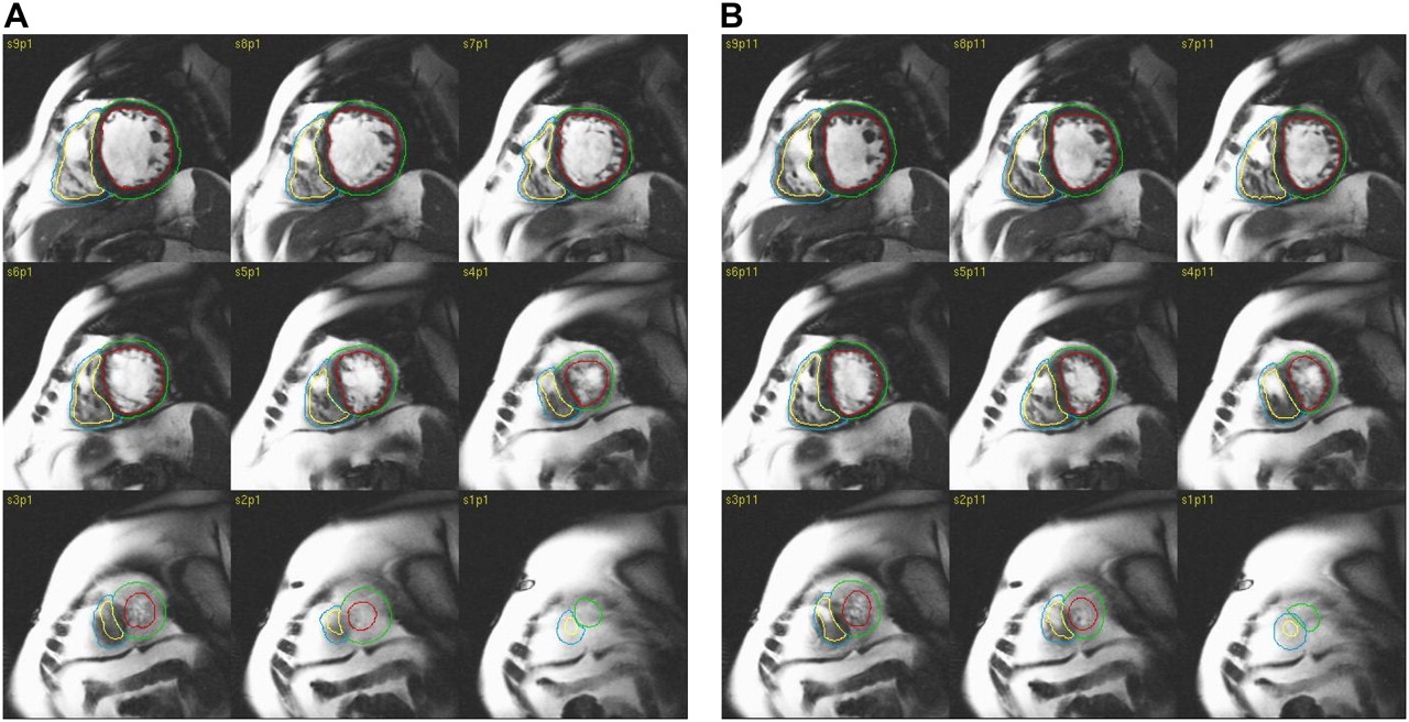
- FIGURE 1.
MR images of 75-y-old male patient with history of congestive heart failure and hypertension with severe LV and RV dysfunction and abnormal LV EF (23%) at ED (A) and ES (B). Manually drawn epicardial and endocardial outlines are superimposed on images.
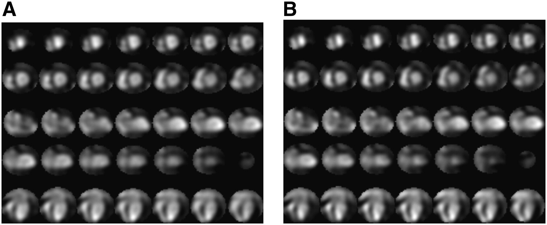
- FIGURE 2.
Screens of cines used to visually analyze BP-gated SPECT regional WM abnormalities for same patient in Figure 1 at ED (A) and ES (B).
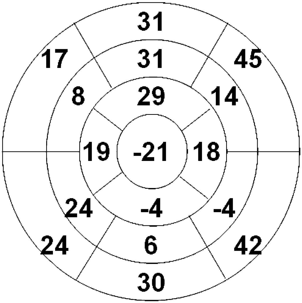
- FIGURE 3.
Polar map of BP-gated SPECT LV regional EF (percentage) for patient in Figure 1.
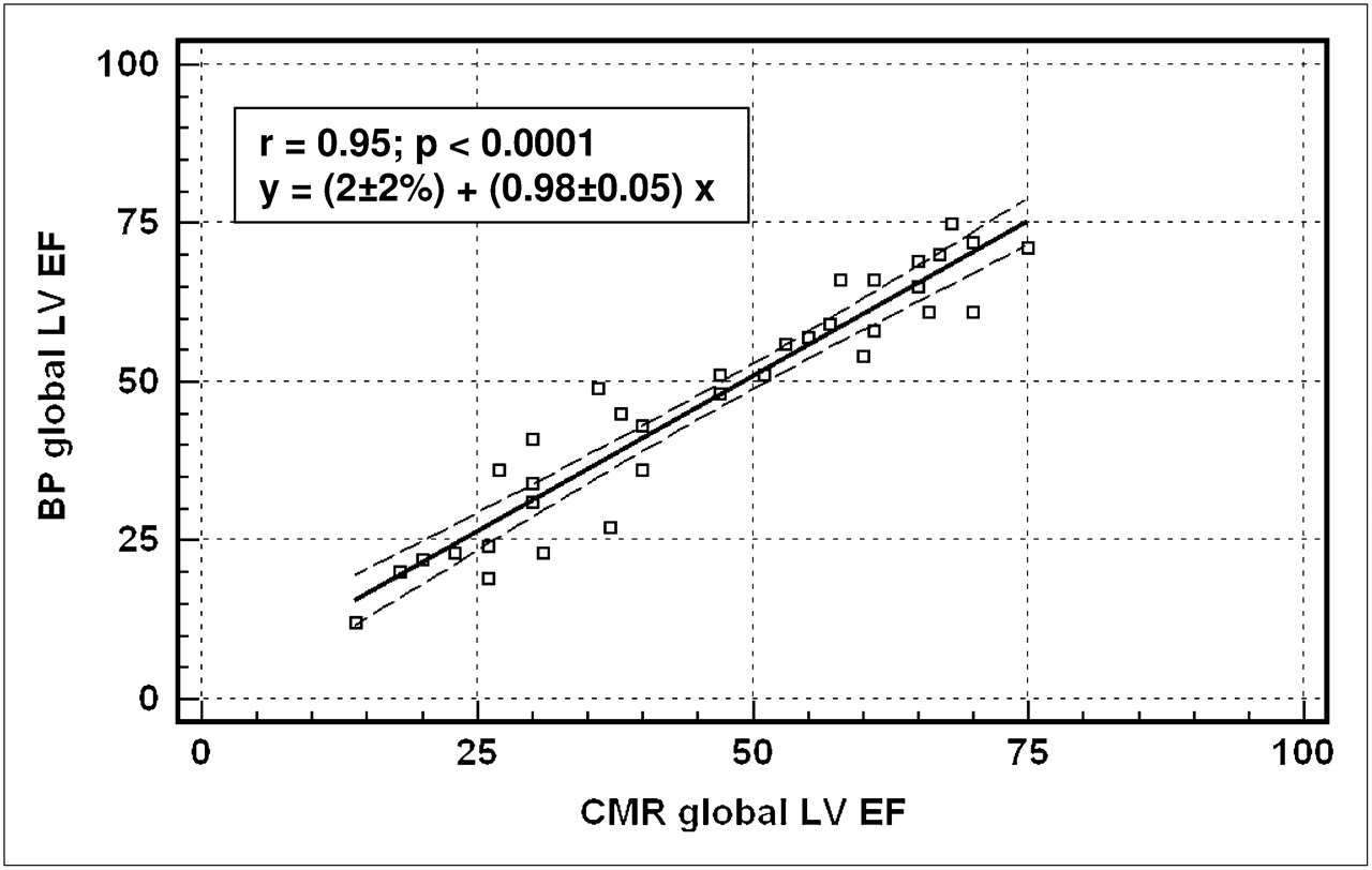
- FIGURE 4.
BP measurement of global LV EF vs. cardiac MRI–quantified global LV EF. CMR = cardiac MRI.
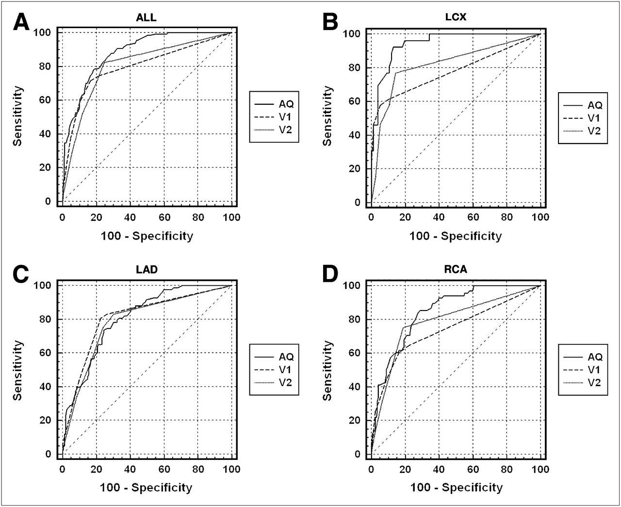
- FIGURE 5.
Receiver-operating-characteristic curves to predict LV segments with abnormal WM as seen on cardiac MRI, for AQ BP-gated SPECT EF, initial visual assessment (V1), and second visual assessment (V2) for all territories (A) and for LCX (B), LAD (C), and RCA (D) territories only.


Tables
- TABLE 1
Strength of Agreement and Significance of Differences of BP AQ with Cardiac MRI in Identifying Patients with Abnormally Low Global EF
Discrimination threshold κ Agreement (14) McNemar difference McNemar P Global EF < 35% 0.94 “Very good” 3.0% 1.00 Global EF < 50% 0.94 “Very good” 3.0% 1.00 - TABLE 2
Accuracy (Receiver-Operating-Characteristic Curve Area) of Segment-by-Segment Identification, Grouped by Coronary Territory
Territory All LCX LAD RCA WM AQ 88% ± 2% 95% ± 2% 86% ± 2% 88% ± 2% VA1 80% ± 2%* 78% ± 6%* 84% ± 3% 74% ± 4%* VA2 81% ± 2%* 82% ± 5%* 80% ± 3% 80% ± 3%* WT AQ 80% ± 2% 84% ± 4% 79% ± 3% 78% ± 3% VA1 73% ± 2% 66% ± 5%* 75% ± 4% 76% ± 3% VA2 74% ± 2% 71% ± 5%* 75% ± 4% 77% ± 3% WV AQ 87% ± 2% 94% ± 3% 84% ± 3% 88% ± 2% VA1 79% ± 3%* 74% ± 6%* 84% ± 4% 74% ± 4%* WV VA2 79% ± 2%* 81% ± 5%* 79% ± 4% 78% ± 4%* ↵* P < 0.05 versus AQ.
All = all coronary artery territories; VA1 = initial visual assessment; VA2 = second visual assessment.
- TABLE 3
Strength of Agreement and Significance of Differences Between BP AQ and BP Visual Assessment with Cardiac MRI in Identifying Territories with Abnormal WM
Territory showing abnormal WM κ Agreement (14) McNemar difference McNemar P AQ LCX 0.71 “Good” 5.6% 0.15 LAD 0.50 “Moderate” 5.0% 0.16 RCA 0.65 “Good” 5.9% 0.08 Visual assessment LCX 0.58 “Moderate” 6.9% 0.12 LAD 0.59 “Moderate” 1.8% 0.75 RCA 0.45 “Moderate” 9.6% 0.0006 - TABLE 4
Strength of Agreement and Significance of Differences Between BP Initial Visual Assessment and BP Second Visual Assessment in Identifying Territories with Abnormal WM
Territory showing abnormal WM κ Agreement (14) McNemar difference McNemar P LCX 0.55 “Moderate” 7.8% 0.07 LAD 0.72 “Good” 5.1% 0.06 RCA 0.58 “Moderate” 3.9% 0.20


Jump to section
Related Articles
Cited By...
- No citing articles found.