
Main menu

  • Home
  • Content
    • Current
    • Ahead of print
    • Past Issues
    • JNM Supplement
    • SNMMI Annual Meeting Abstracts
    • Continuing Education
    • JNM Podcasts
  • Subscriptions
    • Subscribers
    • Institutional and Non-member
    • Rates
    • Journal Claims
    • Corporate & Special Sales
  • Authors
    • Submit to JNM
    • Information for Authors
    • Assignment of Copyright
    • AQARA requirements
  • Info
    • Reviewers
    • Permissions
    • Advertisers
  • About
    • About Us
    • Editorial Board
    • Contact Information
  • More
    • Alerts
    • Feedback
    • Help
    • SNMMI Journals
  • SNMMI
    • JNM
    • JNMT
    • SNMMI Journals
    • SNMMI

User menu

  • Subscribe
  • My alerts
  • Log in
  • My Cart

Search

  • Advanced search
Journal of Nuclear Medicine
  • SNMMI
    • JNM
    • JNMT
    • SNMMI Journals
    • SNMMI
  • Subscribe
  • My alerts
  • Log in
  • My Cart
Journal of Nuclear Medicine

Advanced Search

  • Home
  • Content
    • Current
    • Ahead of print
    • Past Issues
    • JNM Supplement
    • SNMMI Annual Meeting Abstracts
    • Continuing Education
    • JNM Podcasts
  • Subscriptions
    • Subscribers
    • Institutional and Non-member
    • Rates
    • Journal Claims
    • Corporate & Special Sales
  • Authors
    • Submit to JNM
    • Information for Authors
    • Assignment of Copyright
    • AQARA requirements
  • Info
    • Reviewers
    • Permissions
    • Advertisers
  • About
    • About Us
    • Editorial Board
    • Contact Information
  • More
    • Alerts
    • Feedback
    • Help
    • SNMMI Journals
  • View or Listen to JNM Podcast
  • Visit JNM on Facebook
  • Join JNM on LinkedIn
  • Follow JNM on Twitter
  • Subscribe to our RSS feeds
OtherBASIC SCIENCE INVESTIGATIONS

PET/MRI Dual-Modality Tumor Imaging Using Arginine-Glycine-Aspartic (RGD)–Conjugated Radiolabeled Iron Oxide Nanoparticles

Ha-Young Lee, Zibo Li, Kai Chen, Andrew R. Hsu, Chenjie Xu, Jin Xie, Shouheng Sun and Xiaoyuan Chen
Journal of Nuclear Medicine August 2008, 49 (8) 1371-1379; DOI: https://doi.org/10.2967/jnumed.108.051243
Ha-Young Lee
  • Find this author on Google Scholar
  • Find this author on PubMed
  • Search for this author on this site
Zibo Li
  • Find this author on Google Scholar
  • Find this author on PubMed
  • Search for this author on this site
Kai Chen
  • Find this author on Google Scholar
  • Find this author on PubMed
  • Search for this author on this site
Andrew R. Hsu
  • Find this author on Google Scholar
  • Find this author on PubMed
  • Search for this author on this site
Chenjie Xu
  • Find this author on Google Scholar
  • Find this author on PubMed
  • Search for this author on this site
Jin Xie
  • Find this author on Google Scholar
  • Find this author on PubMed
  • Search for this author on this site
Shouheng Sun
  • Find this author on Google Scholar
  • Find this author on PubMed
  • Search for this author on this site
Xiaoyuan Chen
  • Find this author on Google Scholar
  • Find this author on PubMed
  • Search for this author on this site
  • Article
  • Figures & Data
  • Info & Metrics
  • PDF
Loading

Article Figures & Data

Figures

  • FIGURE 1. 
    • Download figure
    • Open in new tab
    • Download powerpoint
    FIGURE 1. 

    (A) Synthesis of PET/MRI dual functional probe DOTA-IO-RGD. DOTA-IO was prepared similarly except that no RGD peptide was used. (B) Illustration of PET/MRI probe based on IO nanoparticle.
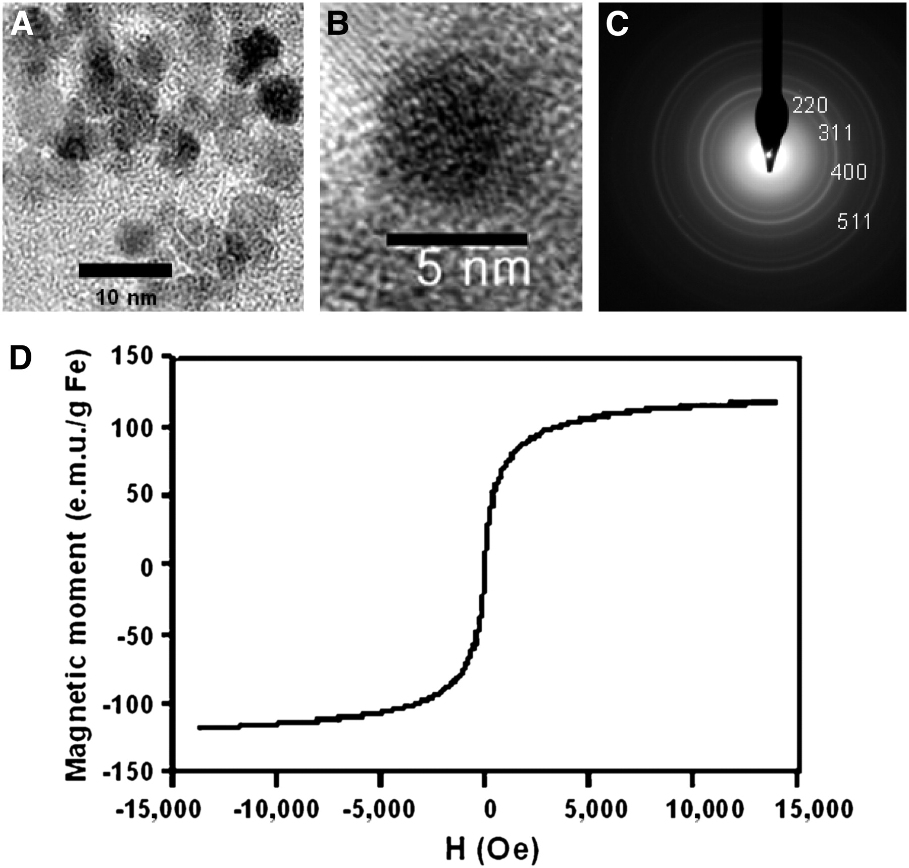
  • FIGURE 2. 
    • Download figure
    • Open in new tab
    • Download powerpoint
    FIGURE 2. 

    (A) TEM image of PASP-coated IO nanoparticle. (B) Selected-area electron diffraction pattern of PASP-coated IO nanoparticle. (C) High-resolution TEM image of PASP-coated IO nanoparticle. (D) Magnetization curve of PASP-coated IO nanoparticle.

  • FIGURE 3. 
    • Download figure
    • Open in new tab
    • Download powerpoint
    FIGURE 3. 

    (A) Phantom image acquired from T1-weighted MRI scan (top) and T2-weighted MRI scan (bottom) for ferumoxide and PASP-IO at different iron concentrations. (B) 1/T2 (top) and 1/T2* (bottom) vs. Fe concentration for PASP-IO and ferumoxide. Relaxivity values r2 and r2* were obtained from slopes of linear fits of experimental data.
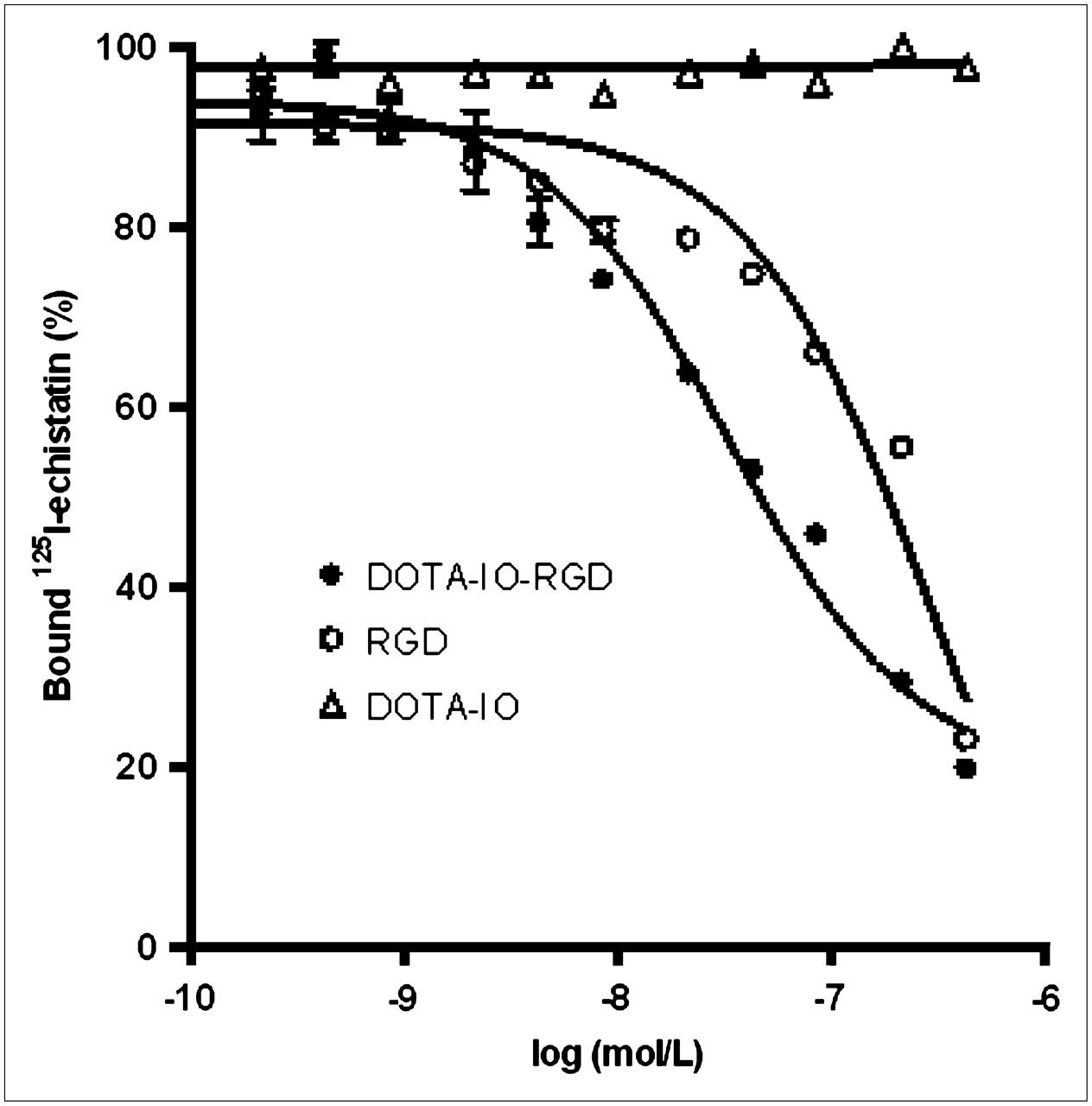
  • FIGURE 4. 
    • Download figure
    • Open in new tab
    • Download powerpoint
    FIGURE 4. 

    Inhibition of 125I-echistatin (integrin αvβ3–specific) binding to integrin αvβ3 on U87MG cells by DOTA-IO-RGD, c(RGDyK), and DOTA-IO (n = 3, mean ± SD).

  • FIGURE 5. 
    • Download figure
    • Open in new tab
    • Download powerpoint
    FIGURE 5. 

    (A) Decay-corrected whole-body coronal PET images of nude mouse bearing human U87MG tumor at 1, 4, and 21 h after injection of 3.7 MBq of 64Cu-DOTA-IO, 64Cu-DOTA-IO-RGD, or 64Cu-DOTA-IO-RGD with 10 mg of c(RGDyK) peptide per kilogram (300 μg of iron-equivalent IO particles per mouse). (B) Time–activity curves of U87MG tumors after injection of 3.7 MBq of 64Cu-DOTA-IO, 64Cu-DOTA-IO-RGD, or 64Cu-DOTA-IO-RGD with blocking dose of c(RGDyK) (n = 3/group).
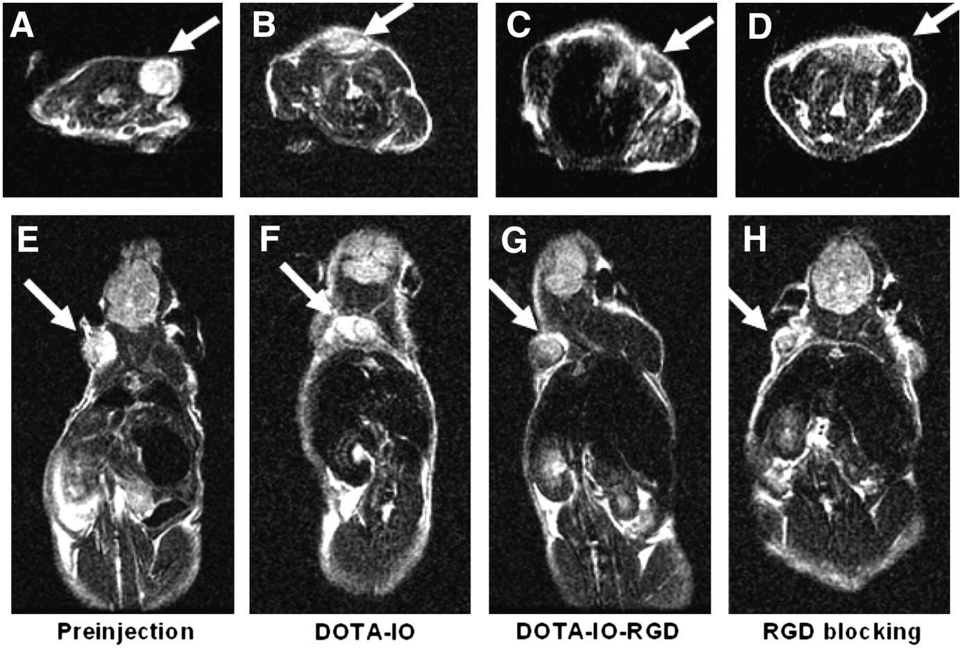
  • FIGURE 6. 
    • Download figure
    • Open in new tab
    • Download powerpoint
    FIGURE 6. 

    T2-weighted MR images of nude mice bearing U87MG tumor before injection of IO nanoparticles (A and E) and at 4 h after tail-vein injection of DOTA-IO (B and F), DOTA-IO-RGD (C and G), and DOTA-IO-RGD with blocking dose of c(RGDyK) (D and H).

  • FIGURE 7. 
    • Download figure
    • Open in new tab
    • Download powerpoint
    FIGURE 7. 

    Prussian blue–stained U87MG tumor, liver, and spleen sections after injection of 64Cu-DOTA-IO-RGD, 64Cu-DOTA-IO, and 64Cu-DOTA-IO-RGD with blocking dose of c(RGDyK). IO was stained as blue spots in figure (magnification, 200×).

PreviousNext
Back to top

In this issue

Journal of Nuclear Medicine: 49 (8)
Journal of Nuclear Medicine
Vol. 49, Issue 8
August 2008
  • Table of Contents
  • Table of Contents (PDF)
  • About the Cover
  • Index by author
Print
Download PDF
Article Alerts
Sign In to Email Alerts with your Email Address
Email Article

Thank you for your interest in spreading the word on Journal of Nuclear Medicine.

NOTE: We only request your email address so that the person you are recommending the page to knows that you wanted them to see it, and that it is not junk mail. We do not capture any email address.

Enter multiple addresses on separate lines or separate them with commas.
PET/MRI Dual-Modality Tumor Imaging Using Arginine-Glycine-Aspartic (RGD)–Conjugated Radiolabeled Iron Oxide Nanoparticles
(Your Name) has sent you a message from Journal of Nuclear Medicine
(Your Name) thought you would like to see the Journal of Nuclear Medicine web site.
Citation Tools
PET/MRI Dual-Modality Tumor Imaging Using Arginine-Glycine-Aspartic (RGD)–Conjugated Radiolabeled Iron Oxide Nanoparticles
Ha-Young Lee, Zibo Li, Kai Chen, Andrew R. Hsu, Chenjie Xu, Jin Xie, Shouheng Sun, Xiaoyuan Chen
Journal of Nuclear Medicine Aug 2008, 49 (8) 1371-1379; DOI: 10.2967/jnumed.108.051243

Citation Manager Formats

  • BibTeX
  • Bookends
  • EasyBib
  • EndNote (tagged)
  • EndNote 8 (xml)
  • Medlars
  • Mendeley
  • Papers
  • RefWorks Tagged
  • Ref Manager
  • RIS
  • Zotero
Share
PET/MRI Dual-Modality Tumor Imaging Using Arginine-Glycine-Aspartic (RGD)–Conjugated Radiolabeled Iron Oxide Nanoparticles
Ha-Young Lee, Zibo Li, Kai Chen, Andrew R. Hsu, Chenjie Xu, Jin Xie, Shouheng Sun, Xiaoyuan Chen
Journal of Nuclear Medicine Aug 2008, 49 (8) 1371-1379; DOI: 10.2967/jnumed.108.051243
Twitter logo Facebook logo LinkedIn logo Mendeley logo
  • Tweet Widget
  • Facebook Like
  • Google Plus One
Bookmark this article

Jump to section

  • Article
    • Abstract
    • MATERIALS AND METHODS
    • RESULTS
    • DISCUSSION
    • CONCLUSION
    • Acknowledgments
    • Footnotes
    • References
  • Figures & Data
  • Info & Metrics
  • PDF

Related Articles

  • This Month in JNM
  • PubMed
  • Google Scholar

Cited By...

  • A Bridge Not Too Far: Linking Disciplines Through Molecular Imaging Probes
  • Simultaneous Multiparametric PET/MRI with Silicon Photomultiplier PET and Ultra-High-Field MRI for Small-Animal Imaging
  • PET and MR Imaging: The Odd Couple or a Match Made in Heaven?
  • RGD Peptide-Conjugated Multimodal NaGdF4:Yb3+/Er3+ Nanophosphors for Upconversion Luminescence, MR, and PET Imaging of Tumor Angiogenesis
  • Effects of Photoacoustic Imaging and Photothermal Ablation Therapy Mediated by Targeted Hollow Gold Nanospheres in an Orthotopic Mouse Xenograft Model of Glioma
  • A Bridge Not Too Far: Linking Disciplines Through Molecular Imaging Probes
  • Design of Targeted Cardiovascular Molecular Imaging Probes
  • A Nucleolin-Targeted Multimodal Nanoparticle Imaging Probe for Tracking Cancer Cells Using an Aptamer
  • The Advantages of Nanoparticles for PET
  • Cancer Nanotargeted Radiopharmaceuticals for Tumor Imaging and Therapy
  • Google Scholar

More in this TOC Section

  • Counting Rate Characteristics and Image Distortion in Preclinical PET Imaging During Radiopharmaceutical Therapy
  • Design and Fabrication of Kidney Phantoms for Internal Radiation Dosimetry Using 3D Printing Technology
  • Synthesis and Biologic Evaluation of Novel 18F-Labeled Probes Targeting Prostate-Specific Membrane Antigen for PET of Prostate Cancer
Show more Basic Science Investigations

Similar Articles

SNMMI

© 2025 SNMMI

Powered by HighWire