
Main menu

  • Home
  • Content
    • Current
    • Ahead of print
    • Past Issues
    • JNM Supplement
    • SNMMI Annual Meeting Abstracts
    • Continuing Education
    • JNM Podcasts
  • Subscriptions
    • Subscribers
    • Institutional and Non-member
    • Rates
    • Journal Claims
    • Corporate & Special Sales
  • Authors
    • Submit to JNM
    • Information for Authors
    • Assignment of Copyright
    • AQARA requirements
  • Info
    • Reviewers
    • Permissions
    • Advertisers
  • About
    • About Us
    • Editorial Board
    • Contact Information
  • More
    • Alerts
    • Feedback
    • Help
    • SNMMI Journals
  • SNMMI
    • JNM
    • JNMT
    • SNMMI Journals
    • SNMMI

User menu

  • Subscribe
  • My alerts
  • Log in
  • My Cart

Search

  • Advanced search
Journal of Nuclear Medicine
  • SNMMI
    • JNM
    • JNMT
    • SNMMI Journals
    • SNMMI
  • Subscribe
  • My alerts
  • Log in
  • My Cart
Journal of Nuclear Medicine

Advanced Search

  • Home
  • Content
    • Current
    • Ahead of print
    • Past Issues
    • JNM Supplement
    • SNMMI Annual Meeting Abstracts
    • Continuing Education
    • JNM Podcasts
  • Subscriptions
    • Subscribers
    • Institutional and Non-member
    • Rates
    • Journal Claims
    • Corporate & Special Sales
  • Authors
    • Submit to JNM
    • Information for Authors
    • Assignment of Copyright
    • AQARA requirements
  • Info
    • Reviewers
    • Permissions
    • Advertisers
  • About
    • About Us
    • Editorial Board
    • Contact Information
  • More
    • Alerts
    • Feedback
    • Help
    • SNMMI Journals
  • View or Listen to JNM Podcast
  • Visit JNM on Facebook
  • Join JNM on LinkedIn
  • Follow JNM on Twitter
  • Subscribe to our RSS feeds
OtherBASIC SCIENCE INVESTIGATIONS

Relationship Between Induction of Phosphorylated H2AX and Survival in Breast Cancer Cells Exposed to 111In-DTPA-hEGF

Zhongli Cai, Zhuo Chen, Kristy E. Bailey, Deborah A. Scollard, Raymond M. Reilly and Katherine A. Vallis
Journal of Nuclear Medicine August 2008, 49 (8) 1353-1361; DOI: https://doi.org/10.2967/jnumed.108.051805
Zhongli Cai
  • Find this author on Google Scholar
  • Find this author on PubMed
  • Search for this author on this site
Zhuo Chen
  • Find this author on Google Scholar
  • Find this author on PubMed
  • Search for this author on this site
Kristy E. Bailey
  • Find this author on Google Scholar
  • Find this author on PubMed
  • Search for this author on this site
Deborah A. Scollard
  • Find this author on Google Scholar
  • Find this author on PubMed
  • Search for this author on this site
Raymond M. Reilly
  • Find this author on Google Scholar
  • Find this author on PubMed
  • Search for this author on this site
Katherine A. Vallis
  • Find this author on Google Scholar
  • Find this author on PubMed
  • Search for this author on this site
  • Article
  • Figures & Data
  • Info & Metrics
  • PDF
Loading

Article Figures & Data

Figures

  • FIGURE 1. 
    • Download figure
    • Open in new tab
    • Download powerpoint
    FIGURE 1. 

    Induction of γ-H2AX foci by 111In-DTPA-hEGF in breast cancer cell lines. (A) Cells were incubated with DTPA-hEGF (21 nM), 111In-acetate (3.2 MBq/mL), or 111In-DTPA-hEGF (3.2 MBq/mL, 21 nM) for 1 h; were exposed to γ-irradiation (1 Gy); or were untreated (control). (B) Cells were incubated with DTPA-hEGF (43 nM), 111In-acetate (5.2 MBq/mL), or 111In-DTPA-hEGF (5.2 MBq/mL, 43 nM) for 20 h; were exposed to γ-irradiation (10 Gy); or were untreated (controls). (C) Confocal microscopy of MCF-7, MDA-MB-231, and MDA-MB-468 cells exposed to DTPA-hEGF (21 nM), 111In-acetate (3.2 MBq/mL), or 111In-DTPA-hEGF (3.2 MBq/mL, 21 nM) for 1 h; exposed to γ-irradiation (1 Gy); or untreated (control). Cells were immunostained for γ-H2AX. 4′,6-diamidino-2-phenylindole was used to visualize cell nucleus. Exposure to 111In-DTPA-hEGF caused increase in number of γ-H2AX foci in MDA-MB-468 and MDA-MB-231 cells but not in MCF-7 cells.

  • FIGURE 2. 
    • Download figure
    • Open in new tab
    • Download powerpoint
    FIGURE 2. 

    MDA-MB-468, MDA-MB-231, and MCF-7 cells were treated with 111In-DTPA-hEGF (3.2 MBq/mL, 21 nM) for 1 h (A) or 111In-DTPA-hEGF (5.2 MBq/mL, 43 nM) for 20 h (B). Amount of 111In in cell membrane, cytoplasm, and nucleus was measured in cell fractionation experiments. Amount of 111In per cell decreased in order of MDA-MB-468 > MDA-MB-231 > MCF-7. (C) Amount of 111In that localizes in cytoplasm and nucleus is linearly dependent on amount of 111In that binds to cell membrane.
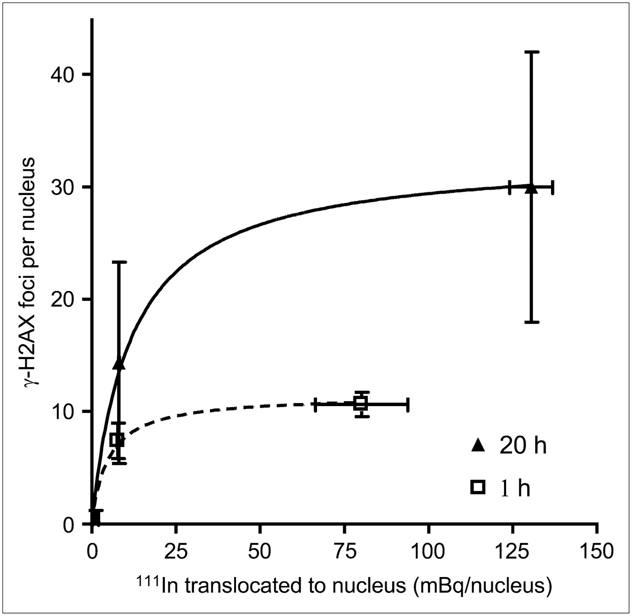
  • FIGURE 3. 
    • Download figure
    • Open in new tab
    • Download powerpoint
    FIGURE 3. 

    MCF-7, MDA-MB-468, and MDA-MB 231 cells were exposed to 111In-DTPA-hEGF (3.2 MBq/mL, 21 nM) for 1 h or 111In-DTPA-hEGF (5.2 MBq/mL, 43 nM) for 20 h. Relationship between amount of nuclear 111In and induction of γ-H2AX foci is shown. Data were fitted to 1-site binding equation.
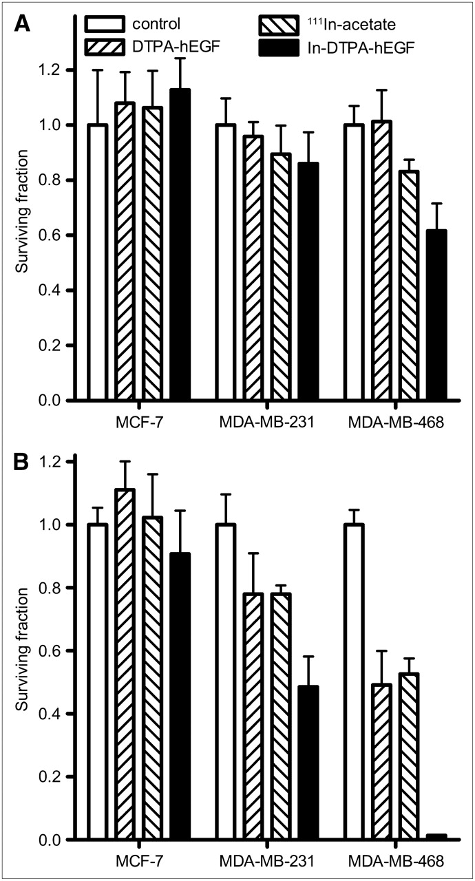
  • FIGURE 4. 
    • Download figure
    • Open in new tab
    • Download powerpoint
    FIGURE 4. 

    Clonogenic assays were performed using MDA-MB-468, MDA-MB-231, and MCF-7 cells incubated with DTPA-hEGF (21 nM), 111In-acetate (3.2 MBq/mL), or 111In-DTPA-hEGF (3.2 MBq/mL, 21 nM) for 1 h (A) or DTPA-hEGF (43 nM), 111In-acetate (5.2 MBq/mL), or 111In-DTPA-hEGF (5.2 MBq/mL, 43 nM) for 20 h (B). After exposure to 111In-DTPA-hEGF, significant decrease in SF was observed in MDA-MB-468 (0.6 ± 0.1 at 1 h and 0.013 ± 0.001 at 20 h, P < 0.0001) and MDA-MB-231 cells (0.5 ± 0.1 at 20 h, P = 0.0001) but not in MCF-7 cells, compared with untreated cells. Error bars represent SD of mean SF, calculated from 3 experiments.

  • FIGURE 5. 
    • Download figure
    • Open in new tab
    • Download powerpoint
    FIGURE 5. 

    Dependence of SF on γ-absorbed dose. Clonogenic assays were generated for MDA-MB-468, MDA-MB-231, and MCF-7 cells. Curves are nonlinear regression fits to experimental data, using linear quadratic function for MDA-MB-468 and first-order exponential function for other cell lines.

  • FIGURE 6. 
    • Download figure
    • Open in new tab
    • Download powerpoint
    FIGURE 6. 

    Induction of γ-H2AX foci in MDA-MB-468 cells depends on concentration of 111In-DTPA-hEGF (specific activity, 20 MBq/μg) (A), specific activity of 111In-DTPA-hEGF (hEGF concentration, 17 nM) (B), and incubation time of 111In-DTPA-hEGF (1.9 MBq/mL, 11 nM) (C).

  • FIGURE 7. 
    • Download figure
    • Open in new tab
    • Download powerpoint
    FIGURE 7. 

    (A) Linear dependence of membrane-bound 111In on number of EGFR per cell. (B) Correlation of γ-H2AX foci formation (difference between foci number in 111In-DTPA-hEGF–treated and control cells) with EGFR number per cell. Data are fitted to 1-site binding equation. (C) Correlation of γ-H2AX foci formation with SF. Cells were treated with 111In-DTPA-hEGF (3.2 MBq/mL, 21 nM) for 1 h or 111In-DTPA-hEGF (5.2 MBq/mL, 43 nM) for 20 h.

PreviousNext
Back to top

In this issue

Journal of Nuclear Medicine: 49 (8)
Journal of Nuclear Medicine
Vol. 49, Issue 8
August 2008
  • Table of Contents
  • Table of Contents (PDF)
  • About the Cover
  • Index by author
Print
Download PDF
Article Alerts
Sign In to Email Alerts with your Email Address
Email Article

Thank you for your interest in spreading the word on Journal of Nuclear Medicine.

NOTE: We only request your email address so that the person you are recommending the page to knows that you wanted them to see it, and that it is not junk mail. We do not capture any email address.

Enter multiple addresses on separate lines or separate them with commas.
Relationship Between Induction of Phosphorylated H2AX and Survival in Breast Cancer Cells Exposed to 111In-DTPA-hEGF
(Your Name) has sent you a message from Journal of Nuclear Medicine
(Your Name) thought you would like to see the Journal of Nuclear Medicine web site.
Citation Tools
Relationship Between Induction of Phosphorylated H2AX and Survival in Breast Cancer Cells Exposed to 111In-DTPA-hEGF
Zhongli Cai, Zhuo Chen, Kristy E. Bailey, Deborah A. Scollard, Raymond M. Reilly, Katherine A. Vallis
Journal of Nuclear Medicine Aug 2008, 49 (8) 1353-1361; DOI: 10.2967/jnumed.108.051805

Citation Manager Formats

  • BibTeX
  • Bookends
  • EasyBib
  • EndNote (tagged)
  • EndNote 8 (xml)
  • Medlars
  • Mendeley
  • Papers
  • RefWorks Tagged
  • Ref Manager
  • RIS
  • Zotero
Share
Relationship Between Induction of Phosphorylated H2AX and Survival in Breast Cancer Cells Exposed to 111In-DTPA-hEGF
Zhongli Cai, Zhuo Chen, Kristy E. Bailey, Deborah A. Scollard, Raymond M. Reilly, Katherine A. Vallis
Journal of Nuclear Medicine Aug 2008, 49 (8) 1353-1361; DOI: 10.2967/jnumed.108.051805
Twitter logo Facebook logo LinkedIn logo Mendeley logo
  • Tweet Widget
  • Facebook Like
  • Google Plus One
Bookmark this article

Jump to section

  • Article
    • Abstract
    • MATERIALS AND METHODS
    • RESULTS
    • DISCUSSION
    • CONCLUSION
    • Acknowledgments
    • Footnotes
    • References
  • Figures & Data
  • Info & Metrics
  • PDF

Related Articles

  • This Month in JNM
  • PubMed
  • Google Scholar

Cited By...

  • Early Radiation Therapy Response Assessment using Multi-scale Photoacoustic Imaging
  • Molecular Radiotherapy Using Cleavable Radioimmunoconjugates That Target EGFR and {gamma}H2AX
  • Inactivation of HNSCC Cells by 90Y-Labeled Cetuximab Strictly Depends on the Number of Induced DNA Double-Strand Breaks
  • Auger Electron Radioimmunotherapeutic Agent Specific for the CD123+/CD131- Phenotype of the Leukemia Stem Cell Population
  • Imaging DNA Damage In Vivo Using {gamma}H2AX-Targeted Immunoconjugates
  • ErbB-2 Blockade and Prenyltransferase Inhibition Alter Epidermal Growth Factor and Epidermal Growth Factor Receptor Trafficking and Enhance 111In-DTPA-hEGF Auger Electron Radiation Therapy
  • Cellular Dosimetry of 111In Using Monte Carlo N-Particle Computer Code: Comparison with Analytic Methods and Correlation with In Vitro Cytotoxicity
  • Monitoring Drug-Induced {gamma}H2AX as a Pharmacodynamic Biomarker in Individual Circulating Tumor Cells
  • Google Scholar

More in this TOC Section

  • PET/MRI of Hypoxic Atherosclerosis Using 64Cu-ATSM in a Rabbit Model
  • Tumor Uptake of Anti-CD20 Fabs Depends on Tumor Perfusion
  • How Sensitive Is the Upper Gastrointestinal Tract to 90Y Radioembolization? A Histologic and Dosimetric Analysis in a Porcine Model
Show more Basic Science Investigations

Similar Articles

SNMMI

© 2025 SNMMI

Powered by HighWire