


Article Figures & Data
Figures
- FIGURE 1.
Schematic display of neuronal pathways tracked by 3 different sympathoneuronal PET tracers.
- FIGURE 2.
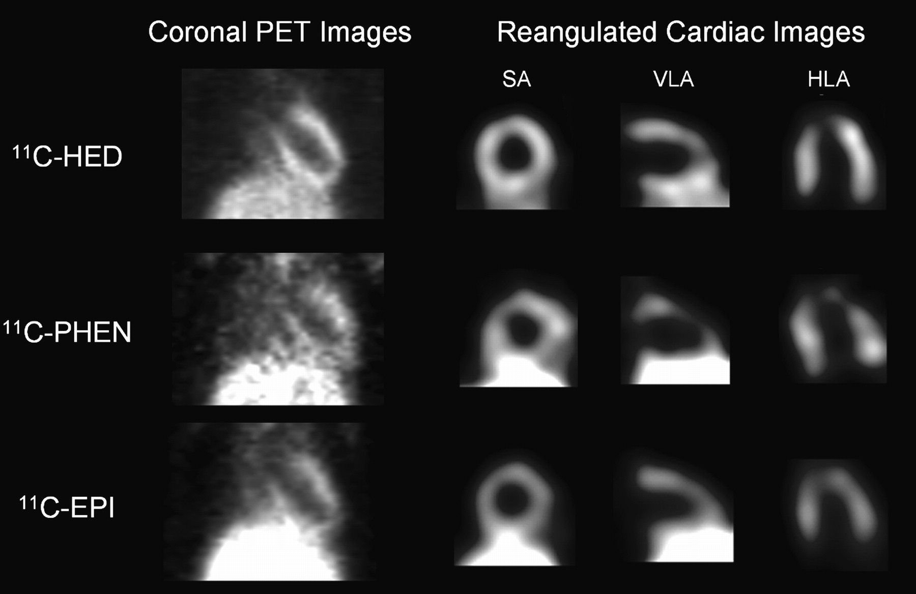
Representative static coronal PET images and reangulated, smoothed cardiac images of rats, acquired between 50 and 60 min after intravenous injection of HED, PHEN, or EPI. Shown on right are midventricular short-axis (SA), vertical long-axis (VLA), and horizontal long-axis slices (HLA). Liver is visualized inferior to left ventricle.
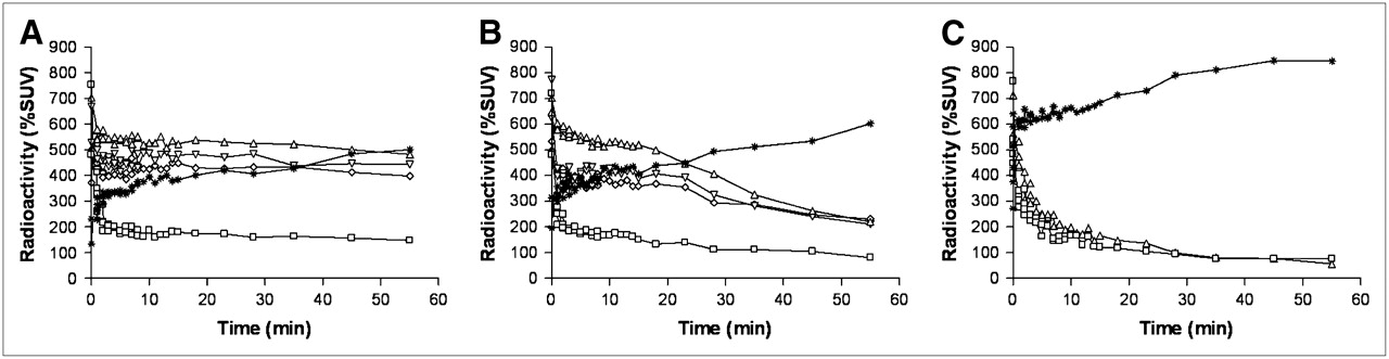
- FIGURE 3.
Myocardial time–activity curves for LV chamber (□), LV lateral wall (▵), LV septum (▿), LV apex (⋄), and liver (*) in rats after intravenous injection of HED, without pharmacologic intervention (A), with intraperitoneal administration of 2 mg of DMI solution per kilogram 15 min after tracer injection (DMI chase) (B), and after intraperitoneal administration of 2 mg of DMI solution per kilogram 15 min before tracer injection (DMI block) (C). A and B show data averaged from 4 rats, and C shows observation from 1 rat (LV septum and LV apex were included in FOV but not detectable in C; kinetics in anterior wall were also analyzed but did not differ from other myocardial regions and are therefore not shown).
- FIGURE 4.
Myocardial time–activity curves of radioactivity in LV chamber (□), LV lateral wall (▵), LV septum (▿), LV apex (⋄), and liver (*) in rats after intravenous injection of PHEN, without pharmacologic intervention (A), with intraperitoneal administration of 2 mg of DMI solution per kilogram 15 min after tracer injection (DMI chase) (B), and after intraperitoneal administration of 2 mg of DMI solution per kilogram 15 min before tracer injection (DMI block) (C). A and B show data averaged from 4 rats, and C shows observation from 1 rat (LV septum and LV apex were included in FOV but not detectable in C; kinetics in anterior wall were also analyzed but did not differ from other myocardial regions and are therefore not shown).
- FIGURE 5.
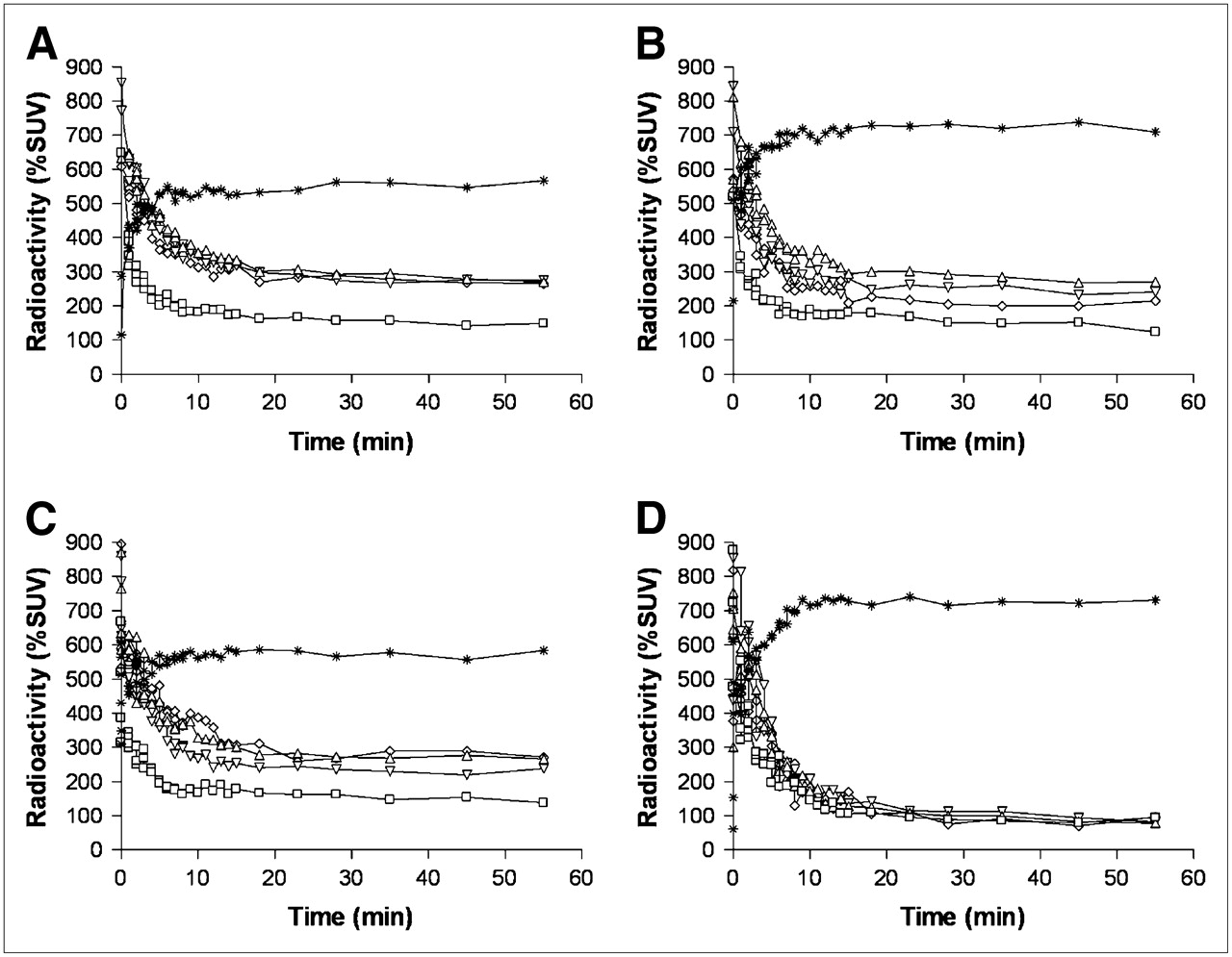
Myocardial time–activity curves of radioactivity in LV chamber (□), LV lateral wall (▵), LV septum (▿), LV apex (⋄), and liver (*) in rats after intravenous injection of EPI, without pharmacologic intervention (A), with lower specific radioactivity without pharmacologic intervention (B), after intraperitoneal administration of 2 mg of DMI solution per kilogram 15 min after tracer injection (DMI chase) (C), and after intraperitoneal administration of 2 mg of DMI solution pre kilogram 15 min before tracer injection (DMI block) (D). A, B, and C show data averaged from 4 rats, and D shows observation from 1 rat (LV septum and LV apex were included in FOV but not detectable in D; kinetics in anterior wall were also analyzed but did not differ from other myocardial regions and are therefore not shown).


Tables
- TABLE 1
Non–Metabolite-Corrected Myocardial Retention Index and Washout Rate of Radiotracers With and Without Treatment With DMI
Retention index (%/min at 40 min) Washout rate (%/min between 10 and 60 min) Radiotracer Without intervention (n = 4) DMI chase (n = 4) DMI pretreatment (n = 1) Without intervention (n = 4) DMI chase (n = 4) DMI pretreatment (n = 1) HED 7.38 ± 0.82 3.28 ± 0.60* 0.21 ± 0.35† 0.13 ± 0.23 1.86 ± 0.57* ‡ PHEN 3.43 ± 0.45 2.95 ± 0.51* 0.24 ± 0.25† 1.13 ± 0.35 1.51 ± 0.16 ‡ EPI 4.24 ± 0.59 4.02 ± 0.99 0.22 ± 0.30† 0.50 ± 0.24 0.61 ± 0.28 ‡ EPI (low specific radioactivity) 3.87 ± 0.81 — — 0.69 ± 0.17 — — ↵* P < 0.05 vs. animals without intervention.
↵† Single-case observation precludes statistical testing, but values are markedly reduced.
↵‡ Complete washout of radiotracer from myocardium in first 10 min in DMI pretreated rats.
Values are average ± SD from septal, lateral, inferior, anterior, and apex regions of left ventricle.


Jump to section
Related Articles
Cited By...
- Cardiac Presynaptic Sympathetic Nervous Function Evaluated by Cardiac PET in Patients with Chronotropic Incompetence Without Heart Failure
- Nuclear Imaging of the Cardiac Sympathetic Nervous System: A Disease-Specific Interpretation in Heart Failure
- Impaired Myocardial Sympathetic Innervation Is Associated with Diastolic Dysfunction in Heart Failure with Preserved Ejection Fraction: 11C-Hydroxyephedrine PET Study
- Retention Kinetics of the 18F-Labeled Sympathetic Nerve PET Tracer LMI1195: Comparison with 11C-Hydroxyephedrine and 123I-MIBG
- Multiparametric Molecular Imaging Provides Mechanistic Insights into Sympathetic Innervation Impairment in the Viable Infarct Border Zone
- Imaging Targets of the Sympathetic Nervous System of the Heart: Translational Considerations
- Molecular Imaging of Cardiac Sympathetic Innervation by 11C-mHED and PET: From Man to Mouse?