
Main menu

  • Home
  • Content
    • Current
    • Ahead of print
    • Past Issues
    • JNM Supplement
    • SNMMI Annual Meeting Abstracts
    • Continuing Education
    • JNM Podcasts
  • Subscriptions
    • Subscribers
    • Institutional and Non-member
    • Rates
    • Journal Claims
    • Corporate & Special Sales
  • Authors
    • Submit to JNM
    • Information for Authors
    • Assignment of Copyright
    • AQARA requirements
  • Info
    • Reviewers
    • Permissions
    • Advertisers
  • About
    • About Us
    • Editorial Board
    • Contact Information
  • More
    • Alerts
    • Feedback
    • Help
    • SNMMI Journals
  • SNMMI
    • JNM
    • JNMT
    • SNMMI Journals
    • SNMMI

User menu

  • Subscribe
  • My alerts
  • Log in
  • My Cart

Search

  • Advanced search
Journal of Nuclear Medicine
  • SNMMI
    • JNM
    • JNMT
    • SNMMI Journals
    • SNMMI
  • Subscribe
  • My alerts
  • Log in
  • My Cart
Journal of Nuclear Medicine

Advanced Search

  • Home
  • Content
    • Current
    • Ahead of print
    • Past Issues
    • JNM Supplement
    • SNMMI Annual Meeting Abstracts
    • Continuing Education
    • JNM Podcasts
  • Subscriptions
    • Subscribers
    • Institutional and Non-member
    • Rates
    • Journal Claims
    • Corporate & Special Sales
  • Authors
    • Submit to JNM
    • Information for Authors
    • Assignment of Copyright
    • AQARA requirements
  • Info
    • Reviewers
    • Permissions
    • Advertisers
  • About
    • About Us
    • Editorial Board
    • Contact Information
  • More
    • Alerts
    • Feedback
    • Help
    • SNMMI Journals
  • View or Listen to JNM Podcast
  • Visit JNM on Facebook
  • Join JNM on LinkedIn
  • Follow JNM on Twitter
  • Subscribe to our RSS feeds
Research ArticleCLINICAL INVESTIGATIONS

Assessment of Large-Vessel Involvement in Giant Cell Arteritis with 18F-FDG PET: Introducing an ROC-Analysis–Based Cutoff Ratio

Hubertus Hautzel, Oliver Sander, Alexander Heinzel, Matthias Schneider and Hans-Wilhelm Müller
Journal of Nuclear Medicine July 2008, 49 (7) 1107-1113; DOI: https://doi.org/10.2967/jnumed.108.051920
Hubertus Hautzel
  • Find this author on Google Scholar
  • Find this author on PubMed
  • Search for this author on this site
Oliver Sander
  • Find this author on Google Scholar
  • Find this author on PubMed
  • Search for this author on this site
Alexander Heinzel
  • Find this author on Google Scholar
  • Find this author on PubMed
  • Search for this author on this site
Matthias Schneider
  • Find this author on Google Scholar
  • Find this author on PubMed
  • Search for this author on this site
Hans-Wilhelm Müller
  • Find this author on Google Scholar
  • Find this author on PubMed
  • Search for this author on this site
  • Article
  • Figures & Data
  • Info & Metrics
  • PDF
Loading

Article Figures & Data

Figures

  • Tables
  • FIGURE 1. 
    • Download figure
    • Open in new tab
    • Download powerpoint
    FIGURE 1. 

    18F-FDG PET images and SUVmax ratios of selected patients: patient from control group I with no history of atherosclerosis (A), patient from control group I with atherosclerosis of aorta and iliac arteries (confirmed by CT) (B), and GCA patients 3 (C) and 13 (D), both of whom had histologically proven GCA. On axial views, ROIs (black lines) were drawn over aortic arch (A–C) and ascending and descending parts of aorta (D). On coronal and sagittal views, arrows indicate 18F-FDG uptake into aortic vessel wall. Coronal view of B additionally shows uptake into iliac vessel wall.
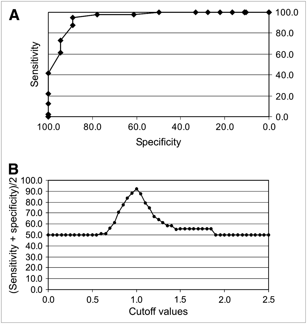
  • FIGURE 2. 
    • Download figure
    • Open in new tab
    • Download powerpoint
    FIGURE 2. 

    (A) ROC analysis comparing GCA group with control group I: at cutoff of 1.0, sensitivity was 88.9% and specificity was 95.1%. (B) Cutoff optimization according to CLSI/NCCLS guidelines.

Tables

  • Figures
    • View popup
    TABLE 1

    GCA Group: Detailed Clinical Patient Data

    Patient no.Age (y)SexVessel-to- liver ratioPositive for ACR criteriaHistologyDuplex sonographyAnemiaLiver enzymes (GGT, AST, or ALT)Final diagnosis
    GCA true-positive patients
     174F0.845Pos.Pos. (scl.)YesElevated
     261M0.934NDPos. (scl.)YesElevated
     361F1.004Pos.Pos. (ax.; br.)NoNormal
     457F1.034NDPos. (scl.; ax.; br.)YesNormal
     574F1.054NDPos. (scl.; ax.; br.)NDElevated
     664F1.093NDNDYesElevated
     767F1.093NDPos. (car.; scl.; ax.)YesNormal
     864F1.124Pos.Pos. (scl.; ax.)YesElevated
     962F1.145Pos.Pos. (scl.; ax.)NoNormal
     1062F1.154NDPos. (temp.)NoNormal
     1174F1.185Pos.Pos. (temp.)NoNormal
     1264F1.195Pos.Neg.NoElevated
     1356F1.244Pos.Pos. (car.)YesNormal
     1467F1.263NDNeg.YesNormal
     1565F1.335Pos.Pos. (car.; scl.; ax.)YesElevated
     1665M1.433NDNDYesNormal
     1763M1.873NDPos. (ax.; br.)YesNormal
     1857F1.883NDPos. (scl.)NDNormal
    GCA true-negative patients
     1960F0.793NDNeg.YesElevatedRheumatoid arthritis
     2067M0.833NDNeg.YesNormalParaneoplastic syndrome
     2176F0.843NDNeg.YesElevatedNo diagnosis
     2259F0.853NDNeg.NoNormalNo diagnosis
     2359F0.883NDNeg.NoNormalPolymyalgia rheumatica
    • pos. = positive; neg. = negative; ND = not done; scl. = arteria subclavia; ax. = arteria axillaris; br. = arteria brachialis. car. = arteria carotis; temp. = arteria temporalis.

    • All patients had elevated erythrocyte sedimentation rate/C-reactive protein.

PreviousNext
Back to top

In this issue

Journal of Nuclear Medicine: 49 (7)
Journal of Nuclear Medicine
Vol. 49, Issue 7
July 2008
  • Table of Contents
  • Table of Contents (PDF)
  • About the Cover
  • Index by author
Print
Download PDF
Article Alerts
Sign In to Email Alerts with your Email Address
Email Article

Thank you for your interest in spreading the word on Journal of Nuclear Medicine.

NOTE: We only request your email address so that the person you are recommending the page to knows that you wanted them to see it, and that it is not junk mail. We do not capture any email address.

Enter multiple addresses on separate lines or separate them with commas.
Assessment of Large-Vessel Involvement in Giant Cell Arteritis with 18F-FDG PET: Introducing an ROC-Analysis–Based Cutoff Ratio
(Your Name) has sent you a message from Journal of Nuclear Medicine
(Your Name) thought you would like to see the Journal of Nuclear Medicine web site.
Citation Tools
Assessment of Large-Vessel Involvement in Giant Cell Arteritis with 18F-FDG PET: Introducing an ROC-Analysis–Based Cutoff Ratio
Hubertus Hautzel, Oliver Sander, Alexander Heinzel, Matthias Schneider, Hans-Wilhelm Müller
Journal of Nuclear Medicine Jul 2008, 49 (7) 1107-1113; DOI: 10.2967/jnumed.108.051920

Citation Manager Formats

  • BibTeX
  • Bookends
  • EasyBib
  • EndNote (tagged)
  • EndNote 8 (xml)
  • Medlars
  • Mendeley
  • Papers
  • RefWorks Tagged
  • Ref Manager
  • RIS
  • Zotero
Share
Assessment of Large-Vessel Involvement in Giant Cell Arteritis with 18F-FDG PET: Introducing an ROC-Analysis–Based Cutoff Ratio
Hubertus Hautzel, Oliver Sander, Alexander Heinzel, Matthias Schneider, Hans-Wilhelm Müller
Journal of Nuclear Medicine Jul 2008, 49 (7) 1107-1113; DOI: 10.2967/jnumed.108.051920
Twitter logo Facebook logo LinkedIn logo Mendeley logo
  • Tweet Widget
  • Facebook Like
  • Google Plus One
Bookmark this article

Jump to section

  • Article
    • Abstract
    • MATERIALS AND METHODS
    • RESULTS
    • DISCUSSION
    • CONCLUSION
    • Footnotes
    • References
  • Figures & Data
  • Info & Metrics
  • PDF

Related Articles

  • This Month in JNM
  • PubMed
  • Google Scholar

Cited By...

  • Comparing Semiquantitative and Qualitative Methods of Vascular 18F-FDG PET Activity Measurement in Large-Vessel Vasculitis
  • Imaging in diagnosis, outcome prediction and monitoring of large vessel vasculitis: a systematic literature review and meta-analysis informing the EULAR recommendations
  • Positron Emission Tomography/Computerized Tomography in Newly Diagnosed Patients with Giant Cell Arteritis Who Are Taking Glucocorticoids
  • Positron emission tomography assessment of large vessel inflammation in patients with newly diagnosed, biopsy-proven giant cell arteritis: a prospective, case-control study
  • Imaging of Inflammation by PET, Conventional Scintigraphy, and Other Imaging Techniques
  • Letter by Lensen et al Regarding Article, "Anti-Tumor Necrosis Factor-{alpha} Therapy Reduces Aortic Inflammation and Stiffness in Patients With Rheumatoid Arthritis"
  • Increased Metabolic Activity Highlighted by Positron Emission Tomography/Computed Tomography in the Wall of the Dissected Ascending Aorta in a Patient With Horton Disease
  • EANM/SNMMI Guideline for 18F-FDG Use in Inflammation and Infection
  • 18F-Fludeoxyglucose PET/CT in the evaluation of large-vessel vasculitis: diagnostic performance and correlation with clinical and laboratory parameters
  • Imaging of Inflammation by PET, Conventional Scintigraphy, and Other Imaging Techniques
  • Imaging Atherosclerotic Plaque Inflammation by Fluorodeoxyglucose With Positron Emission Tomography: Ready for Prime Time?
  • Google Scholar

More in this TOC Section

  • Feasibility of Ultra-Low-Activity 18F-FDG PET/CT Imaging Using a Long–Axial-Field-of-View PET/CT System
  • Cardiac Presynaptic Sympathetic Nervous Function Evaluated by Cardiac PET in Patients with Chronotropic Incompetence Without Heart Failure
  • Validation and Evaluation of a Vendor-Provided Head Motion Correction Algorithm on the uMI Panorama PET/CT System
Show more Clinical Investigations

Similar Articles

SNMMI

© 2025 SNMMI

Powered by HighWire