
Main menu

  • Home
  • Content
    • Current
    • Ahead of print
    • Past Issues
    • JNM Supplement
    • SNMMI Annual Meeting Abstracts
    • Continuing Education
    • JNM Podcasts
  • Subscriptions
    • Subscribers
    • Institutional and Non-member
    • Rates
    • Journal Claims
    • Corporate & Special Sales
  • Authors
    • Submit to JNM
    • Information for Authors
    • Assignment of Copyright
    • AQARA requirements
  • Info
    • Reviewers
    • Permissions
    • Advertisers
  • About
    • About Us
    • Editorial Board
    • Contact Information
  • More
    • Alerts
    • Feedback
    • Help
    • SNMMI Journals
  • SNMMI
    • JNM
    • JNMT
    • SNMMI Journals
    • SNMMI

User menu

  • Subscribe
  • My alerts
  • Log in
  • Log out
  • My Cart

Search

  • Advanced search
Journal of Nuclear Medicine
  • SNMMI
    • JNM
    • JNMT
    • SNMMI Journals
    • SNMMI
  • Subscribe
  • My alerts
  • Log in
  • Log out
  • My Cart
Journal of Nuclear Medicine

Advanced Search

  • Home
  • Content
    • Current
    • Ahead of print
    • Past Issues
    • JNM Supplement
    • SNMMI Annual Meeting Abstracts
    • Continuing Education
    • JNM Podcasts
  • Subscriptions
    • Subscribers
    • Institutional and Non-member
    • Rates
    • Journal Claims
    • Corporate & Special Sales
  • Authors
    • Submit to JNM
    • Information for Authors
    • Assignment of Copyright
    • AQARA requirements
  • Info
    • Reviewers
    • Permissions
    • Advertisers
  • About
    • About Us
    • Editorial Board
    • Contact Information
  • More
    • Alerts
    • Feedback
    • Help
    • SNMMI Journals
  • View or Listen to JNM Podcast
  • Visit JNM on Facebook
  • Join JNM on LinkedIn
  • Follow JNM on Twitter
  • Subscribe to our RSS feeds
OtherBASIC SCIENCE INVESTIGATIONS

Impact of Image-Space Resolution Modeling for Studies with the High-Resolution Research Tomograph

Florent C. Sureau, Andrew J. Reader, Claude Comtat, Claire Leroy, Maria-Joao Ribeiro, Irène Buvat and Régine Trébossen
Journal of Nuclear Medicine June 2008, 49 (6) 1000-1008; DOI: https://doi.org/10.2967/jnumed.107.045351
Florent C. Sureau
  • Find this author on Google Scholar
  • Find this author on PubMed
  • Search for this author on this site
Andrew J. Reader
  • Find this author on Google Scholar
  • Find this author on PubMed
  • Search for this author on this site
Claude Comtat
  • Find this author on Google Scholar
  • Find this author on PubMed
  • Search for this author on this site
Claire Leroy
  • Find this author on Google Scholar
  • Find this author on PubMed
  • Search for this author on this site
Maria-Joao Ribeiro
  • Find this author on Google Scholar
  • Find this author on PubMed
  • Search for this author on this site
Irène Buvat
  • Find this author on Google Scholar
  • Find this author on PubMed
  • Search for this author on this site
Régine Trébossen
  • Find this author on Google Scholar
  • Find this author on PubMed
  • Search for this author on this site
  • Article
  • Figures & Data
  • Info & Metrics
  • PDF
Loading

Article Figures & Data

Figures

  • Tables
  • FIGURE 1. 
    • Download figure
    • Open in new tab
    • Download powerpoint
    FIGURE 1. 

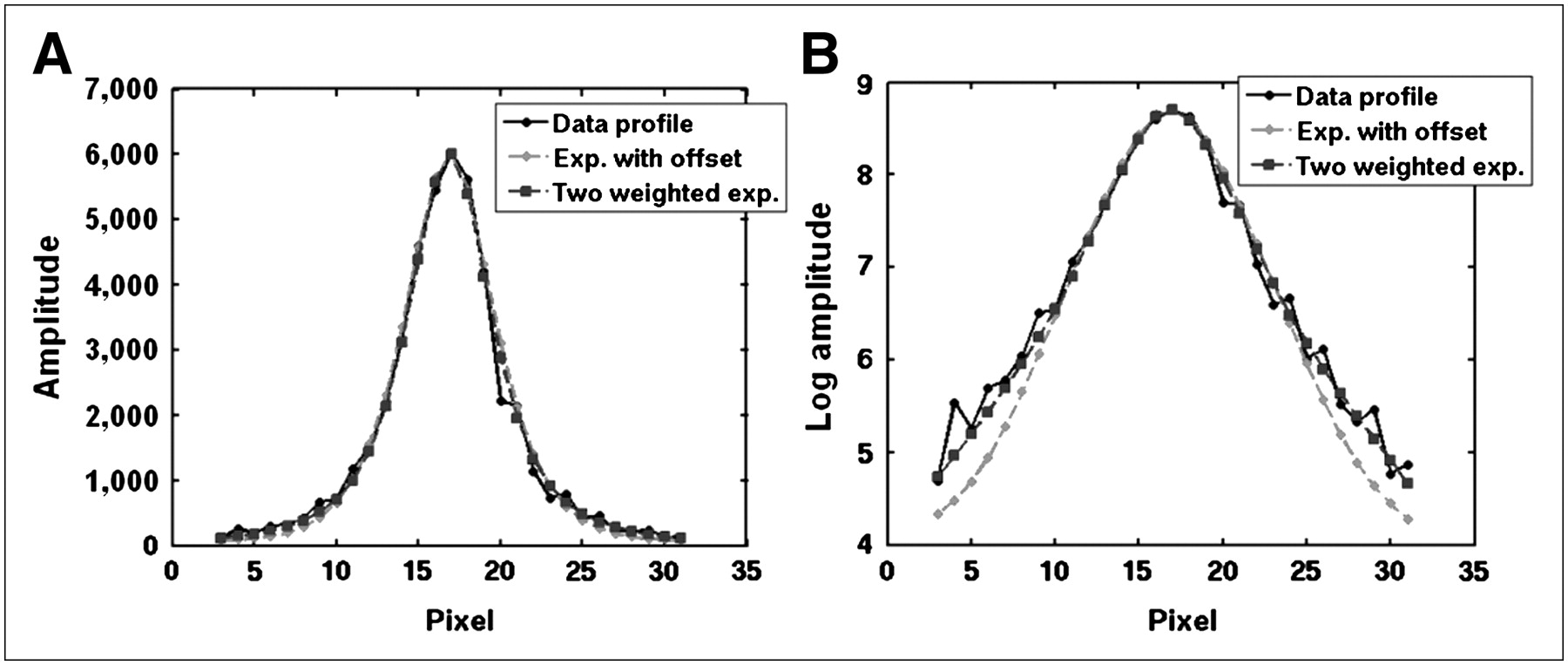
    Fitted profiles after convolution with size of point source (zoom, ×2.5; pixel size, ∼0.5 mm). Kernels are displayed in linear (A) and logarithmic (B) scales.

  • FIGURE 2. 
    • Download figure
    • Open in new tab
    • Download powerpoint
    FIGURE 2. 

    Qualitative impact of RM at 2 axial locations at center (A) and at 4 cm off center of FOV axially (B). Images without RM were reconstructed using 12 iterations of 16 subsets, whereas images with RM were reconstructed using 40 iterations of 16 subsets.
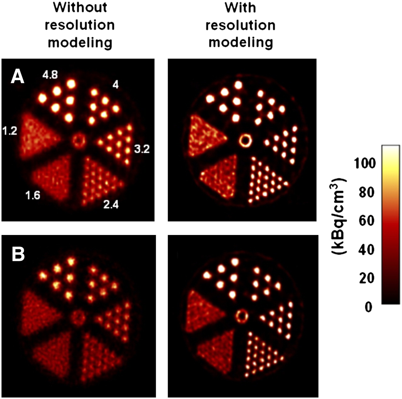
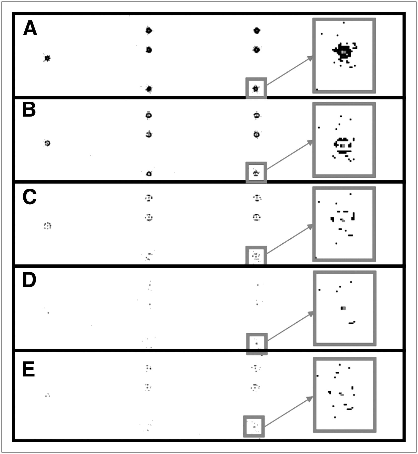
  • FIGURE 3. 
    • Download figure
    • Open in new tab
    • Download powerpoint
    FIGURE 3. 

    Axial, sagittal, and coronal planes of 3 point sources reconstructed with different resolution kernels (using 15 iterations of 16 subsets), with zoom on 1 point source in last column. Panels A, B, C, and D are reconstructed with 3-parameter kernel of size 33, 53, 93, and 153 voxels and E with 2-parameter kernel of size 153 voxels. Note that color scale was stretched to assist visualization of artifacts.
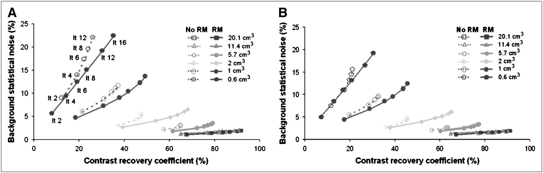
  • FIGURE 4. 
    • Download figure
    • Open in new tab
    • Download powerpoint
    FIGURE 4. 

    Impact of RM on CRC and noise properties, without (A) and with (B) postreconstruction smoothing. Each color corresponds to different sphere. RM-OP-OSEM curves are represented by filled symbols and solid lines and OP-OSEM curve by hollow symbols and dashed lines. Iteration numbers are shown for smallest sphere in A.
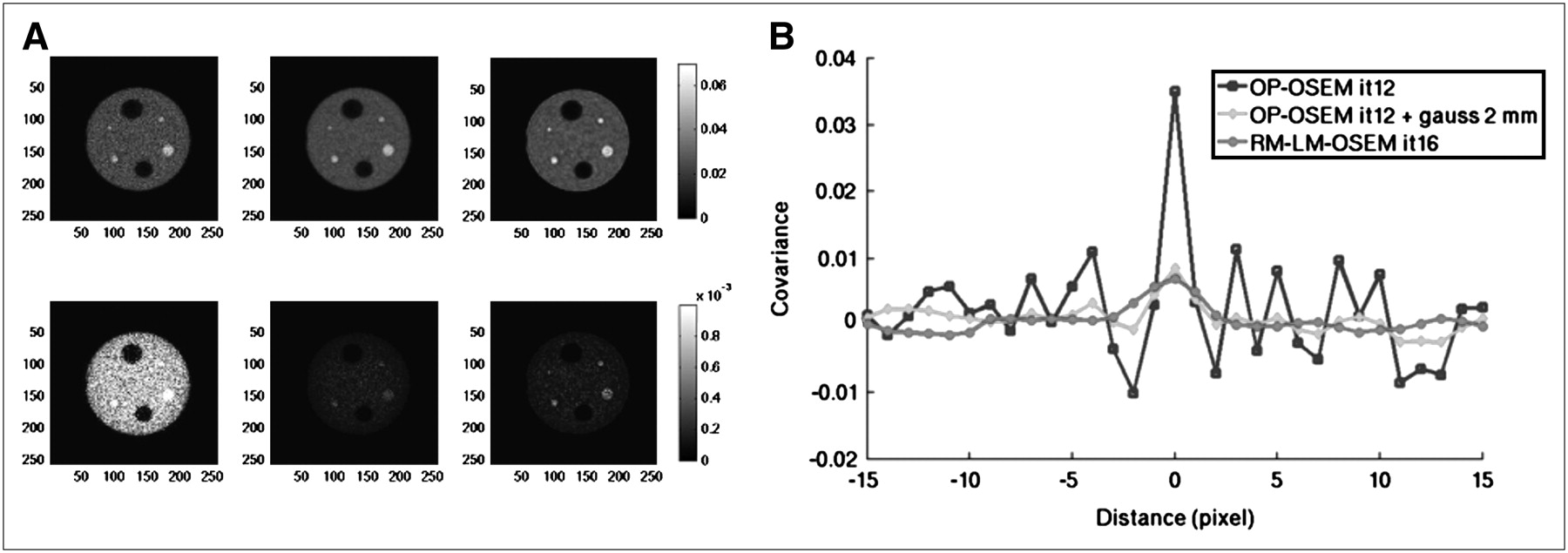
  • FIGURE 5. 
    • Download figure
    • Open in new tab
    • Download powerpoint
    FIGURE 5. 

    Impact of RM on noise properties. (A) Mean (first row) and variance images (second row) over all 36 realizations, for OP-OSEM (first column, 12 iterations of 16 subsets), OP-OSEM (12 iterations of 16 subsets) with 2-mm FWHM gaussian postreconstruction smoothing (second column), and RM-OP-OSEM (last column, 16 iterations of 16 subsets). (B) Covariance in summed planes on homogeneous background region.
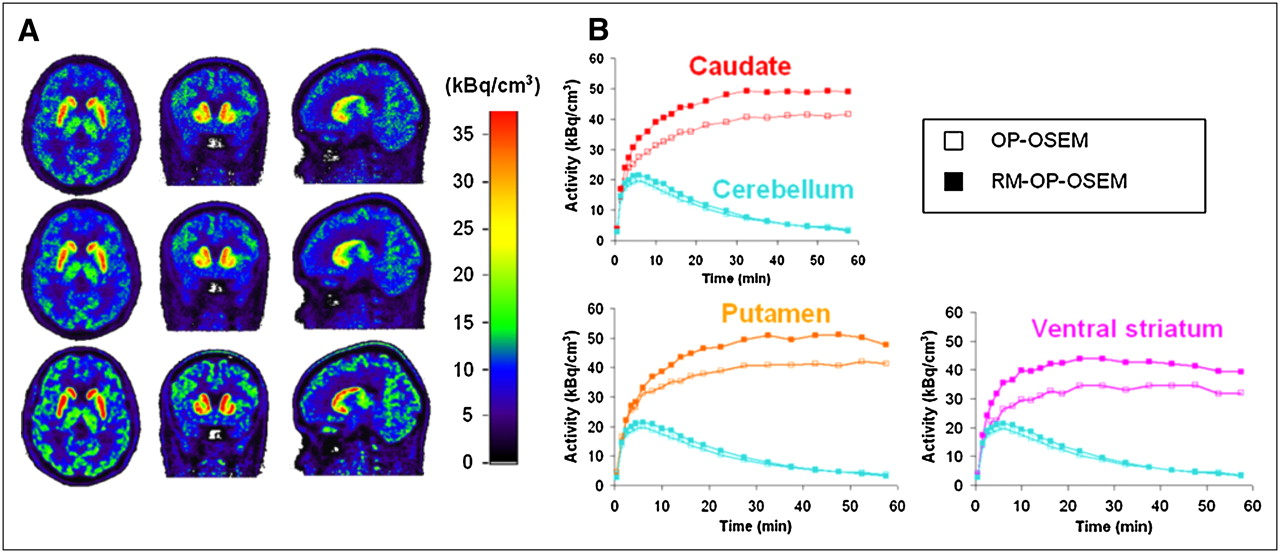
  • FIGURE 6. 
    • Download figure
    • Open in new tab
    • Download powerpoint
    FIGURE 6. 

    Time-averaged images (A) and corresponding VOI time–activity curves (B) for subject. In A, images across striatum were reconstructed with OP-OSEM (first row, 12 iterations of 16 subsets), OP-OSEM (12 iterations of 16 subsets) followed by a postreconstruction smoothing with 2-mm FWHM gaussian kernel (second row), and RM-OP-OSEM (last row, 15 iterations of 16 subsets).

Tables

  • Figures
    • View popup
    TABLE 1

    Periphery-to-Total Activity Ratio (Eq. 10) of Point Source

    Kernel size (offset axially)2 weighted exponentialsSingle exponential
    3 × 3 × 35 × 5 × 59 × 9 × 915 × 15 × 1515 × 15 × 15
    1 cm0.790.290.080.010.04
    5 cm0.780.280.080.010.04
    10 cm0.910.320.090.020.04
    • View popup
    TABLE 2

    BP Values in Anatomic Structures for All 5 Subjects

    ParameterCaudatePutamenVentral striatum
    Without RM9.11 (0.6)10.25 (1.3)7.1 (0.8)
    With RM11.34 (0.7)12.60 (1.7)7.8 (1.1)
    • Corresponding SDs are in parentheses.

PreviousNext
Back to top

In this issue

Journal of Nuclear Medicine: 49 (6)
Journal of Nuclear Medicine
Vol. 49, Issue 6
June 2008
  • Table of Contents
  • Table of Contents (PDF)
  • About the Cover
  • Index by author
Print
Download PDF
Article Alerts
Sign In to Email Alerts with your Email Address
Email Article

Thank you for your interest in spreading the word on Journal of Nuclear Medicine.

NOTE: We only request your email address so that the person you are recommending the page to knows that you wanted them to see it, and that it is not junk mail. We do not capture any email address.

Enter multiple addresses on separate lines or separate them with commas.
Impact of Image-Space Resolution Modeling for Studies with the High-Resolution Research Tomograph
(Your Name) has sent you a message from Journal of Nuclear Medicine
(Your Name) thought you would like to see the Journal of Nuclear Medicine web site.
Citation Tools
Impact of Image-Space Resolution Modeling for Studies with the High-Resolution Research Tomograph
Florent C. Sureau, Andrew J. Reader, Claude Comtat, Claire Leroy, Maria-Joao Ribeiro, Irène Buvat, Régine Trébossen
Journal of Nuclear Medicine Jun 2008, 49 (6) 1000-1008; DOI: 10.2967/jnumed.107.045351

Citation Manager Formats

  • BibTeX
  • Bookends
  • EasyBib
  • EndNote (tagged)
  • EndNote 8 (xml)
  • Medlars
  • Mendeley
  • Papers
  • RefWorks Tagged
  • Ref Manager
  • RIS
  • Zotero
Share
Impact of Image-Space Resolution Modeling for Studies with the High-Resolution Research Tomograph
Florent C. Sureau, Andrew J. Reader, Claude Comtat, Claire Leroy, Maria-Joao Ribeiro, Irène Buvat, Régine Trébossen
Journal of Nuclear Medicine Jun 2008, 49 (6) 1000-1008; DOI: 10.2967/jnumed.107.045351
Twitter logo Facebook logo LinkedIn logo Mendeley logo
  • Tweet Widget
  • Facebook Like
  • Google Plus One
Bookmark this article

Jump to section

  • Article
    • Abstract
    • MATERIALS AND METHODS
    • RESULTS
    • DISCUSSION
    • CONCLUSION
    • Acknowledgments
    • Footnotes
    • References
  • Figures & Data
  • Info & Metrics
  • PDF

Related Articles

  • This Month in JNM
  • PubMed
  • Google Scholar

Cited By...

  • Pitfalls of mapping functional and molecular human brain imaging data from separate cohorts
  • An In Vivo High-Resolution Human Brain Atlas of Synaptic Density
  • Kinetic models for PET displacement studies
  • Tau-PET imaging predicts cognitive decline and brain atrophy progression in early Alzheimers disease
  • Evaluation of the {alpha}-synuclein PET radiotracer (d3)-[11C]MODAG-001 in pigs
  • An in vivo pig model for testing novel PET radioligands targeting cerebral protein aggregates
  • Improved Detection of Postoperative Residual Meningioma with [68Ga]Ga-DOTA-TOC PET Imaging Using a High-resolution Research Tomograph PET Scanner
  • A High-Resolution In Vivo Atlas of the Human Brains Benzodiazepine Binding Site of GABAA Receptors
  • A High-Resolution In Vivo Atlas of the Human Brain's Serotonin System
  • Variability and Uncertainty of 18F-FDG PET Imaging Protocols for Assessing Inflammation in Atherosclerosis: Suggestions for Improvement
  • Methods for Motion Correction Evaluation Using 18F-FDG Human Brain Scans on a High-Resolution PET Scanner
  • 11C-NS14492 as a Novel PET Radioligand for Imaging Cerebral {alpha}7 Nicotinic Acetylcholine Receptors: In Vivo Evaluation and Drug Occupancy Measurements
  • Google Scholar

More in this TOC Section

  • Design and Fabrication of Kidney Phantoms for Internal Radiation Dosimetry Using 3D Printing Technology
  • Synthesis and Biologic Evaluation of Novel 18F-Labeled Probes Targeting Prostate-Specific Membrane Antigen for PET of Prostate Cancer
  • Tumor-Specific Binding of Radiolabeled PEGylated GIRLRG Peptide: A Novel Agent for Targeting Cancers
Show more Basic Science Investigations

Similar Articles

SNMMI

© 2025 SNMMI

Powered by HighWire