
Main menu

  • Home
  • Content
    • Current
    • Ahead of print
    • Past Issues
    • JNM Supplement
    • SNMMI Annual Meeting Abstracts
    • Continuing Education
    • JNM Podcasts
  • Subscriptions
    • Subscribers
    • Institutional and Non-member
    • Rates
    • Journal Claims
    • Corporate & Special Sales
  • Authors
    • Submit to JNM
    • Information for Authors
    • Assignment of Copyright
    • AQARA requirements
  • Info
    • Reviewers
    • Permissions
    • Advertisers
  • About
    • About Us
    • Editorial Board
    • Contact Information
  • More
    • Alerts
    • Feedback
    • Help
    • SNMMI Journals
  • SNMMI
    • JNM
    • JNMT
    • SNMMI Journals
    • SNMMI

User menu

  • Subscribe
  • My alerts
  • Log in
  • My Cart

Search

  • Advanced search
Journal of Nuclear Medicine
  • SNMMI
    • JNM
    • JNMT
    • SNMMI Journals
    • SNMMI
  • Subscribe
  • My alerts
  • Log in
  • My Cart
Journal of Nuclear Medicine

Advanced Search

  • Home
  • Content
    • Current
    • Ahead of print
    • Past Issues
    • JNM Supplement
    • SNMMI Annual Meeting Abstracts
    • Continuing Education
    • JNM Podcasts
  • Subscriptions
    • Subscribers
    • Institutional and Non-member
    • Rates
    • Journal Claims
    • Corporate & Special Sales
  • Authors
    • Submit to JNM
    • Information for Authors
    • Assignment of Copyright
    • AQARA requirements
  • Info
    • Reviewers
    • Permissions
    • Advertisers
  • About
    • About Us
    • Editorial Board
    • Contact Information
  • More
    • Alerts
    • Feedback
    • Help
    • SNMMI Journals
  • View or Listen to JNM Podcast
  • Visit JNM on Facebook
  • Join JNM on LinkedIn
  • Follow JNM on Twitter
  • Subscribe to our RSS feeds
OtherBASIC SCIENCE INVESTIGATIONS

Molecular Imaging of bcl-2 Expression in Small Lymphocytic Lymphoma Using 111In-Labeled PNA–Peptide Conjugates

Fang Jia, Said Daibes Figueroa, Fabio Gallazzi, Baghavathy S. Balaji, Mark Hannink, Susan Z. Lever, Timothy J. Hoffman and Michael R. Lewis
Journal of Nuclear Medicine March 2008, 49 (3) 430-438; DOI: https://doi.org/10.2967/jnumed.107.045138
Fang Jia
  • Find this author on Google Scholar
  • Find this author on PubMed
  • Search for this author on this site
Said Daibes Figueroa
  • Find this author on Google Scholar
  • Find this author on PubMed
  • Search for this author on this site
Fabio Gallazzi
  • Find this author on Google Scholar
  • Find this author on PubMed
  • Search for this author on this site
Baghavathy S. Balaji
  • Find this author on Google Scholar
  • Find this author on PubMed
  • Search for this author on this site
Mark Hannink
  • Find this author on Google Scholar
  • Find this author on PubMed
  • Search for this author on this site
Susan Z. Lever
  • Find this author on Google Scholar
  • Find this author on PubMed
  • Search for this author on this site
Timothy J. Hoffman
  • Find this author on Google Scholar
  • Find this author on PubMed
  • Search for this author on this site
Michael R. Lewis
  • Find this author on Google Scholar
  • Find this author on PubMed
  • Search for this author on this site
  • Article
  • Figures & Data
  • Info & Metrics
  • PDF
Loading

Article Figures & Data

Figures

  • Tables
  • FIGURE 1. 
    • Download figure
    • Open in new tab
    • Download powerpoint
    FIGURE 1. 

    Structures and synthesis of DOTA–PNA–peptide conjugates.
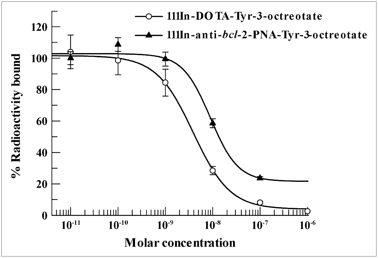
  • FIGURE 2. 
    • Download figure
    • Open in new tab
    • Download powerpoint
    FIGURE 2. 

    Somatostatin receptor binding of 111In-DOTA-anti-bcl-2-PNA-Tyr3-octreotate and 111In-DOTA-Tyr3-octreotate in Mec-1 cells.
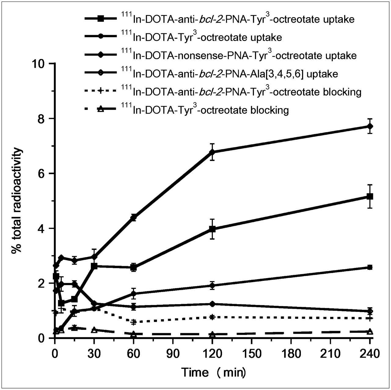
  • FIGURE 3. 
    • Download figure
    • Open in new tab
    • Download powerpoint
    FIGURE 3. 

    Uptake and blocking of 111In-DOTA–PNA–peptide conjugates in Mec-1 cells (n = 3).

  • FIGURE 4. 
    • Download figure
    • Open in new tab
    • Download powerpoint
    FIGURE 4. 

    Internalization of 111In-DOTA–PNA–peptide conjugates in Mec-1 cells (n = 3).
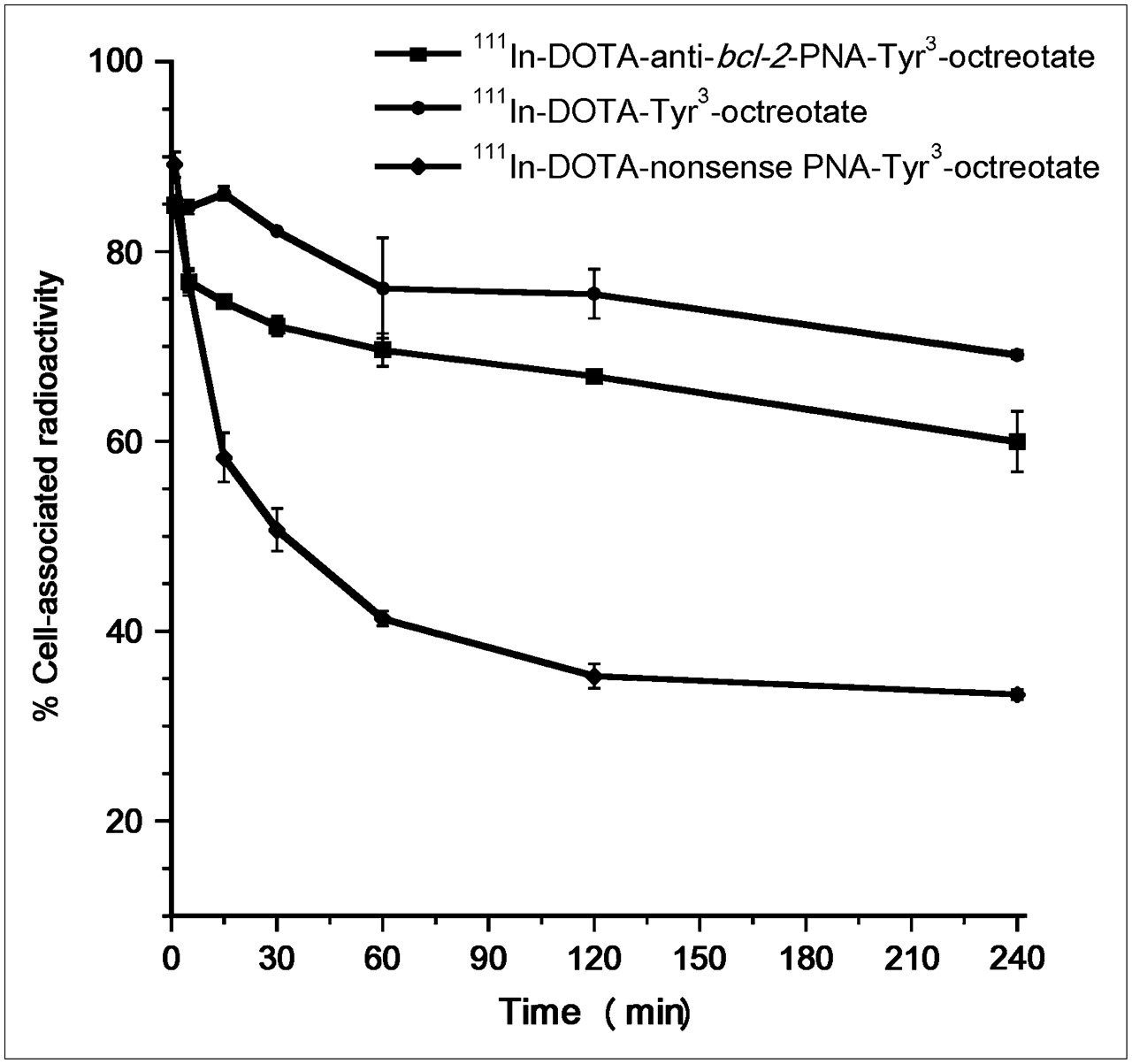
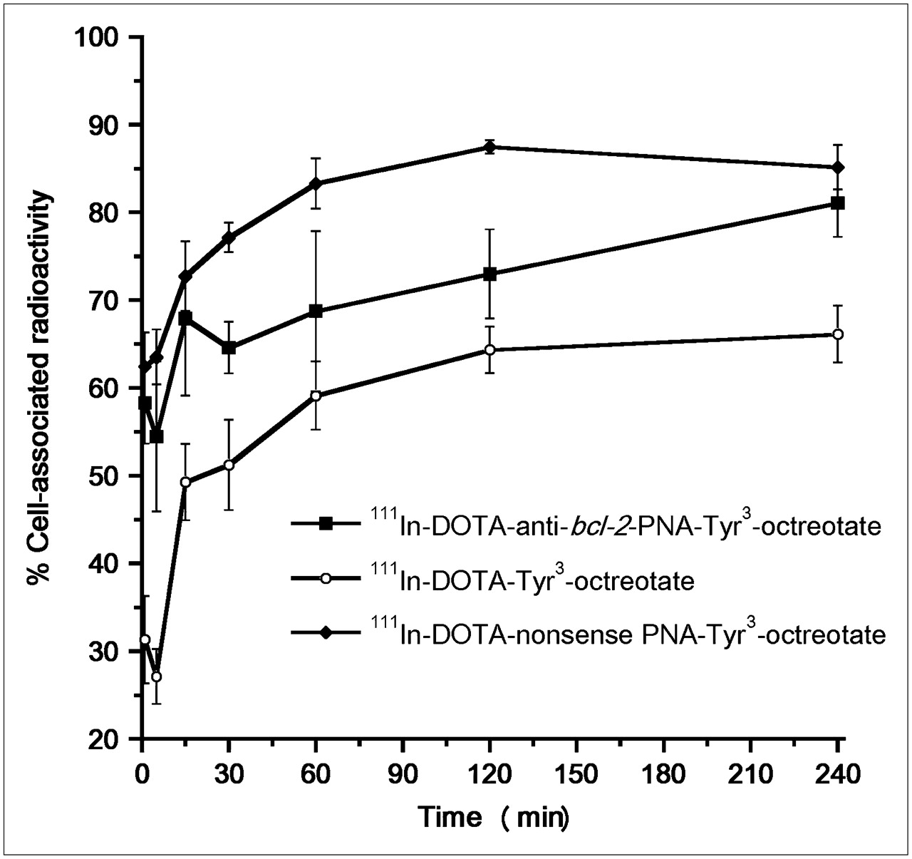
  • FIGURE 5. 
    • Download figure
    • Open in new tab
    • Download powerpoint
    FIGURE 5. 

    Efflux of 111In-DOTA–PNA–peptide conjugates in Mec-1 cells (n = 3).
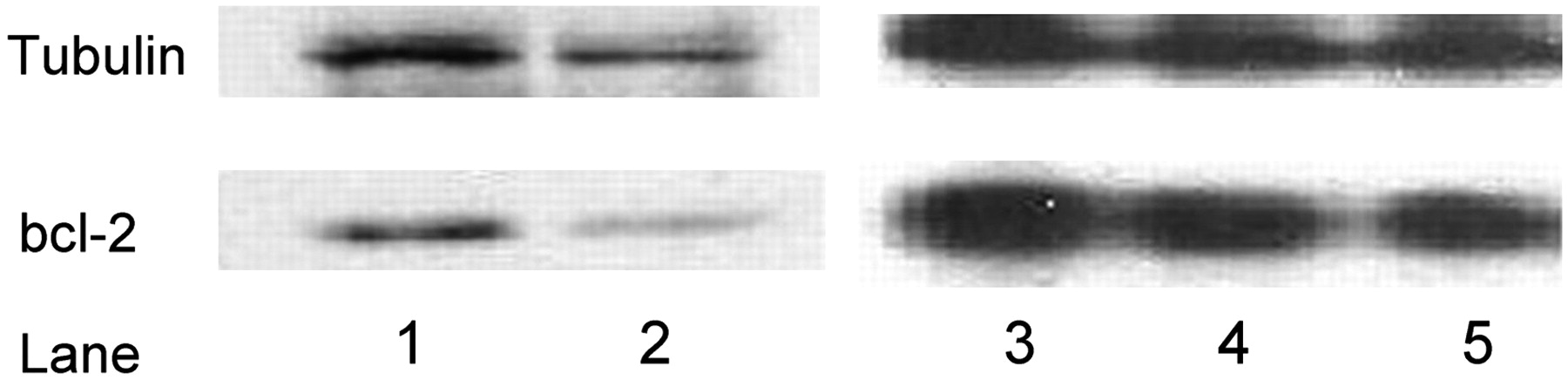
  • FIGURE 6. 
    • Download figure
    • Open in new tab
    • Download powerpoint
    FIGURE 6. 

    Western blot analysis of bcl-2 protein synthesis. Lane 1, untreated control; lane 2, cells treated with compound 1; lane 3, untreated control; lane 4, cells treated with compound 2; lane 5, cells treated with compound 3.
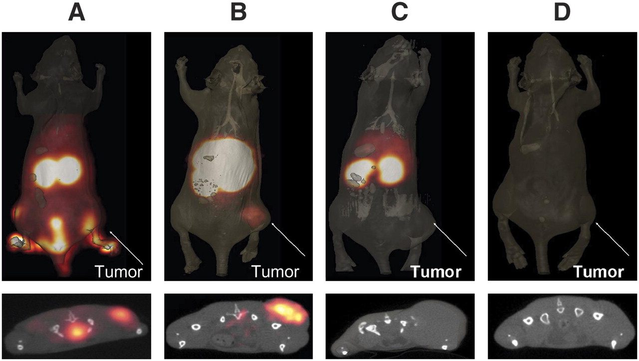
  • FIGURE 7. 
    • Download figure
    • Open in new tab
    • Download powerpoint
    FIGURE 7. 

    (Top) MicroSPECT/CT images of 111In-DOTA–PNA–peptide conjugates in Mec-1–bearing SCID mice (n = 3). (A) 111In-DOTA-Tyr3-octreotate (1 h). (B) 111In-DOTA-anti-bcl-2-PNA-Tyr3-octreotate (48 h). (C) 111In-DOTA-nonsense-PNA-Tyr3-octreotate (48 h). (D) 111In-DOTA-anti-bcl-2-PNA-Ala[3,4,5,6] (48 h). (Bottom) Corresponding transaxial slices through the centers of the tumors.

Tables

  • Figures
    • View popup
    TABLE 1

    Biodistribution of 111In-DOTA-Tyr3-Octreotate in SCID Mice Bearing Mec-1 SLL Xenografts (n = 5)

    Biodistribution (%ID/g ± SD)
    Tissue1 h4 h24 h
    Blood0.31 ± 0.070.06 ± 0.010.02 ± 0.003
    Lung8.27 ± 1.886.87 ± 0.544.12 ± 0.84
    Liver0.32 ± 0.070.29 ± 0.060.20 ± 0.04
    Spleen0.63 ± 0.140.48 ± 0.100.44 ± 0.10
    Kidney11.4 ± 1.7011.0 ± 1.466.39 ± 0.73
    Bladder5.18 ± 1.510.59 ± 0.210.22 ± 0.03
    Muscle0.04 ± 0.010.02 ± 0.0020.01 ± 0.001
    Fat0.08 ± 0.0260.03 ± 0.0080.01 ± 0.003
    Heart0.26 ± 0.030.12 ± 0.020.07 ± 0.02
    Bone0.52 ± 0.100.57 ± 0.100.22 ± 0.05
    Stomach5.80 ± 1.475.14 ± 1.261.87 ± 0.31
    Small intestine1.70 ± 0.310.96 ± 0.0090.40 ± 0.06
    Large intestine2.06 ± 0.335.25 ± 0.841.09 ± 0.25
    Tumor3.24 ± 0.773.24 ± 0.851.49 ± 0.29
    • View popup
    TABLE 2

    Biodistribution of 111In-DOTA-Anti-bcl-2-PNA-Tyr3-Octreotate in SCID Mice Bearing Mec-1 SLL Xenografts (n = 5)

    Biodistribution (%ID/g ± SD)
    Tissue1 h4 h24 h48 h
    Blood2.56 ± 0.212.38 ± 0.460.57 ± 0.120.19 ± 0.02
    Lung6.06 ± 1.202.56 ± 0.311.36 ± 0.291.97 ± 0.04
    Liver12.6 ± 2.036.70 ± 1.676.17 ± 1.366.12 ± 1.50
    Spleen2.75 ± 0.511.34 ± 0.041.36 ± 0.281.05 ± 0.06
    Kidney77.1 ± 16.5129.4 ± 25.770.2 ± 5.7332.8 ± 6.38
    Bladder1.71 ± 0.931.34 ± 0.290.84 ± 0.180.71 ± 0.18
    Muscle0.32 ± 0.020.20 ± 0.040.12 ± 0.020.12 ± 0.02
    Fat0.21 ± 0.040.29 ± 0.060.23 ± 0.050.16 ± 0.03
    Heart0.93 ± 0.120.99 ± 0.130.61 ± 0.120.34 ± 0.04
    Bone0.82 ± 0.130.41 ± 0.050.42 ± 0.080.35 ± 0.09
    Stomach2.12 ± 0.702.33 ± 0.791.02 ± 0.180.51 ± 0.14
    Small intestine1.24 ± 0.190.79 ± 0.210.32 ± 0.040.24 ± 0.04
    Large intestine1.19 ± 0.122.26 ± 0.470.48 ± 0.070.41 ± 0.10
    Tumor1.32 ± 0.081.40 ± 0.321.02 ± 0.180.88 ± 0.22
    • View popup
    TABLE 3

    Biodistribution of 111In-DOTA-Nonsense-PNA-Tyr3- Octreotate in SCID Mice Bearing Mec-1 SLL Xenografts (n = 5)

    Biodistribution (%ID/g ± SD)
    Tissue4 h24 h48 h
    Blood1.27 ± 0.300.25 ± 0.020.09 ± 0.01
    Lung1.23 ± 0.210.77 ± 0.150.51 ± 0.07
    Liver6.34 ± 1.195.74 ± 0.595.61 ± 0.70
    Spleen1.56 ± 0.231.70 ± 0.361.32 ± 0.16
    Kidney100.7 ± 23.376.1 ± 9.8253.3 ± 6.06
    Bladder0.48 ± 0.010.57 ± 0.140.20 ± 0.06
    Muscle0.13 ± 0.030.08 ± 0.010.07 ± 0.01
    Fat0.14 ± 0.020.17 ± 0.040.07 ± 0.01
    Heart0.50 ± 0.080.26 ± 0.010.19 ± 0.01
    Bone0.30 ± 0.030.24 ± 0.050.18 ± 0.05
    Stomach0.17 ± 0.090.28 ± 0.050.20 ± 0.05
    Small intestine0.56 ± 0.130.18 ± 0.030.13 ± 0.02
    Large intestine2.15 ± 0.570.27 ± 0.050.18 ± 0.04
    Tumor0.89 ± 0.210.75 ± 0.130.57 ± 0.08
    • View popup
    TABLE 4

    Biodistribution of 111In-DOTA-Anti-bcl-2-PNA-Ala[3,4,5,6] in SCID Mice Bearing Mec-1 SLL Xenografts (n = 5)

    Biodistribution (%ID/g ± SD)
    Tissue4 h24 h48 h
    Blood0.14 ± 0.010.04 ± 0.0060.02 ± 0.003
    Lung0.13 ± 0.020.08 ± 0.010.06 ± 0.01
    Liver0.24 ± 0.020.27 ± 0.030.23 ± 0.02
    Spleen0.09 ± 0.020.11 ± 0.010.12 ± 0.005
    Kidney7.06 ± 1.836.02 ± 0.882.26 ± 0.52
    Bladder0.06 ± 0.010.17 ± 0.030.10 ± 0.02
    Muscle0.02 ± 0.0020.02 ± 0.0040.01 ± 0.002
    Fat0.03 ± 0.0060.02 ± 0.0050.02 ± 0.005
    Heart0.06 ± 0.010.04 ± 0.010.03 ± 0.005
    Bone0.03 ± 0.0030.02 ± 0.0060.01 ± 0.001
    Stomach0.10 ± 0.0020.03 ± 0.0070.02 ± 0.001
    Small intestine0.06 ± 0.0050.04 ± 0.0040.02 ± 0.003
    Large intestine0.19 ± 0.010.05 ± 0.010.02 ± 0.005
    Tumor0.12 ± 0.010.07 ± 0.020.06 ± 0.01
PreviousNext
Back to top

In this issue

Journal of Nuclear Medicine: 49 (3)
Journal of Nuclear Medicine
Vol. 49, Issue 3
March 2008
  • Table of Contents
  • Table of Contents (PDF)
  • About the Cover
  • Index by author
Print
Download PDF
Article Alerts
Sign In to Email Alerts with your Email Address
Email Article

Thank you for your interest in spreading the word on Journal of Nuclear Medicine.

NOTE: We only request your email address so that the person you are recommending the page to knows that you wanted them to see it, and that it is not junk mail. We do not capture any email address.

Enter multiple addresses on separate lines or separate them with commas.
Molecular Imaging of bcl-2 Expression in Small Lymphocytic Lymphoma Using 111In-Labeled PNA–Peptide Conjugates
(Your Name) has sent you a message from Journal of Nuclear Medicine
(Your Name) thought you would like to see the Journal of Nuclear Medicine web site.
Citation Tools
Molecular Imaging of bcl-2 Expression in Small Lymphocytic Lymphoma Using 111In-Labeled PNA–Peptide Conjugates
Fang Jia, Said Daibes Figueroa, Fabio Gallazzi, Baghavathy S. Balaji, Mark Hannink, Susan Z. Lever, Timothy J. Hoffman, Michael R. Lewis
Journal of Nuclear Medicine Mar 2008, 49 (3) 430-438; DOI: 10.2967/jnumed.107.045138

Citation Manager Formats

  • BibTeX
  • Bookends
  • EasyBib
  • EndNote (tagged)
  • EndNote 8 (xml)
  • Medlars
  • Mendeley
  • Papers
  • RefWorks Tagged
  • Ref Manager
  • RIS
  • Zotero
Share
Molecular Imaging of bcl-2 Expression in Small Lymphocytic Lymphoma Using 111In-Labeled PNA–Peptide Conjugates
Fang Jia, Said Daibes Figueroa, Fabio Gallazzi, Baghavathy S. Balaji, Mark Hannink, Susan Z. Lever, Timothy J. Hoffman, Michael R. Lewis
Journal of Nuclear Medicine Mar 2008, 49 (3) 430-438; DOI: 10.2967/jnumed.107.045138
Twitter logo Facebook logo LinkedIn logo Mendeley logo
  • Tweet Widget
  • Facebook Like
  • Google Plus One
Bookmark this article

Jump to section

  • Article
    • Abstract
    • MATERIALS AND METHODS
    • RESULTS
    • DISCUSSION
    • CONCLUSION
    • Acknowledgments
    • Footnotes
    • References
  • Figures & Data
  • Info & Metrics
  • PDF

Related Articles

  • This Month in JNM
  • PubMed
  • Google Scholar

Cited By...

  • In Vitro Evaluation of Targeted Antisense 177Lu Radiotherapy
  • Google Scholar

More in this TOC Section

  • How Sensitive Is the Upper Gastrointestinal Tract to 90Y Radioembolization? A Histologic and Dosimetric Analysis in a Porcine Model
  • 11C-Methionine PET of Myocardial Inflammation in a Rat Model of Experimental Autoimmune Myocarditis
  • Counting Rate Characteristics and Image Distortion in Preclinical PET Imaging During Radiopharmaceutical Therapy
Show more Basic Science Investigations

Similar Articles

SNMMI

© 2025 SNMMI

Powered by HighWire