Main menu

  • Home
  • Content
    • Current
    • Ahead of print
    • Past Issues
    • JNM Supplement
    • SNMMI Annual Meeting Abstracts
    • Continuing Education
    • JNM Podcasts
  • Subscriptions
    • Subscribers
    • Institutional and Non-member
    • Rates
    • Journal Claims
    • Corporate & Special Sales
  • Authors
    • Submit to JNM
    • Information for Authors
    • Assignment of Copyright
    • AQARA requirements
  • Info
    • Reviewers
    • Permissions
    • Advertisers
  • About
    • About Us
    • Editorial Board
    • Contact Information
  • More
    • Alerts
    • Feedback
    • Help
    • SNMMI Journals
  • SNMMI
    • JNM
    • JNMT
    • SNMMI Journals
    • SNMMI

User menu

  • Subscribe
  • My alerts
  • Log in
  • Log out
  • My Cart

Search

  • Advanced search
Journal of Nuclear Medicine
  • SNMMI
    • JNM
    • JNMT
    • SNMMI Journals
    • SNMMI
  • Subscribe
  • My alerts
  • Log in
  • Log out
  • My Cart
Journal of Nuclear Medicine

Advanced Search

  • Home
  • Content
    • Current
    • Ahead of print
    • Past Issues
    • JNM Supplement
    • SNMMI Annual Meeting Abstracts
    • Continuing Education
    • JNM Podcasts
  • Subscriptions
    • Subscribers
    • Institutional and Non-member
    • Rates
    • Journal Claims
    • Corporate & Special Sales
  • Authors
    • Submit to JNM
    • Information for Authors
    • Assignment of Copyright
    • AQARA requirements
  • Info
    • Reviewers
    • Permissions
    • Advertisers
  • About
    • About Us
    • Editorial Board
    • Contact Information
  • More
    • Alerts
    • Feedback
    • Help
    • SNMMI Journals
  • View or Listen to JNM Podcast
  • Visit JNM on Facebook
  • Join JNM on LinkedIn
  • Follow JNM on Twitter
  • Subscribe to our RSS feeds

About the Cover

March 2008; Volume 49,Issue 3

Cover image

Cover image expansion

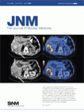
Data suggest that contrast-enhanced PET/CT might be considered the first-line restaging tool for suspected recurrence of colorectal cancer. At top, unenhanced PET/CT reveals a large, hypodense 18F-FDG-avid lesion in the right liver lobe. At bottom, enhanced PET/CT clearly reveals the liver vessels and their relationship to the lesion and clearly identifies the lesion margins. This patient, who had been treated earlier for sigmoidal carcinoma, was referred because of an increasing carcinoembryonic antigen level. The patient underwent surgery, and the lesions were confirmed histologically.
See page 354.

Back to top
PreviousNext

In this issue

Journal of Nuclear Medicine: 49 (3)
Journal of Nuclear Medicine
Vol. 49, Issue 3
March 2008
  • Table of Contents
  • Table of Contents (PDF)
  • About the Cover
  • Index by author
Sign up for alerts

Jump to

  • This Month in JNM
  • SNM Newsline
  • Focus on Molecular Imaging
  • Comments and Perspectives
  • Invited Perspectives
  • Clinical Investigations
  • Continuing Education
  • Erratum
  • Basic Science Investigations
  • Special Contributions
  • Book Review
  • Most Cited
  • Most Read
Loading
  • MIRD Pamphlet No. 28, Part 1: MIRDcalc—A Software Tool for Medical Internal Radiation Dosimetry
  • Absorbed Dose–Response Relationship in Patients with Gastroenteropancreatic Neuroendocrine Tumors Treated with [177Lu]Lu-DOTATATE: One Step Closer to Personalized Medicine
  • Prediction of 177Lu-DOTATATE PRRT Outcome Using Multimodality Imaging in Patients with Gastroenteropancreatic Neuroendocrine Tumors: Results from a Prospective Phase II LUMEN Study
  • Renal and Multiorgan Safety of 177Lu-PSMA-617 in Patients with Metastatic Castration-Resistant Prostate Cancer in the VISION Dosimetry Substudy
  • High-Temporal-Resolution Lung Kinetic Modeling Using Total-Body Dynamic PET with Time-Delay and Dispersion Corrections
More...
SNMMI

© 2025 SNMMI

Powered by HighWire