


Article Figures & Data
Figures
- FIGURE 1.
Protocol for cardiac rest–stress 82Rb PET/CT study with CT-based and TCT-based AC. Total imaging time is approximately 23 min.
- FIGURE 2.
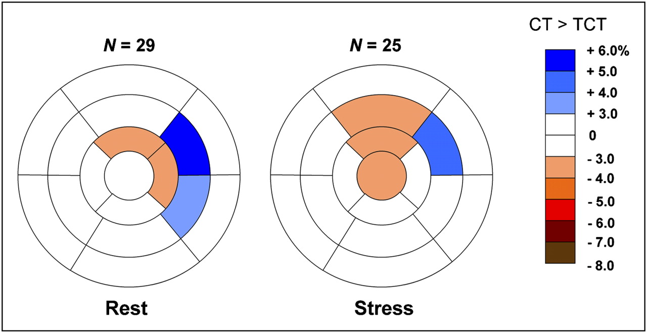
Stress and rest American Heart Association 17-segment representation of average differences between 82Rb PET data attenuation-corrected with TCT and with CT for studies with no misregistration or with misregistration less than 5 mm. Changes are expressed as percentage of all counts in myocardium (all changes are P < 0.05). Negative change (red) implies that CT-corrected value is smaller.
- FIGURE 3.
Stress and rest American Heart Association 17-segment representation of average differences between 82Rb PET data attenuation, corrected with TCT and with CT for studies with misregistration greater than 5 mm before (A) and after (B) alignment correction.
- FIGURE 4.
Frequency and magnitude of misalignment as judged by 2 observers for rest (A) and stress (B) studies.
- FIGURE 5.
Example of misalignment in rest 82Rb study of female patient (weight, 54.4 kg [122 lb]; age, 76 y; body mass index, 20.9). Images are before alignment correction (A) and attenuation-corrected with aligned CT (aCT) (B). From left to right, figure shows TCT- and CT-corrected original images, positive and negative change images with overlaid contours, raw polar maps, change polar maps, and PET/CT fusion images with blue arrows showing identified misalignment. TCT > CT changes are seen in septal wall (white arrows). Differences between CT- and TCT-corrected studies changed after alignment correction of CT. Two observers identified displacements of x = 10, y = 4, and z = 6 mm and x = 6, y = 11, and z = −1 mm.
- FIGURE 6.
Example of misalignment in stress study of male patient (weight, 75.9 kg [167 lb]; age, 67 y; body mass index, 25.4), in which AC by aligned CT (aCT) did not eliminate differences between CT- and TCT-corrected images. TCT > aCT changes remain in anterior and septal walls (white arrow), and TCT < aCT changes are shown in inferior wall (yellow arrow), despite manual alignment. Note that irregular heart structure on CT maps (green arrows), which is likely due to respiratory and cardiac motion effects during normal breathing CT, could be likely cause of remaining differences.


Tables
Direction (mm) Magnitude (mm) Study x y z Rest 2.16 ± 2.06 3.24 ± 3.50 2.16 ± 2.97 3.18 ± 3.52 Stress 2.49 ± 2.91 2.91 ± 3.28 2.35 ± 3.14 3.61 ± 3.95 Rest and Stress 2.33 ± 2.51 3.07 ± 3.38 2.25 ± 3.05 3.39 ± 3.71 Data are average differences between 2 observers in visual PET/CT alignment verification in all 3 directions.


Jump to section
Related Articles
Cited By...
- Dual-Gated Motion-Frozen Cardiac PET with Flurpiridaz F 18
- Coronary Arterial 18F-FDG Uptake by Fusion of PET and Coronary CT Angiography at Sites of Percutaneous Stenting for Acute Myocardial Infarction and Stable Coronary Artery Disease
- Comparison of Clinical Tools for Measurements of Regional Stress and Rest Myocardial Blood Flow Assessed with 13N-Ammonia PET/CT
- Agreement of Visual Estimation of Coronary Artery Calcium From Low-Dose CT Attenuation Correction Scans in Hybrid PET/CT and SPECT/CT With Standard Agatston Score
- Low-Dose Quantitative Myocardial Blood Flow Imaging Using 15O-Water and PET Without Attenuation Correction