
Main menu

  • Home
  • Content
    • Current
    • Ahead of print
    • Past Issues
    • JNM Supplement
    • SNMMI Annual Meeting Abstracts
    • Continuing Education
    • JNM Podcasts
  • Subscriptions
    • Subscribers
    • Institutional and Non-member
    • Rates
    • Journal Claims
    • Corporate & Special Sales
  • Authors
    • Submit to JNM
    • Information for Authors
    • Assignment of Copyright
    • AQARA requirements
  • Info
    • Reviewers
    • Permissions
    • Advertisers
  • About
    • About Us
    • Editorial Board
    • Contact Information
  • More
    • Alerts
    • Feedback
    • Help
    • SNMMI Journals
  • SNMMI
    • JNM
    • JNMT
    • SNMMI Journals
    • SNMMI

User menu

  • Subscribe
  • My alerts
  • Log in
  • My Cart

Search

  • Advanced search
Journal of Nuclear Medicine
  • SNMMI
    • JNM
    • JNMT
    • SNMMI Journals
    • SNMMI
  • Subscribe
  • My alerts
  • Log in
  • My Cart
Journal of Nuclear Medicine

Advanced Search

  • Home
  • Content
    • Current
    • Ahead of print
    • Past Issues
    • JNM Supplement
    • SNMMI Annual Meeting Abstracts
    • Continuing Education
    • JNM Podcasts
  • Subscriptions
    • Subscribers
    • Institutional and Non-member
    • Rates
    • Journal Claims
    • Corporate & Special Sales
  • Authors
    • Submit to JNM
    • Information for Authors
    • Assignment of Copyright
    • AQARA requirements
  • Info
    • Reviewers
    • Permissions
    • Advertisers
  • About
    • About Us
    • Editorial Board
    • Contact Information
  • More
    • Alerts
    • Feedback
    • Help
    • SNMMI Journals
  • View or Listen to JNM Podcast
  • Visit JNM on Facebook
  • Join JNM on LinkedIn
  • Follow JNM on Twitter
  • Subscribe to our RSS feeds
OtherBASIC SCIENCE INVESTIGATIONS

Usefulness of 11C-Methionine for Differentiating Tumors from Granulomas in Experimental Rat Models: A Comparison with 18F-FDG and 18F-FLT

Songji Zhao, Yuji Kuge, Masashi Kohanawa, Toshiyuki Takahashi, Yan Zhao, Min Yi, Kakuko Kanegae, Koh-ichi Seki and Nagara Tamaki
Journal of Nuclear Medicine January 2008, 49 (1) 135-141; DOI: https://doi.org/10.2967/jnumed.107.044578
Songji Zhao
  • Find this author on Google Scholar
  • Find this author on PubMed
  • Search for this author on this site
Yuji Kuge
  • Find this author on Google Scholar
  • Find this author on PubMed
  • Search for this author on this site
Masashi Kohanawa
  • Find this author on Google Scholar
  • Find this author on PubMed
  • Search for this author on this site
Toshiyuki Takahashi
  • Find this author on Google Scholar
  • Find this author on PubMed
  • Search for this author on this site
Yan Zhao
  • Find this author on Google Scholar
  • Find this author on PubMed
  • Search for this author on this site
Min Yi
  • Find this author on Google Scholar
  • Find this author on PubMed
  • Search for this author on this site
Kakuko Kanegae
  • Find this author on Google Scholar
  • Find this author on PubMed
  • Search for this author on this site
Koh-ichi Seki
  • Find this author on Google Scholar
  • Find this author on PubMed
  • Search for this author on this site
Nagara Tamaki
  • Find this author on Google Scholar
  • Find this author on PubMed
  • Search for this author on this site
  • Article
  • Figures & Data
  • Info & Metrics
  • PDF
Loading

Article Figures & Data

Figures

  • Tables
  • FIGURE 1. 
    • Download figure
    • Open in new tab
    • Download powerpoint
    FIGURE 1. 

    Experimental protocols of this study. LSC = liquid scintillation counter.

  • FIGURE 2. 
    • Download figure
    • Open in new tab
    • Download powerpoint
    FIGURE 2. 

    Microscopy images (×400) of hematoxylin-and-eosin staining (granuloma [A], tumor [C], and turpentine-induced inflammation [D]) and immunostaining for Ia antigen (granuloma [B]). (A) Intramuscular granuloma induced by BCG shows mature epithelioid cell granuloma formation and massive lymphocyte infiltration around granuloma. (B) Immunostaining for Ia antigen shows infiltrations of Ia-positive epithelioid cells and macrophages in granuloma and Ia-positive lymphocytes in periphery of granuloma. In A and B, arrowhead indicates epithelioid cell granuloma; white arrow, lymphocyte infiltration; and black arrow, macrophage infiltration. (C) Massive viable and proliferating cancer cells in tumor tissue. Black arrow indicates viable cancer cell; white arrow, proliferating cancer cell (mitotic division). (D) Massive neutrophil infiltration and ambient connective tissue formation were observed around site of turpentine oil injection. White arrow indicates neutrophil infiltration; black arrow, connective tissue.
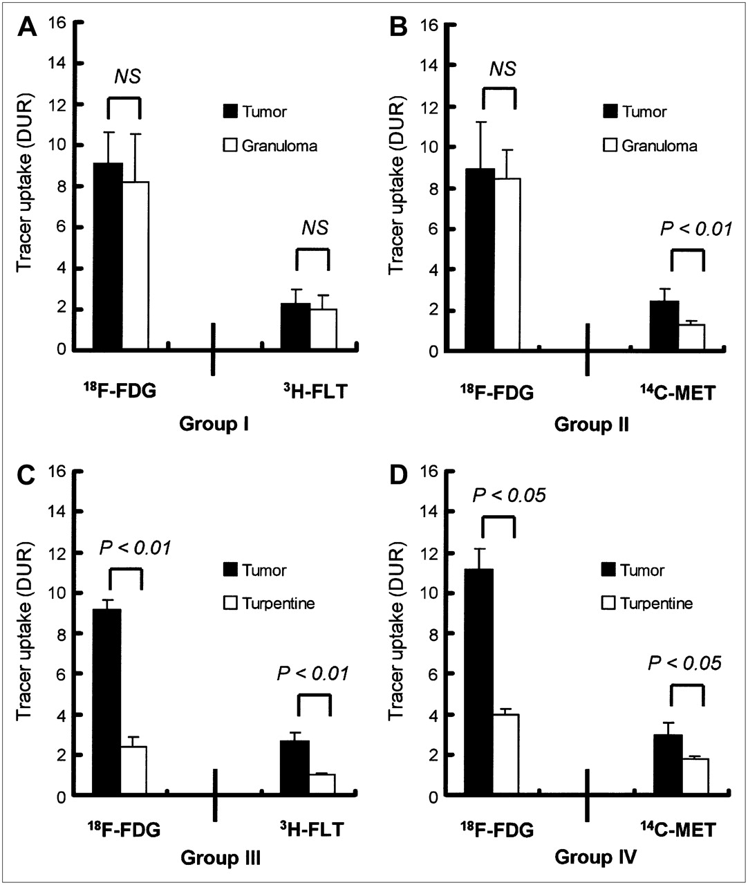
  • FIGURE 3. 
    • Download figure
    • Open in new tab
    • Download powerpoint
    FIGURE 3. 

    18F-FDG, 3H-FLT, and 14C-methionine uptake in tumor, granuloma, and turpentine oil–induced inflammation. (A) Group I: 18F-FDG and 3H-FLT uptake in tumor and granuloma. (B) Group II: 18F-FDG and 14C-methionine uptake in tumor and granuloma. (C) Group III: 18F-FDG and 3H-FLT uptake in tumor and turpentine oil–induced inflammation. (D) Group IV: 18F-FDG and 14C-methionine uptake in turpentine oil–induced inflammation. Values are mean ± SD. NS = not statistically significant.

Tables

  • Figures
    • View popup
    TABLE 1

    Blood Glucose Levels (mg/dL)

    TimeGroup I (n = 7): 18F-FDG + 3H-FLTGroup II (n = 6): 18F-FDG + 14C-methionineGroup III (n = 5): 18F-FDG + 3H-FLTGroup IV (n = 4): 18F-FDG + 14C-methionine
    At tracer injection87.7 ± 7.493.2 ± 6.584.2 ± 8.587.3 ± 5.2
    At sacrifice88.0 ± 6.786.0 ± 5.791.0 ± 8.085.3 ± 2.2
    • Data are mean ± SD.

    • View popup
    TABLE 2

    Uptake Levels, L/M, and L/B 60 Minutes After Injection of 18F-FDG, 3H-FLT, and 14C-Methionine (DUR)

    KDH-8 and BCGKDH-8 and turpentine oil
    Group I (n = 7)Group II (n = 6)Group III (n = 5)Group IV (n = 4)
    Parameter18F-FDG3H-FLT18F-FDG14C-Methionine18F-FDG3H-FLT18F-FDG14C-Methionine
    Blood0.69 ± 0.140.91 ± 0.150.60 ± 0.110.71 ± 0.040.57 ± 0.080.72 ± 0.110.77 ± 0.100.99 ± 0.03
    Plasma0.95 ± 0.250.85 ± 0.120.59 ± 0.130.95 ± 0.040.66 ± 0.110.74 ± 0.080.79 ± 0.101.37 ± 0.08
    Muscle0.28 ± 0.050.85 ± 0.090.31 ± 0.110.45 ± 0.060.20 ± 0.030.74 ± 0.070.30 ± 0.030.41 ± 0.04
    Brown fat0.52 ± 0.480.48 ± 0.250.33 ± 0.060.42 ± 0.070.38 ± 0.080.43 ± 0.050.44 ± 0.030.50 ± 0.03
    White fat0.18 ± 0.030.16 ± 0.050.18 ± 0.020.11 ± 0.020.21 ± 0.080.22 ± 0.090.19 ± 0.020.13 ± 0.01
    Heart1.04 ± 0.600.86 ± 0.100.62 ± 0.270.83 ± 0.050.54 ± 0.050.72 ± 0.090.60 ± 0.130.97 ± 0.03
    Brain3.05 ± 0.240.15 ± 0.022.97 ± 0.150.48 ± 0.032.69 ± 0.160.16 ± 0.073.49 ± 0.460.64 ± 0.04
    Lung2.35 ± 0.701.51 ± 0.271.16 ± 0.131.30 ± 0.032.51 ± 0.331.72 ± 0.251.52 ± 0.081.63 ± 0.06
    Thymus1.89 ± 0.331.03 ± 0.392.16 ± 0.231.57 ± 0.111.20 ± 0.240.84 ± 0.462.46 ± 0.201.85 ± 0.30
    Spleen3.26 ± 0.824.20 ± 0.732.34 ± 0.262.16 ± 0.412.75 ± 0.305.06 ± 1.093.19 ± 0.203.03 ± 0.14
    Liver2.37 ± 0.641.71 ± 0.340.89 ± 0.227.15 ± 0.752.10 ± 0.401.42 ± 0.091.24 ± 0.117.97 ± 0.83
    Kidney2.06 ± 0.922.37 ± 0.381.31 ± 0.303.76 ± 0.141.36 ± 0.241.92 ± 0.152.98 ± 1.313.97 ± 0.23
    Bone marrow2.50 ± 0.359.42 ± 0.992.37 ± 0.243.33 ± 0.242.07 ± 0.3611.93 ± 2.182.92 ± 0.144.69 ± 0.26
    Tumor9.13 ± 1.522.30 ± 0.678.91 ± 2.322.47 ± 0.609.13 ± 0.502.66 ± 0.4111.14 ± 1.032.96 ± 0.57
    Granuloma or turpentine8.18 ± 2.401.98 ± 0.708.43 ± 1.451.31 ± 0.22*2.42 ± 0.43*0.99 ± 0.13*3.99 ± 0.22*1.77 ± 0.18†
    Ratio
     L (tumor)/M34.2 ± 9.82.7 ± 0.733.0 ± 16.85.7 ± 1.945.3 ± 6.53.6 ± 0.537.6 ± 1.57.2 ± 1.6
     L (granuloma)/M or L (turpentine)/M30.0 ± 9.32.3 ± 0.829.7 ± 9.63.0 ± 0.6*11.9 ± 2.4*1.3 ± 0.2*13.6 ± 1.9†4.3 ± 0.8†
     L (tumor)/B13.9 ± 4.42.6 ± 0.814.9 ± 2.73.5 ± 1.016.2 ± 1.73.8 ± 0.914.6 ± 1.73.0 ± 0.6
     L (granuloma)/B or L (turpentine)/B12.4 ± 5.12.2 ± 0.514.3 ± 2.51.8 ± 0.3*4.3 ± 0.9*1.4 ± 0.2*5.3 ± 0.9*1.8 ± 0.1†
    • ↵* P < 0.01, tumor vs. granuloma or tumor vs. turpentine-induced inflammation.

    • ↵† P < 0.05.

PreviousNext
Back to top

In this issue

Journal of Nuclear Medicine: 49 (1)
Journal of Nuclear Medicine
Vol. 49, Issue 1
January 2008
  • Table of Contents
  • Table of Contents (PDF)
  • About the Cover
  • Index by author
Print
Download PDF
Article Alerts
Sign In to Email Alerts with your Email Address
Email Article

Thank you for your interest in spreading the word on Journal of Nuclear Medicine.

NOTE: We only request your email address so that the person you are recommending the page to knows that you wanted them to see it, and that it is not junk mail. We do not capture any email address.

Enter multiple addresses on separate lines or separate them with commas.
Usefulness of 11C-Methionine for Differentiating Tumors from Granulomas in Experimental Rat Models: A Comparison with 18F-FDG and 18F-FLT
(Your Name) has sent you a message from Journal of Nuclear Medicine
(Your Name) thought you would like to see the Journal of Nuclear Medicine web site.
Citation Tools
Usefulness of 11C-Methionine for Differentiating Tumors from Granulomas in Experimental Rat Models: A Comparison with 18F-FDG and 18F-FLT
Songji Zhao, Yuji Kuge, Masashi Kohanawa, Toshiyuki Takahashi, Yan Zhao, Min Yi, Kakuko Kanegae, Koh-ichi Seki, Nagara Tamaki
Journal of Nuclear Medicine Jan 2008, 49 (1) 135-141; DOI: 10.2967/jnumed.107.044578

Citation Manager Formats

  • BibTeX
  • Bookends
  • EasyBib
  • EndNote (tagged)
  • EndNote 8 (xml)
  • Medlars
  • Mendeley
  • Papers
  • RefWorks Tagged
  • Ref Manager
  • RIS
  • Zotero
Share
Usefulness of 11C-Methionine for Differentiating Tumors from Granulomas in Experimental Rat Models: A Comparison with 18F-FDG and 18F-FLT
Songji Zhao, Yuji Kuge, Masashi Kohanawa, Toshiyuki Takahashi, Yan Zhao, Min Yi, Kakuko Kanegae, Koh-ichi Seki, Nagara Tamaki
Journal of Nuclear Medicine Jan 2008, 49 (1) 135-141; DOI: 10.2967/jnumed.107.044578
Twitter logo Facebook logo LinkedIn logo Mendeley logo
  • Tweet Widget
  • Facebook Like
  • Google Plus One
Bookmark this article

Jump to section

  • Article
    • Abstract
    • MATERIALS AND METHODS
    • RESULTS
    • DISCUSSION
    • CONCLUSION
    • Acknowledgments
    • Footnotes
    • References
  • Figures & Data
  • Info & Metrics
  • PDF

Related Articles

  • This Month in JNM
  • PubMed
  • Google Scholar

Cited By...

  • Nononcologic Applications of PET/CT and PET/MRI in Musculoskeletal, Orthopedic, and Rheumatologic Imaging: General Considerations, Techniques, and Radiopharmaceuticals
  • PET Findings of Intramedullary Tumors of the Spinal Cord Using [18F] FDG and [11C] Methionine
  • Evaluation of 4'-[Methyl-11C]Thiothymidine in a Rodent Tumor and Inflammation Model
  • Can Evaluation of Targeted Therapy in Oncology Be Improved by Means of 18F-FLT?
  • Google Scholar

More in this TOC Section

  • PET/MRI of Hypoxic Atherosclerosis Using 64Cu-ATSM in a Rabbit Model
  • Tumor Uptake of Anti-CD20 Fabs Depends on Tumor Perfusion
  • How Sensitive Is the Upper Gastrointestinal Tract to 90Y Radioembolization? A Histologic and Dosimetric Analysis in a Porcine Model
Show more Basic Science Investigations

Similar Articles

SNMMI

© 2025 SNMMI

Powered by HighWire