
Main menu

  • Home
  • Content
    • Current
    • Ahead of print
    • Past Issues
    • JNM Supplement
    • SNMMI Annual Meeting Abstracts
    • Continuing Education
    • JNM Podcasts
  • Subscriptions
    • Subscribers
    • Institutional and Non-member
    • Rates
    • Journal Claims
    • Corporate & Special Sales
  • Authors
    • Submit to JNM
    • Information for Authors
    • Assignment of Copyright
    • AQARA requirements
  • Info
    • Reviewers
    • Permissions
    • Advertisers
  • About
    • About Us
    • Editorial Board
    • Contact Information
  • More
    • Alerts
    • Feedback
    • Help
    • SNMMI Journals
  • SNMMI
    • JNM
    • JNMT
    • SNMMI Journals
    • SNMMI

User menu

  • Subscribe
  • My alerts
  • Log in
  • Log out
  • My Cart

Search

  • Advanced search
Journal of Nuclear Medicine
  • SNMMI
    • JNM
    • JNMT
    • SNMMI Journals
    • SNMMI
  • Subscribe
  • My alerts
  • Log in
  • Log out
  • My Cart
Journal of Nuclear Medicine

Advanced Search

  • Home
  • Content
    • Current
    • Ahead of print
    • Past Issues
    • JNM Supplement
    • SNMMI Annual Meeting Abstracts
    • Continuing Education
    • JNM Podcasts
  • Subscriptions
    • Subscribers
    • Institutional and Non-member
    • Rates
    • Journal Claims
    • Corporate & Special Sales
  • Authors
    • Submit to JNM
    • Information for Authors
    • Assignment of Copyright
    • AQARA requirements
  • Info
    • Reviewers
    • Permissions
    • Advertisers
  • About
    • About Us
    • Editorial Board
    • Contact Information
  • More
    • Alerts
    • Feedback
    • Help
    • SNMMI Journals
  • View or Listen to JNM Podcast
  • Visit JNM on Facebook
  • Join JNM on LinkedIn
  • Follow JNM on Twitter
  • Subscribe to our RSS feeds
OtherBASIC SCIENCE INVESTIGATIONS

In Vivo Evaluation and Small-Animal PET/CT of a Prostate Cancer Mouse Model Using 64Cu Bombesin Analogs: Side-by-Side Comparison of the CB-TE2A and DOTA Chelation Systems

Jered C. Garrison, Tammy L. Rold, Gary L. Sieckman, Said Daibes Figueroa, Wynn A. Volkert, Silvia S. Jurisson and Timothy J. Hoffman
Journal of Nuclear Medicine August 2007, 48 (8) 1327-1337; DOI: https://doi.org/10.2967/jnumed.107.039487
Jered C. Garrison
  • Find this author on Google Scholar
  • Find this author on PubMed
  • Search for this author on this site
Tammy L. Rold
  • Find this author on Google Scholar
  • Find this author on PubMed
  • Search for this author on this site
Gary L. Sieckman
  • Find this author on Google Scholar
  • Find this author on PubMed
  • Search for this author on this site
Said Daibes Figueroa
  • Find this author on Google Scholar
  • Find this author on PubMed
  • Search for this author on this site
Wynn A. Volkert
  • Find this author on Google Scholar
  • Find this author on PubMed
  • Search for this author on this site
Silvia S. Jurisson
  • Find this author on Google Scholar
  • Find this author on PubMed
  • Search for this author on this site
Timothy J. Hoffman
  • Find this author on Google Scholar
  • Find this author on PubMed
  • Search for this author on this site
  • Article
  • Figures & Data
  • Info & Metrics
  • PDF
Loading

Article Figures & Data

Figures

  • Tables
  • FIGURE 1. 
    • Download figure
    • Open in new tab
    • Download powerpoint
    FIGURE 1. 

    Structure of bombesin and 64Cu-labeled bombesin analogs.

  • FIGURE 2. 
    • Download figure
    • Open in new tab
    • Download powerpoint
    FIGURE 2. 

    Competitive binding assay of Cu-DOTA-8-AOC-BBN(7–14)NH2 and Cu-CB-TE2A-8-AOC-BBN(7–14)NH2 analogs vs. [125I-Tyr4]BBN using PC-3 cells (n = 3).
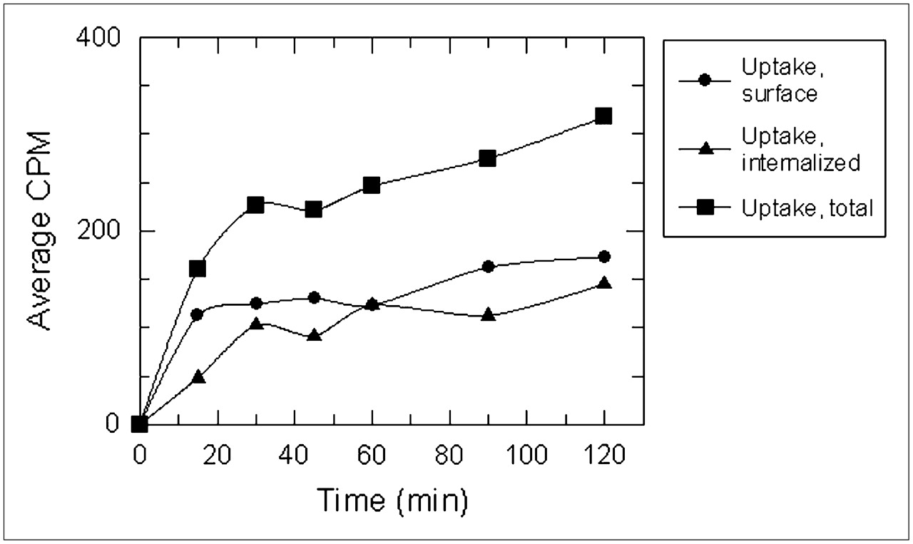
  • FIGURE 3. 
    • Download figure
    • Open in new tab
    • Download powerpoint
    FIGURE 3. 

    Internalization of 64Cu-CB-TE2A-8-AOC-BBN(7–14)NH2 using PC-3 cells (n = 6).

  • FIGURE 4. 
    • Download figure
    • Open in new tab
    • Download powerpoint
    FIGURE 4. 

    Internalization of 64Cu-DOTA-8-AOC-BBN(7–14)NH2 using PC-3 cells (n = 2).
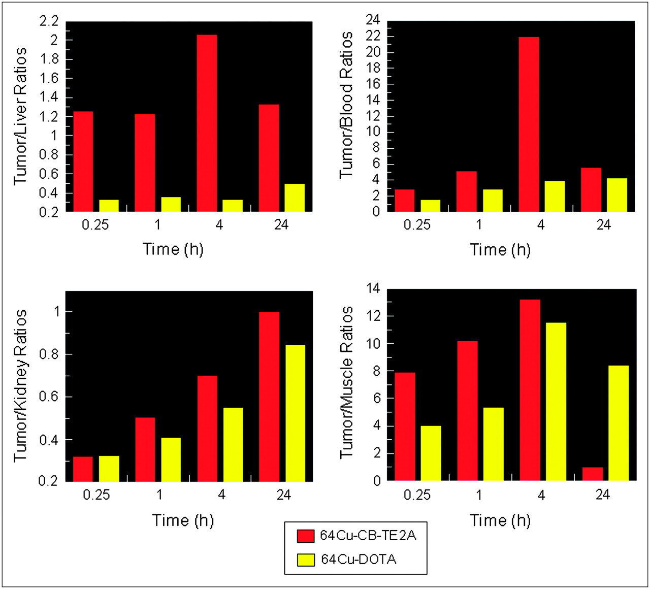
  • FIGURE 5. 
    • Download figure
    • Open in new tab
    • Download powerpoint
    FIGURE 5. 

    Comparison of 64Cu-CB-TE2A-8-AOC-BBN(7–14)NH2 (red) and 64Cu-DOTA-8-AOC-BBN(7–14)NH2 (yellow) using a radiolocalization index ([RI] %ID/g of tumor/%ID/g of nontarget tissue).

  • FIGURE 6. 
    • Download figure
    • Open in new tab
    • Download powerpoint
    FIGURE 6. 

    mircoPET, fused microPET/CT, and axial images of 64Cu-DOTA-8-AOC-BBN(7–14)NH2 (A) and 64Cu-CB-TE2A-8-AOC-BBN(7–14)NH2 (B) in PC-3 tumor-bearing SCID mice at 20 h after administration. Lateral and axial projection images have been normalized to the highest pixel intensity for each respective image.

Tables

  • Figures
    • View popup
    TABLE 1

    Mass Spectrometric, RP-HPLC, and In Vitro IC50 Characterization of Conjugates

    AnalogMolecular formulaMS calculatedMS observedRP-HPLC tr (min)*IC50 (nM)†
    DOTA-8-AOC-BBN(7–14)NH2C67H106N18O17S1,467.81,468.013.8
    Cu-DOTA-8-AOC-BBN(7–14)NH2C67H104N18O17SCu1,528.71,528.515.01.44 ± 0.08
    CB-TE2A-8-AOC-BBN(7–14)NH2C67H108N18O13S1,405.81,405.815.80.67 ± 0.07
    Cu-CB-TE2A-8-AOC-BBN(7–14)NH2C67H106N18O13SCu1,465.71,465.717.20.48 ± 0.01
    • ↵* RP-HPLC methods described in Materials and Methods.

    • ↵† In vitro binding assays were performed in triplicate.

    • View popup
    TABLE 2

    In Vivo Pharmacokinetic Studies of 64Cu-DOTA-8-AOC-BBN(7–14)NH2 Using SCID Mice Bearing PC-3 Xenografts

    Tissue/organ15 min1 h4 h24 h
    Blood3.18 ± 0.911.22 ± 0.620.76 ± 0.460.90 ± 0.48
    Heart3.36 ± 1.062.05 ± 1.451.63 ± 0.732.69 ± 1.09
    Lung5.19 ± 1.762.96 ± 1.843.27 ± 1.313.55 ± 1.17
    Liver14.97 ± 3.379.56 ± 5.208.98 ± 2.747.80 ± 1.51
    Stomach3.94 ± 0.702.30 ± 0.962.46 ± 0.951.68 ± 0.59
    Small intestines8.26 ± 3.5513.45 ± 2.085.04 ± 1.072.84 ± 0.48
    Large intestines5.13 ± 3.255.58 ± 2.8416.29 ± 3.813.78 ± 0.85
    Kidneys15.16 ± 2.998.46 ± 2.795.43 ± 0.904.58 ± 0.95
    Spleen2.68 ± 1.602.52 ± 1.593.57 ± 2.233.59 ± 2.48
    Pancreas23.30 ± 4.4313.81 ± 2.6912.16 ± 1.343.65 ± 0.44
    Muscle1.23 ± 0.140.65 ± 0.370.26 ± 0.140.46 ± 0.21
    Bone1.98 ± 0.511.12 ± 0.660.79 ± 0.351.15 ± 0.59
    Tumors4.95 ± 0.913.48 ± 0.903.00 ± 1.103.88 ± 1.40
    Excretion (%ID)19.13 ± 6.9444.63 ± 9.1549.77 ± 8.1967.84 ± 5.43
    • All data, except excretion, are presented as %ID/g ± SD. Excretion data are presented as %ID ± SD (n = 5).

    • View popup
    TABLE 3

    In Vivo Pharmacokinetic Studies of 64Cu-CB-TE2A-8-AOC-BBN(7–14)NH2 Using SCID Mice Bearing PC-3 Xenografts

    Tissue/organ15 min1 h4 h24 h
    Blood2.37 ± 1.470.51 ± 0.140.06 ± 0.050.05 ± 0.03
    Heart1.45 ± 0.670.09 ± 0.080.08 ± 0.080.10 ± 0.09
    Lung2.86 ± 1.120.60 ± 0.190.22 ± 0.150.15 ± 0.08
    Liver5.52 ± 1.322.15 ± 0.260.64 ± 0.080.21 ± 0.06
    Stomach2.56 ± 0.620.94 ± 0.271.65 ± 2.630.05 ± 0.04
    Small intestines12.88 ± 2.0613.17 ± 1.211.78 ± 0.570.11 ± 0.04
    Large intestines4.27 ± 0.973.52 ± 1.1217.70 ± 7.060.20 ± 0.08
    Kidneys21.66 ± 8.585.26 ± 0.581.88 ± 0.530.28 ± 0.22
    Spleen2.96 ± 2.840.68 ± 0.530.16 ± 0.160.56 ± 0.30
    Pancreas31.28 ± 3.2417.66 ± 2.002.18 ± 0.710.18 ± 0.14
    Muscle0.88 ± 0.380.26 ± 0.150.10 ± 0.070.22 ± 0.17
    Bone2.15 ± 2.010.58 ± 0.410.39 ± 0.250.29 ± 0.10
    Tumors6.95 ± 2.272.65 ± 1.051.32 ± 0.490.28 ± 0.21
    Excretion (%ID)26.48 ± 10.1860.38 ± 2.1677.88 ± 6.5898.60 ± 0.28
    • All data, except excretion, are presented as %ID/g ± SD. Excretion data are presented as %ID ± SD (n = 5).

PreviousNext
Back to top

In this issue

Journal of Nuclear Medicine: 48 (8)
Journal of Nuclear Medicine
Vol. 48, Issue 8
August 2007
  • Table of Contents
  • Table of Contents (PDF)
  • About the Cover
  • Index by author
Print
Download PDF
Article Alerts
Sign In to Email Alerts with your Email Address
Email Article

Thank you for your interest in spreading the word on Journal of Nuclear Medicine.

NOTE: We only request your email address so that the person you are recommending the page to knows that you wanted them to see it, and that it is not junk mail. We do not capture any email address.

Enter multiple addresses on separate lines or separate them with commas.
In Vivo Evaluation and Small-Animal PET/CT of a Prostate Cancer Mouse Model Using 64Cu Bombesin Analogs: Side-by-Side Comparison of the CB-TE2A and DOTA Chelation Systems
(Your Name) has sent you a message from Journal of Nuclear Medicine
(Your Name) thought you would like to see the Journal of Nuclear Medicine web site.
Citation Tools
In Vivo Evaluation and Small-Animal PET/CT of a Prostate Cancer Mouse Model Using 64Cu Bombesin Analogs: Side-by-Side Comparison of the CB-TE2A and DOTA Chelation Systems
Jered C. Garrison, Tammy L. Rold, Gary L. Sieckman, Said Daibes Figueroa, Wynn A. Volkert, Silvia S. Jurisson, Timothy J. Hoffman
Journal of Nuclear Medicine Aug 2007, 48 (8) 1327-1337; DOI: 10.2967/jnumed.107.039487

Citation Manager Formats

  • BibTeX
  • Bookends
  • EasyBib
  • EndNote (tagged)
  • EndNote 8 (xml)
  • Medlars
  • Mendeley
  • Papers
  • RefWorks Tagged
  • Ref Manager
  • RIS
  • Zotero
Share
In Vivo Evaluation and Small-Animal PET/CT of a Prostate Cancer Mouse Model Using 64Cu Bombesin Analogs: Side-by-Side Comparison of the CB-TE2A and DOTA Chelation Systems
Jered C. Garrison, Tammy L. Rold, Gary L. Sieckman, Said Daibes Figueroa, Wynn A. Volkert, Silvia S. Jurisson, Timothy J. Hoffman
Journal of Nuclear Medicine Aug 2007, 48 (8) 1327-1337; DOI: 10.2967/jnumed.107.039487
Twitter logo Facebook logo LinkedIn logo Mendeley logo
  • Tweet Widget
  • Facebook Like
  • Google Plus One
Bookmark this article

Jump to section

  • Article
    • Abstract
    • MATERIALS AND METHODS
    • RESULTS
    • DISCUSSION
    • CONCLUSION
    • Acknowledgments
    • Footnotes
    • References
  • Figures & Data
  • Info & Metrics
  • PDF

Related Articles

  • This Month in JNM
  • PubMed
  • Google Scholar

Cited By...

  • Improving Contrast and Detectability: Imaging with [55Co]Co-DOTATATE in Comparison with [64Cu]Cu-DOTATATE and [68Ga]Ga-DOTATATE
  • Novel "Add-On" Molecule Based on Evans Blue Confers Superior Pharmacokinetics and Transforms Drugs to Theranostic Agents
  • Clinical Translation of a Dual Integrin {alpha}v{beta}3- and Gastrin-Releasing Peptide Receptor-Targeting PET Radiotracer, 68Ga-BBN-RGD
  • 68Ga-NOTA-Aca-BBN(7-14) PET/CT in Healthy Volunteers and Glioma Patients
  • In Vivo Molecular Imaging of Thrombosis and Thrombolysis Using a Fibrin-Binding Positron Emission Tomographic Probe
  • Synthesis and In Vitro and In Vivo Evaluation of Hypoxia-Enhanced 111In-Bombesin Conjugates for Prostate Cancer Imaging
  • Targeted Radiotherapy of Prostate Cancer with a Gastrin-Releasing Peptide Receptor Antagonist Is Effective as Monotherapy and in Combination with Rapamycin
  • Positron-emission Tomography (PET) Imaging Agents for Diagnosis of Human Prostate Cancer: Agonist vs. Antagonist Ligands
  • Exploring molecular genetics of bladder cancer: lessons learned from mouse models
  • Radiopeptide Imaging and Therapy in Europe
  • Bombesin Antagonist-Based Radioligands for Translational Nuclear Imaging of Gastrin-Releasing Peptide Receptor-Positive Tumors
  • In Vitro and In Vivo Evaluation of 64Cu-Labeled SarAr-Bombesin Analogs in Gastrin-Releasing Peptide Receptor-Expressing Prostate Cancer
  • Small-Animal PET of Tumors with 64Cu-Labeled RGD-Bombesin Heterodimer
  • Humanized Radioiodinated Minibody For Imaging of Prostate Stem Cell Antigen-Expressing Tumors
  • International Union of Pharmacology. LXVIII. Mammalian Bombesin Receptors: Nomenclature, Distribution, Pharmacology, Signaling, and Functions in Normal and Disease States
  • Google Scholar

More in this TOC Section

  • 11C-Methionine PET of Myocardial Inflammation in a Rat Model of Experimental Autoimmune Myocarditis
  • Counting Rate Characteristics and Image Distortion in Preclinical PET Imaging During Radiopharmaceutical Therapy
  • Design and Fabrication of Kidney Phantoms for Internal Radiation Dosimetry Using 3D Printing Technology
Show more Basic Science Investigations

Similar Articles

SNMMI

© 2025 SNMMI

Powered by HighWire