


Article Figures & Data
Figures
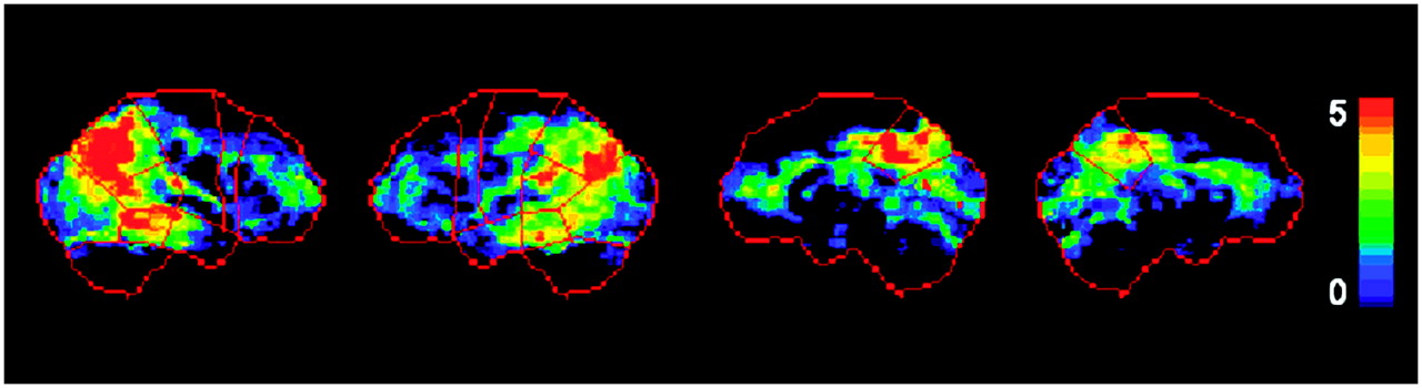
- FIGURE 1.
Standardized ROIs. Predefined anatomic surface ROIs (in red) are superimposed onto (from left to right) right and left lateral and right and left medial views of standardized brain template showing surface projection maps of statistical abnormalities in AD patients as compared with NLs. z scores are represented on color-coded scale ranging from 0 (black) to 5 (red). Typical AD-related CMRglc reductions in parietotemporal and posterior cingulate cortices are evident in ROIs.
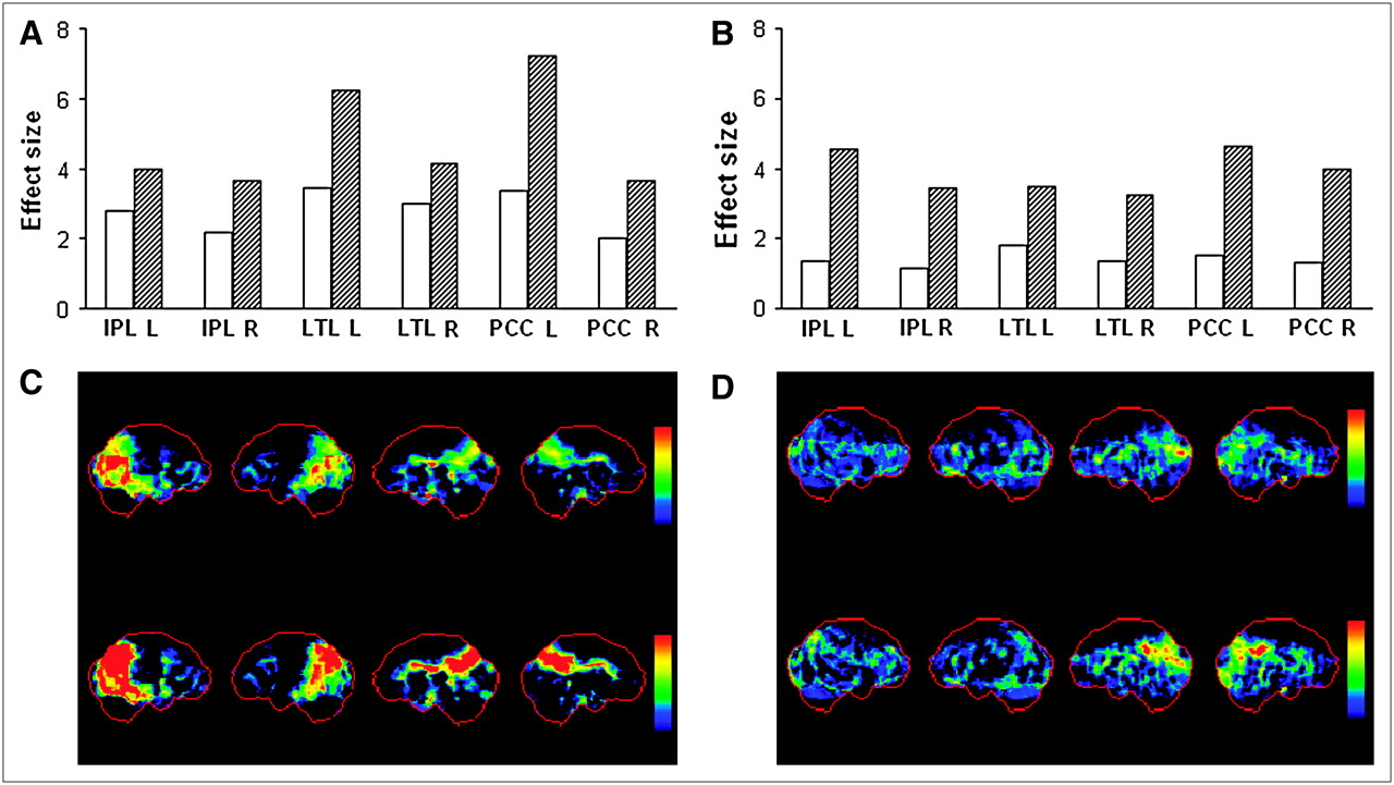
- FIGURE 2.
Database effects on 18F-FDG PET diagnostic accuracy. (A and B) Effects in AD-affected brain regions discriminating AD patients (A) and MCI patients (B) from NLs using DB− (white bars) and DB+ (hatched bars). (C and D) Effects of using DB− (top row) and DB+ (bottom row) to create 3D-SSP maps are depicted in 2 representative AD (C) and MCI (D) patients. 3D-SSP maps showing CMRglc reductions in AD and MCI patients are displayed on same color-coded z score scale. IPL = inferior parietal lobe; L = left hemisphere; LTL = lateral temporal lobe; PCC = posterior cingulate cortex; R = right hemisphere.
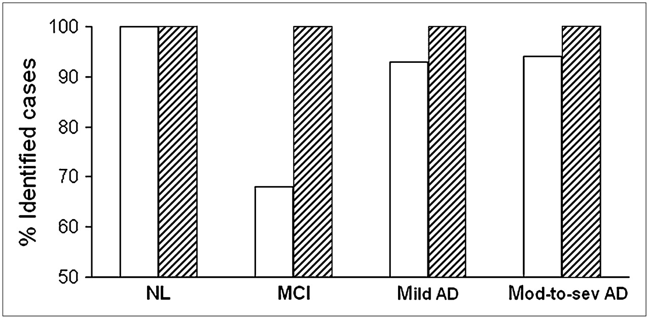
- FIGURE 3.
18F-FDG PET diagnostic accuracy. Percentage of NLs (n = 19), MCI patients (n = 37), patients with mild AD (n = 15), and patients with moderate to severe (mod-to-sev) AD (n = 18) correctly identified using DB− (white bars) and DB+ (hatched bars).
- FIGURE 4.
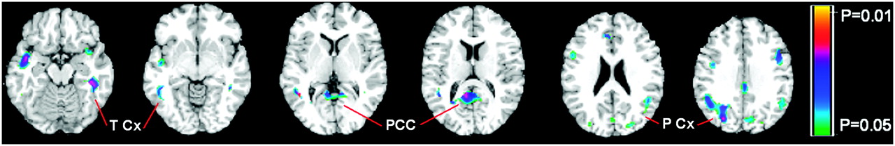
Comparison between NL-MCI patients and NL-NLs. Brain regions showing baseline CMRglc reductions in 22 NL-MCI patients and 22 age-matched NL-NLs are displayed on standardized T1-weighted MRI template in axial view (from left to right: z = 29 to z = 64 mm, every 5 mm, relative to anterior commissure–posterior commissure line). Voxelwise group comparison of spatially normalized 18F-FDG PET scans was performed with t test for independent samples accounting for global CMRglc (3,5). CMRglc reductions in NL-MCI patients compared with NL-NLs were evident mainly in temporal, posterior cingulate, and parietal cortices. Color scale indicate P values corresponding to significance of CMRglc reductions, ranging from P = 0.05 to P = 0.01, uncorrected for multiple comparisons. PCC = posterior cingulate cortex; P Cx = parietal cortex; T Cx = temporal cortex.


Tables
Reference database Clinical groups Characteristic DB− DB+ NL MCI Mild AD Moderate-to-severe AD n 55 55 19 37 15 18 Age* (y) 69 (7), 50–82 68 (7), 50–80 68 (4), 55–80 70 (7), 50–82 69 (8), 50–83 65 (7), 51–80 Education* (y) 15 (2), 12–18 15 (2), 12–18 14 (3), 11–18 11 (4), 8–18† 12 (3), 9–18† 10 (3), 8–18† % Female 55 56 58 60 53 67 MMSE* 29 (1), 28–30 29 (1), 28–30 29 (1), 28–30 27 (2), 24–30 26 (2), 24–28 19 (4), 10–23†‡§ NL MCI AD ROI DB− DB+ DB− DB+ DB− DB+ IPL L 0.47 (0.23) 0.49 (0.21) 1.04 (0.93) 1.56 (0.66)* 1.48 (0.46) 2.49 (0.83)* IPL R 0.44 (0.24) 0.48 (0.22) 0.87 (0.87) 1.25 (0.89)* 1.52 (0.91) 2.43 (0.93)* LTL L 0.47 (0.42) 0.41 (0.23) 1.26 (0.85) 1.32 (0.91) 2.13 (0.79) 2.93 (0.35)* LTL R 0.43 (0.38) 0.51 (0.38) 1.11 (1.08) 1.27 (0.94)* 2.16 (1.03) 2.74 (0.93)* OCC L 0.35 (0.42) 0.32 (0.31) 0.97 (0.40) 0.05 (0.51) 1.35 (0.56) 1.40 (0.72) OCC R 0.48 (0.61) 0.44 (0.50) 0.78 (0.65) 0.85 (0.57) 1.12 (0.66) 1.34 (0.52) PCC L 0.71 (0.62) 0.82 (0.86) 1.10 (0.86) 1.86 (0.82)* 1.81 (0.53) 3.51 (0.45)* PCC R 0.62 (0.76) 0.74 (0.67) 0.98 (0.89) 1.72 (0.98)* 1.50 (1.02) 2.72 (1.13)* PFC L 0.85 (0.84) 0.73 (0.65) 1.13 (0.85) 1.10 (0.95) 1.92 (0.44) 2.28 (0.60) PFC R 1.02 (0.75) 0.85 (0.92) 1.15 (0.79) 1.09 (0.94) 1.54 (1.08) 1.80 (1.12) S-M L 0.40 (0.41) 0.34 (0.23) 0.44 (0.31) 0.54 (0.54) 0.47 (0.24) 0.51 (0.39) S-M R 0.33 (0.35) 0.32 (0.33) 0.32 (0.52) 0.43 (0.57) 0.35 (0.53) 0.53 (0.50) ↵* DB+ higher than DB−, P ≤ 0.05.
IPL = inferior parietal lobe; LTL = lateral temporal lobe; OCC = occipital cortex; PCC = posterior cingulate cortex; PFC = prefrontal cortex; S-M = sensorimotor cortex; L = left hemisphere, R = right hemisphere.
Values in parentheses are SDs.


Jump to section
Related Articles
Cited By...
- Modeling the Effects of Age and Sex on Normal Pediatric Brain Metabolism Using 18F-FDG PET/CT
- Effectiveness and Safety of 18F-FDG PET in the Evaluation of Dementia: A Review of the Recent Literature
- Molecular imaging in the diagnosis of Alzheimer's disease: visual assessment of [11C]PIB and [18F]FDDNP PET images
- Declining brain glucose metabolism in normal individuals with a maternal history of Alzheimer disease
- Multicenter Standardized 18F-FDG PET Diagnosis of Mild Cognitive Impairment, Alzheimer's Disease, and Other Dementias