


Article Figures & Data
Figures
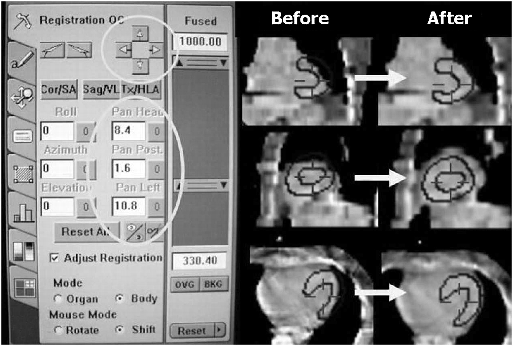
- FIGURE 1.
Software tool for realignment of misregistered CT and SPECT data. Using arrows of toolbar (left), CT can be moved in x-, y-, and z-axes relative to SPECT in order to match borders of left ventricle. In fusion images on right, dark lines depict myocardial borders of SPECT scan. In present case, CT was moved left and cranially to obtain alignment as illustrated by fused images in coronal, sagittal, and transaxial planes before and after realignment. Shifts are displayed in millimeters on toolbar and recorded for further analysis.
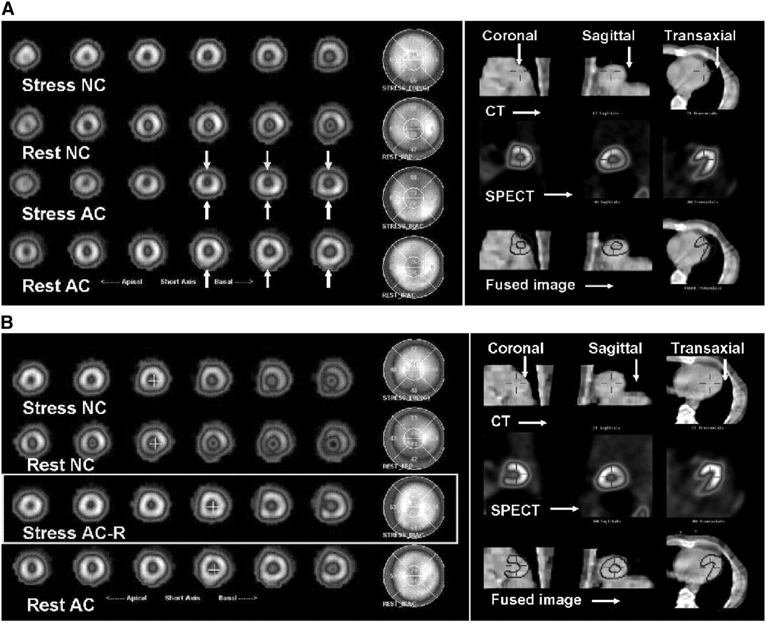
- FIGURE 2.
Representative case of SPECT/CT misalignment before (A) and after (B) reregistration. Stress and rest short-axis slices and polar maps are displayed on left. (A) Noncorrected images (NC) show decreased radiotracer uptake in inferior wall, compared with attenuation-corrected images (AC). Inferior wall uptake is improved, suggesting attenuation artifact on noncorrected images. Anterior wall, however, shows reduced uptake on attenuation-corrected vs. noncorrected images. Significant misalignment of SPECT and CT is shown in fusion images on right. (B) After best possible realignment (right), tracer distribution in attenuation-corrected images at stress and rest is homogeneous (left). AC-R = attenuation-corrected images after reregistration.
Tables
- TABLE 1
Regional Radiotracer Uptake for Noncorrected and Attenuation-Corrected Myocardial Perfusion SPECT Images
Mean ± SD NC to AC Area NC AC Difference 95% confidence interval Anterior 61.5 ± 7.1 64.1 ± 6.4 2.57 1.86, 3.28 Lateral 64.6 ± 6.2 68.4 ± 5.3 3.82 3.06, 4.59 Septal 58.9 ± 7.6 62.5 ± 8.1 3.59 2.72, 4.47 Inferior 55.0 ± 9.0 64.5 ± 7.8 9.45 8.53, 10.4 Anteroapical 77.0 ± 6.9 71.7 ± 7.0 −5.33 6.17, −4.49 Inferoapical 77.3 ± 7.4 73.9 ± 6.9 −3.44 4.40. −2.49 AC = attenuation-corrected; NC = noncorrected.
Data are percentage maximal pixel intensity for each of 6 evaluated segments of left ventricle, with NC-to-AC differences and 95% confidence intervals.
- TABLE 2
Regional Radiotracer Uptake for Attenuation-Corrected Myocardial Perfusion SPECT Images Before and After Reregistration
Mean ± SD AC to RR Area AC RR Difference 95% confidence interval Anterior 64.1 ± 6.4 63.3 ± 6.8 −0.81 1.13, −0.43 Lateral 68.4 ± 5.3 67.7 ± 5.7 −0.69 1.03, −0.35 Septal 62.5 ± 8.1 61.1 ± 8.4 −1.40 1.76, −1.03 Inferior 64.5 ± 7.8 62.7 ± 8.0 −1.71 2.10, −1.32 Anteroapical 71.7 ± 7.0 73.0 ± 7.0 1.33 0.90, 1.75 Inferoapical 73.9 ± 6.9 74.4 ± 6.5 0.52 0.07, 0.97 AC = attenuation-corrected before reregistration; RR = attenuation-corrected after reregistration.
Data are percentage maximal pixel intensity for each of 6 evaluated segments of left ventricle, with AC-to-RR differences and 95% confidence intervals.


Jump to section
Related Articles
Cited By...
- Study of Attenuation Correction Using a Cardiac Dynamic Phantom: Synchronized Time-Phase-Gated Attenuation Correction Method
- Targeting Cardiovascular Implant Infection: Multimodality and Molecular Imaging
- Definition of Vascular Territories on Myocardial Perfusion Images by Integration with True Coronary Anatomy: A Hybrid PET/CT Analysis
- Directions and Magnitudes of Misregistration of CT Attenuation-Corrected Myocardial Perfusion Studies: Incidence, Impact on Image Quality, and Guidance for Reregistration
- SPECT/CT
- Cardiovascular nuclear imaging: from perfusion to molecular function