


Article Figures & Data
Figures
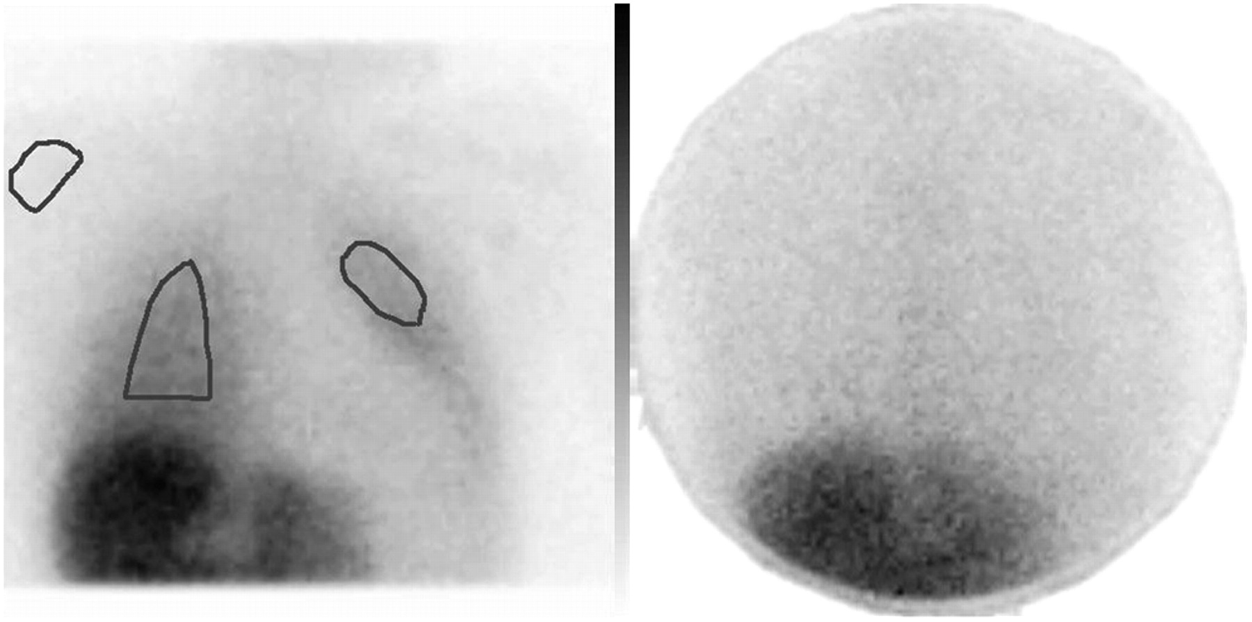
- FIGURE 1.
Planar scintigraphy of lung uptake of 123I-FP-CIT in healthy young male volunteer, at placebo session (left) and paroxetine session (right). Lung uptake is clearly visible after placebo but not after paroxetine. Activity is encoded from high (black) to low (white). Template with fixed ROIs for lungs and shoulder is shown on the left.
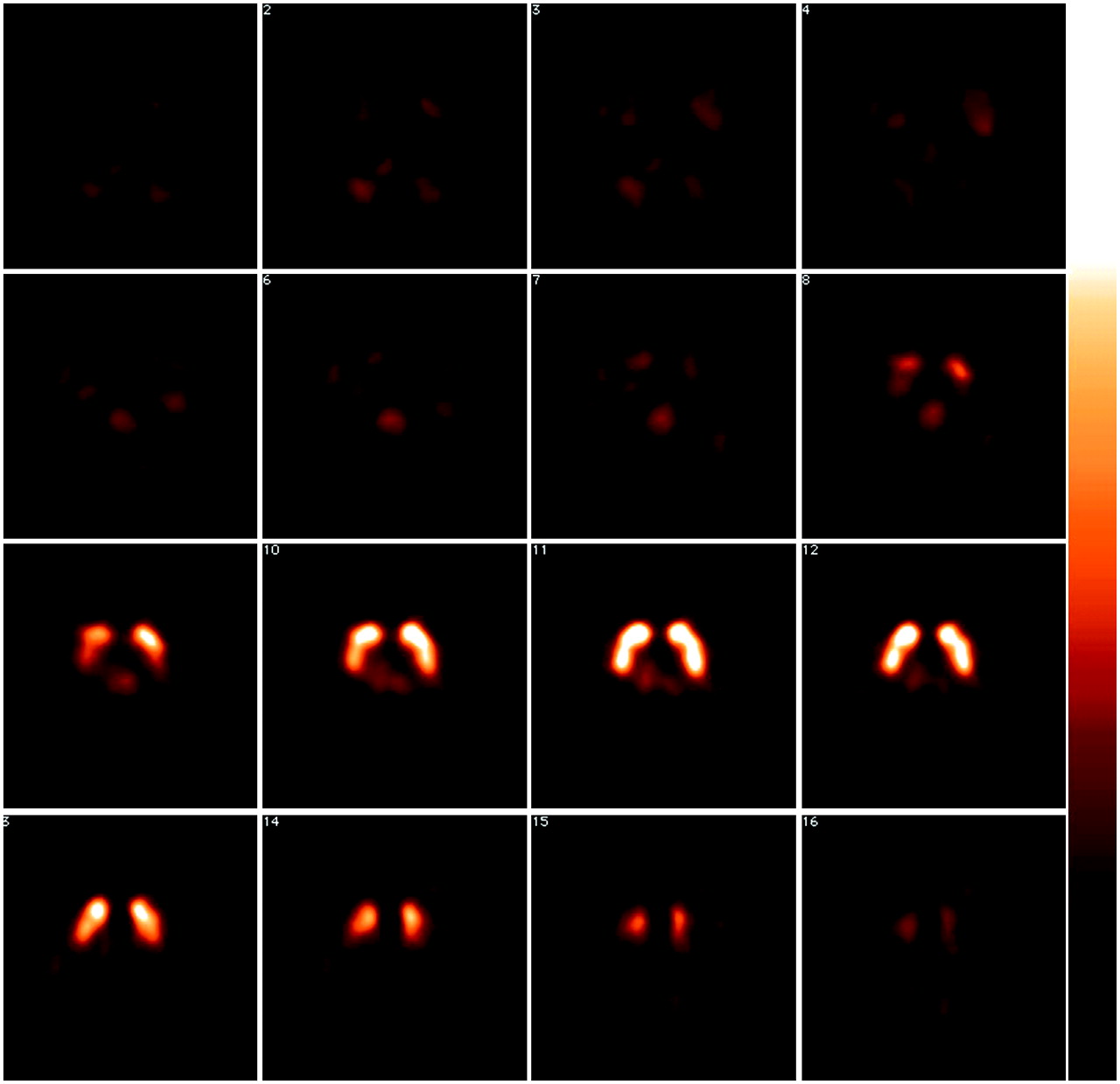
- FIGURE 2.
Transversal slices of 123I-FP-CIT SPECT scan, obtained in healthy young male volunteer 1 h after injection of radiotracer (placebo pretreatment). Intense and symmetric uptake is visualized in striatum. Binding is also visualized in midbrain and diencephalon area, which is higher than activity in cerebellum and cortical areas but much lower than binding in striatum. Activity is color encoded from high (white) to low (black).
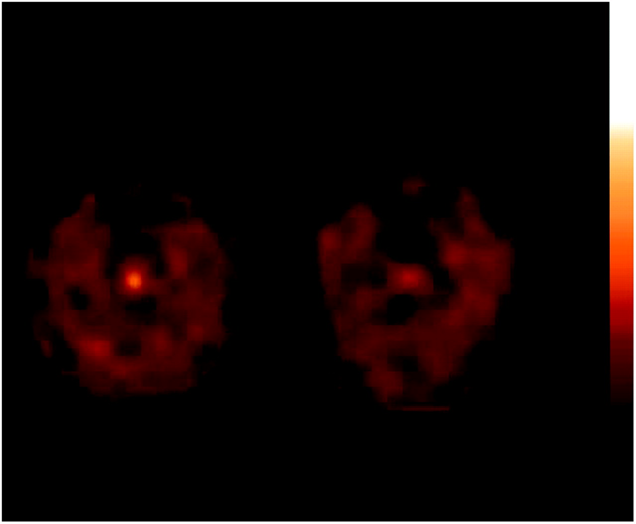
- FIGURE 3.
Transversal slices of 123I-FP-CIT SPECT scan at level of temporal cortex and midbrain, obtained in healthy young male volunteer 3 h after injection of radiotracer. After placebo pretreatment there is clear visualization of activity in midbrain (left), which is much lower after paroxetine pretreatment (right). Activity is color encoded from high (white) to low (black).
Tables
- TABLE 1
Specific-to-Nonspecific 123I-FP-CIT Binding Ratios, Obtained 1 and 3 Hours After Injection of Radiotracer, in a DAT-Rich Brain Area (Striatum) and in SERT-Rich Areas (Midbrain and Diencephalon)
1 h after injection* 3 h after injection 123I-FP-CIT Binding Placebo Paroxetine† Placebo Paroxetine DAT Striatum/cer.‡ 2.23 ± 0.67 2.37 ± 0.55 3.99 ± 0.44 4.12 ± 0.90 Striatum/occ. 2.99 ± 0.75 3.15 ± 0.48 4.20 ± 0.37 4.57 ± 0.53§ SERT Midbrain/cer. 0.10 ± 0.11 0.01 ± 0.15 0.20 ± 0.20 −0.02 ± 0.08§ Midbrain/occ. 0.36 ± 0.08 0.25 ± 0.15§ 0.24 ± 0.16 0.07 ± 0.09§ Diencephalon/cer. 0.23 ± 0.17 0.16 ± 0.17 0.43 ± 0.20 0.26 ± 0.19¶ Diencephalon/occ. 0.53 ± 0.13 0.44 ± 0.18¶ 0.48 ± 0.10 0.37 ± 0.12¶ ↵* In 1 control subject, cerebellum and midbrain area were not adequately scanned 1 h after injection of radiotracer; therefore, ratios of specific over cerebellar (cer.) binding are provided for 7 control subjects instead of 8.
↵† Placebo or paroxetine tablets (20 mg per session) were taken orally approximately 3 and 27 h before injection of radiotracer.
↵‡ Ratios are expressed as specific to nonspecific binding (±SD). Nonspecific binding represents activity in cerebellum (cer.) or occipital cortex (occ.).
↵§ Statistically significantly different from placebo condition.
↵¶ A trend for statistically significant difference (P = 0.054−0.14).
Healthy control subjects (n = 8) received placebo or paroxetine before injection of radiotracer (double-blind, crossover study design).
- TABLE 2
Specific-to-Nonspecific 123I-FP-CIT Binding Ratios, Obtained 1 and 3 Hours After Injection of Radiotracer, in DAT-Rich Brain Area (Striatum) and in SERT-Rich Areas (Midbrain and Diencephalon)
1 h after injection 3 h after injection 123I-FP-CIT Binding Placebo Paroxetine* Placebo Paroxetine DAT Striatum/cer.† 2.30 ± 0.80 2.10 ± 0.27 3.95 ± 0.36 3.71 ± 0.55 Striatum/occ. 2.95 ± 0.97 2.93 ± 0.44 3.97 ± 0.15 4.41 ± 0.43‡ SERT Midbrain/cer. 0.13 ± 0.12 −0.04 ± 0.14‡ 0.21 ± 0.14 −0.04 ± 0.05‡ Midbrain/occ. 0.35 ± 0.10 0.22 ± 0.17‡ 0.21 ± 0.12 0.10 ± 0.06 Diencephalon/cer. 0.29 ± 0.16 0.08 ± 0.13§ 0.51 ± 0.17 0.17 ± 0.15‡ Diencephalon/occ. 0.54 ± 0.15 0.38 ± 0.20§ 0.51 ± 0.10 0.34 ± 0.13‡ ↵* Placebo or paroxetine tablets (20 mg per session) were taken orally approximately 3 and 27 h before injection of radiotracer.
↵† Ratios are expressed as specific to nonspecific binding (±SD). Nonspecific binding represents activity in cerebellum (cer.) or occipital cortex (occ.).
↵‡ Statistically significantly different from placebo condition.
↵§ A trend for statistically significant difference (P = 0.07−0.09).
Healthy control subjects (n = 5) received placebo or paroxetine before injection of radiotracer (double-blind, crossover study design). In these 5 control subjects, at the paroxetine session, paroxetine plasma levels were >5 μg/L, whereas at the placebo session, paroxetine was not detectable in plasma (<5 μg/L).


Jump to section
Related Articles
Cited By...
- Modulation of functional networks related to the serotonin neurotransmitter system by citalopram: evidence from a multimodal neuroimaging study
- Imaging Dopaminergic Neurotransmission in Neurodegenerative Disorders
- Optimization of Parameters for Quantitative Analysis of 123I-Ioflupane SPECT Images for Monitoring Progression of Parkinson Disease
- Serotonin transporter binding and anxiety symptoms in Parkinsons disease
- Analysis of Extrastriatal 123I-FP-CIT Binding Contributes to the Differential Diagnosis of Parkinsonian Diseases
- Relevance of 123I-FP-CIT SPECT brain scans in routine clinical settings
- Use of 11C-PE2I PET in Differential Diagnosis of Parkinsonian Disorders
- Safety Analysis of 10 Clinical Trials and for 13 Years After First Approval of Ioflupane 123I Injection (DaTscan)
- (123I)FP-CIT SPECT in suspected dementia with Lewy bodies: a longitudinal case study
- Receiver-Operating-Characteristic Analysis of an Automated Program for Analyzing Striatal Uptake of 123I-Ioflupane SPECT Images: Calibration Using Visual Reads
- A New Era of Clinical Dopamine Transporter Imaging Using 123I-FP-CIT
- Differentiation of frontotemporal dementia from dementia with Lewy bodies using FP-CIT SPECT
- Assessing the Optimal Time Point for the Measurement of Extrastriatal Serotonin Transporter Binding with 123I-FP-CIT SPECT in Healthy, Male Subjects
- Serotonin Transporters in Dopamine Transporter Imaging: A Head-to-Head Comparison of Dopamine Transporter SPECT Radioligands 123I-FP-CIT and 123I-PE2I
- Molecular Imaging of the Dopamine Transporter