
Main menu

  • Home
  • Content
    • Current
    • Ahead of print
    • Past Issues
    • JNM Supplement
    • SNMMI Annual Meeting Abstracts
    • Continuing Education
    • JNM Podcasts
  • Subscriptions
    • Subscribers
    • Institutional and Non-member
    • Rates
    • Journal Claims
    • Corporate & Special Sales
  • Authors
    • Submit to JNM
    • Information for Authors
    • Assignment of Copyright
    • AQARA requirements
  • Info
    • Reviewers
    • Permissions
    • Advertisers
  • About
    • About Us
    • Editorial Board
    • Contact Information
  • More
    • Alerts
    • Feedback
    • Help
    • SNMMI Journals
  • SNMMI
    • JNM
    • JNMT
    • SNMMI Journals
    • SNMMI

User menu

  • Subscribe
  • My alerts
  • Log in
  • Log out
  • My Cart

Search

  • Advanced search
Journal of Nuclear Medicine
  • SNMMI
    • JNM
    • JNMT
    • SNMMI Journals
    • SNMMI
  • Subscribe
  • My alerts
  • Log in
  • Log out
  • My Cart
Journal of Nuclear Medicine

Advanced Search

  • Home
  • Content
    • Current
    • Ahead of print
    • Past Issues
    • JNM Supplement
    • SNMMI Annual Meeting Abstracts
    • Continuing Education
    • JNM Podcasts
  • Subscriptions
    • Subscribers
    • Institutional and Non-member
    • Rates
    • Journal Claims
    • Corporate & Special Sales
  • Authors
    • Submit to JNM
    • Information for Authors
    • Assignment of Copyright
    • AQARA requirements
  • Info
    • Reviewers
    • Permissions
    • Advertisers
  • About
    • About Us
    • Editorial Board
    • Contact Information
  • More
    • Alerts
    • Feedback
    • Help
    • SNMMI Journals
  • View or Listen to JNM Podcast
  • Visit JNM on Facebook
  • Join JNM on LinkedIn
  • Follow JNM on Twitter
  • Subscribe to our RSS feeds
OtherBASIC SCIENCE INVESTIGATIONS

Noninvasive Imaging of Osteoclasts in Parathyroid Hormone–Induced Osteolysis Using a 64Cu-Labeled RGD Peptide

Jennifer E. Sprague, Hideki Kitaura, Wei Zou, Yunpeng Ye, Samuel Achilefu, Katherine N. Weilbaecher, Steven L. Teitelbaum and Carolyn J. Anderson
Journal of Nuclear Medicine February 2007, 48 (2) 311-318;
Jennifer E. Sprague
  • Find this author on Google Scholar
  • Find this author on PubMed
  • Search for this author on this site
Hideki Kitaura
  • Find this author on Google Scholar
  • Find this author on PubMed
  • Search for this author on this site
Wei Zou
  • Find this author on Google Scholar
  • Find this author on PubMed
  • Search for this author on this site
Yunpeng Ye
  • Find this author on Google Scholar
  • Find this author on PubMed
  • Search for this author on this site
Samuel Achilefu
  • Find this author on Google Scholar
  • Find this author on PubMed
  • Search for this author on this site
Katherine N. Weilbaecher
  • Find this author on Google Scholar
  • Find this author on PubMed
  • Search for this author on this site
Steven L. Teitelbaum
  • Find this author on Google Scholar
  • Find this author on PubMed
  • Search for this author on this site
Carolyn J. Anderson
  • Find this author on Google Scholar
  • Find this author on PubMed
  • Search for this author on this site
  • Article
  • Figures & Data
  • Info & Metrics
  • PDF
Loading

Article Figures & Data

Figures

  • Tables
  • FIGURE 1. 
    • Download figure
    • Open in new tab
    • Download powerpoint
    FIGURE 1. 

    Structure of CB-TE2A-c(RGDyK).
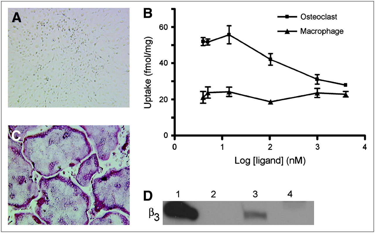
  • FIGURE 2. 
    • Download figure
    • Open in new tab
    • Download powerpoint
    FIGURE 2. 

    Uptake of 64Cu-CB-TE2A-c(RGDyK) by osteoclasts and macrophages. BMMs were harvested from mice and grown in culture. (A) Cells treated with only M-CSF remained TRAP(−) mononuclear macrophages. (C) Cells cultured in presence of MCSF plus RANKL differentiated into large, TRAP(+) osteoclasts (red stain). (B) Cell uptake was determined 2 h after addition of 64Cu-CB-TE2A-c(RGDyK) (4 nM) plus c(RGDyK) (0–4,000 nM) to medium of cells grown in 6-well plates. Each data point represents average of triplicate measurements. (D) Integrin expression was evaluated by Western blot against β3. Lanes: 1 = αvβ3 standard, 10 ng; 2 = empty; 3 = osteoclast lysate, 20 μg; 4 = macrophage lysate, 20 μg.
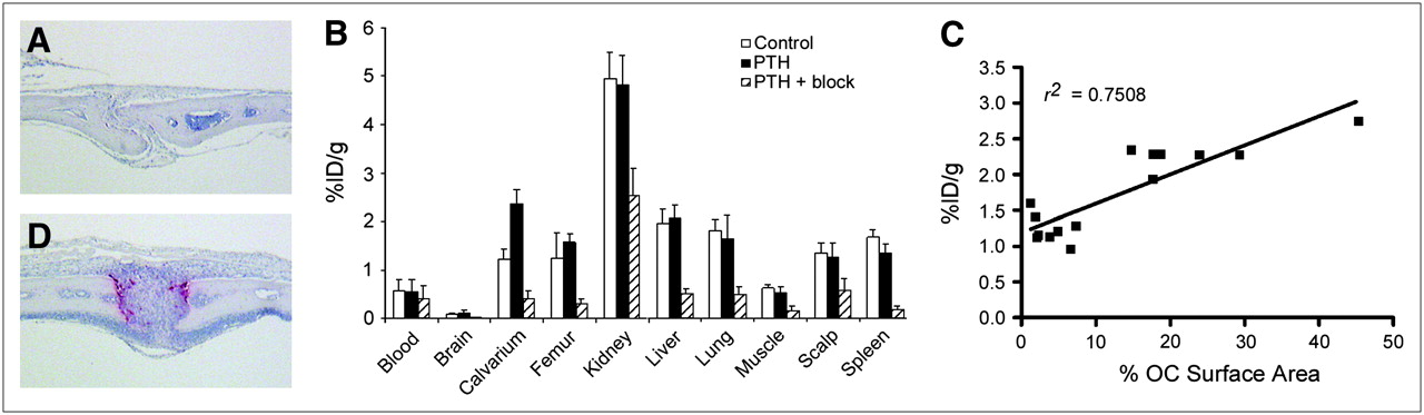
  • FIGURE 3. 
    • Download figure
    • Open in new tab
    • Download powerpoint
    FIGURE 3. 

    PTH induces osteoclasts resulting in increased uptake of 64Cu-CB-TE2A-c(RGDyK) at calvarium. TRAP-stained sections of calvarium of control mice (A) and PTH-treated mice (D) confirm osteoclast induction after PTH treatment. Osteoclasts stain red. (B) Biodistribution (1 h after injection) was performed on control mice (n = 8), PTH-treated mice (n = 8), and PTH-plus-block mice injected with 740 kBq (20 μCi) (10.5–12 ng) of 64Cu-CB-TE2A-c(RGDyK). Note that n = 7 for PTH femur because of contamination of 1 sample by urine. (C) Uptake of 64Cu-CB-TE2A-c(RGDyK) (biodistribution 1 h after injection: control, n = 7; PTH, n = 7) was plotted against ratio of osteoclast surface area to total surface area.
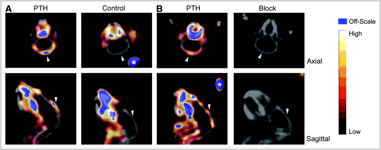
  • FIGURE 4. 
    • Download figure
    • Open in new tab
    • Download powerpoint
    FIGURE 4. 

    Small-animal PET/CT of PTH-treated mice. Calvarium uptake of 64Cu-CB-TE2A-c(RGDyK) was higher in PTH-treated mice (7.4 MBq [199 μCi],115 ng, SUV = 0.53) than in control mice (7.7 MBq [209 μCi], 121 ng, SUV = 0.22) (50- to 60-min summed dynamic image) (A). In PTH-treated mice, uptake was reduced in all tissues, including calvarium, after injection of c(RGDyK) (PTH [left]: 159 μCi, 84 ng, SUV = 0.33; block [right]: 164 μCi, 87 ng, SUV = 0.18) (static image obtained 60 min after injection, 10-min scan) (B). Arrowheads indicate calvarium of each animal. Fiducials (*) are indicated.

Tables

  • Figures
    • View popup
    TABLE 1

    Affinity of c(RGDyK) and Cu(II)-CB-TE2A-c(RGDyK) for Integrins αvβ3 and αvβ5 as Determined in Heterologous Competitive Binding Assay Using Biotinylated Vitronectin

    αvβ3αvβ5
    CompoundIC50 (nM)95% confidence intervalIC50 (nM)95% confidence interval
    Cu(II)-CBTE2A-c(RGDyK)6.03.7–9.6171110–266
    c(RGDyK)3.72.7–5.0194142–266
PreviousNext
Back to top

In this issue

Journal of Nuclear Medicine: 48 (2)
Journal of Nuclear Medicine
Vol. 48, Issue 2
February 2007
  • Table of Contents
  • Table of Contents (PDF)
  • About the Cover
  • Index by author
Print
Download PDF
Article Alerts
Sign In to Email Alerts with your Email Address
Email Article

Thank you for your interest in spreading the word on Journal of Nuclear Medicine.

NOTE: We only request your email address so that the person you are recommending the page to knows that you wanted them to see it, and that it is not junk mail. We do not capture any email address.

Enter multiple addresses on separate lines or separate them with commas.
Noninvasive Imaging of Osteoclasts in Parathyroid Hormone–Induced Osteolysis Using a 64Cu-Labeled RGD Peptide
(Your Name) has sent you a message from Journal of Nuclear Medicine
(Your Name) thought you would like to see the Journal of Nuclear Medicine web site.
Citation Tools
Noninvasive Imaging of Osteoclasts in Parathyroid Hormone–Induced Osteolysis Using a 64Cu-Labeled RGD Peptide
Jennifer E. Sprague, Hideki Kitaura, Wei Zou, Yunpeng Ye, Samuel Achilefu, Katherine N. Weilbaecher, Steven L. Teitelbaum, Carolyn J. Anderson
Journal of Nuclear Medicine Feb 2007, 48 (2) 311-318;

Citation Manager Formats

  • BibTeX
  • Bookends
  • EasyBib
  • EndNote (tagged)
  • EndNote 8 (xml)
  • Medlars
  • Mendeley
  • Papers
  • RefWorks Tagged
  • Ref Manager
  • RIS
  • Zotero
Share
Noninvasive Imaging of Osteoclasts in Parathyroid Hormone–Induced Osteolysis Using a 64Cu-Labeled RGD Peptide
Jennifer E. Sprague, Hideki Kitaura, Wei Zou, Yunpeng Ye, Samuel Achilefu, Katherine N. Weilbaecher, Steven L. Teitelbaum, Carolyn J. Anderson
Journal of Nuclear Medicine Feb 2007, 48 (2) 311-318;
Twitter logo Facebook logo LinkedIn logo Mendeley logo
  • Tweet Widget
  • Facebook Like
  • Google Plus One
Bookmark this article

Jump to section

  • Article
    • Abstract
    • MATERIALS AND METHODS
    • RESULTS
    • DISCUSSION
    • Acknowledgments
    • Footnotes
    • References
  • Figures & Data
  • Info & Metrics
  • PDF

Related Articles

  • This Month in JNM
  • PubMed
  • Google Scholar

Cited By...

  • Radiopeptide Imaging and Therapy in the United States
  • Targeting the {alpha}v{beta}3 Integrin for Small-Animal PET/CT of Osteolytic Bone Metastases
  • Biodegradable dendritic positron-emitting nanoprobes for the noninvasive imaging of angiogenesis
  • Google Scholar

More in this TOC Section

  • Tumor-Specific Binding of Radiolabeled PEGylated GIRLRG Peptide: A Novel Agent for Targeting Cancers
  • PET/MRI of Hypoxic Atherosclerosis Using 64Cu-ATSM in a Rabbit Model
  • Tumor Uptake of Anti-CD20 Fabs Depends on Tumor Perfusion
Show more Basic Science Investigations

Similar Articles

SNMMI

© 2025 SNMMI

Powered by HighWire