Immuno-PET imaging: Shively reviews the promise of routine clinical immuno-PET imaging as an adjunct to therapy and previews a paper in this issue of JNM on the development of diabodies for this purpose.
Page 170
Quantifying tissue ACE activity: Chandrashekhar and Narula review current uses and complications of angiotensin-converting enzyme inhibitors and preview results in this issue of JNM that could lead to routine identification of patients most likely to benefit from such therapy.
Page 173
Microvascular dysfunction in angina: Graf and colleagues use 13N-ammonia PET to investigate the incidence of dysfunctional microcirculation in patients with angina and search for predictive parameters of reduced coronary flow reserve.
Page 175
Imaging tissue ACE: Dilsizian and colleagues explore 18F-FBL binding to tissue angiotensin-converting enzyme (ACE) in explanted hearts and discuss the potential for external imaging in monitoring tissue ACE and ACE-inhibitor therapies in progressive heart failure.
Page 182

Misregistration in cardiac perfusion PET/CT: Martinez-Möller and colleagues assess the clinical impact of emission–transmission misalignment in PET/CT myocardial perfusion imaging and describe promising correction approaches.
Page 188
Evaluating subcortical aphasia: Choi and colleagues examine the relationship between the severity of aphasia and regional cerebral perfusion on brain SPECT using statistical parametric mapping and probabilistic anatomic maps in patients with striatocapsular infarction.
Page 194

SPECT/CT advantages in breast cancer: Lerman and colleagues assess whether the addition of SPECT/CT to lymphoscintigraphy improves sentinel node identification in overweight patients with breast cancer and outline additional benefits of this technique.
Page 201
Imaging opioid receptors in lung cancer: Madar and colleagues use PET to measure human kinetics and distribution of the selective δ-opioid antagonist 11C-MeNTI and the μ-opioid agonist 11C-CFN in the 3 major human lung carcinomas.
Page 207

Sensitive and specific: Kim and colleagues investigate the performance of PET/CT in characterizing solitary pulmonary nodules in a clinical referral setting.
Page 214
Precise anatomic localization and more: Shammas and colleagues evaluate the use of PET/CT in patients with suspected thyroid cancer recurrence and discuss benefits in changed patient management and guidance of therapeutic intervention.
Page 221

Characterization of atherosclerotic plaques: Wu and colleagues examine the correlation between vascular 18F-FDG uptake on PET/CT and circulating matrix metalloproteinase 1 levels in patients with significant carotid stenosis and in healthy volunteers.
Page 227

Variations in cardiac 18F-FDG uptake: Israel and colleagues assess the influence of more than 50 clinical factors on tracer uptake in patients undergoing routine 18F-FDG PET/CT and point to the possible deleterious effects of some pharmaceuticals on image quality.
Page 234
PET in salivary gland cancer: Roh and colleagues evaluate the clinical utility and potential roles of 18F-FDG PET in the management of patients with salivary gland cancers.
Page 240
Novel brain glutamate tracer: Ametamey and colleagues describe the characteristics of an 11C-labeled tracer and assess its suitability as a PET ligand for imaging metabotropic glutamate receptor subtype 5 distribution in humans
Page 247

Scaling dose conversion factors: Siegel and Stabin review and evaluate methods for mass scaling of S values for blood-based estimation of red marrow absorbed dose.
Page 253
Evolution of NM training: Graham and Metter provide an educational overview of the development of nuclear medicine training programs and preview new 2007 requirements as well as the outlook for maintenance-of-certification issues.
Page 257
Reducing RIT toxicity: Mårtensson and colleagues investigate in a rat model the possibility of decreasing the myelotoxicity associated with radioimmunotherapy by extracorporeal depletion of radioimmunoconjugates from the circulation.
Page 269
Small-animal behavioral neuroimaging: Schiffer and colleagues report on a simplified PET imaging approach to animal behavioral models of brain activation using an intraperitoneal administration route and a standard uptake value to reduce scan duration.
Page 277
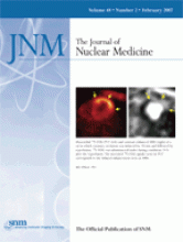
A workable PET/MRI approach: Higuchi and colleagues explore the feasibility of combining data from small-animal PET and a clinical MRI system for the quantification of ischemic injury and infarct size in a rat model.
Page 288
Trimodality imaging: Deroose and colleagues combine small-nimal PET and bioluminescence technology with small-animal CT in a rat model of tumor xenografts and metastases to obtain images with both molecular and anatomic information.
Page 295

Diabody PET for CEA-producing malignancies: Cai and colleagues investigated the utility of a genetically engineered and 18F-labeled anticarcinoembryonic antigen diabody for small-animal PET imaging of CEA expression in colon carcinoma xenograft-bearing mice.
Page 304
64Cu-RGD imaging of osteoclasts: Sprague and colleagues explore the use of radiolabeled RGD peptides in the detection of osteoclasts in lytic bone lesions and discuss the implications for imaging of bone metastasis and inflammatory osteolysis.
Page 311

Low-dose SPECT/CT bone imaging: Even-Sapir and colleagues assess the role of SPECT/multislice low-dose CT in the imaging approach to nononcologic patients referred for 99mTc-MDP bone scintigraphy.
Page 319
ON THE COVER
The combination of PET and MRI may represent an ideal platform for noninvasive imaging in small-animal cardiovascular research. This example in a reperfusion model indicates the potential for delineating the extent of reversibly injured myocardium on 18F-FDG and the extent of necrosis on MRI. By combining both signals, one can follow the extent of ischemic injury and relate it to experimental therapies. Additionally, molecular tracers that are more specific may be substituted for 18F-FDG to explore the full potential of integrating MRI-defined morphology with PET-derived biologic signals.

SEE PAGE 293


