
Main menu

  • Home
  • Content
    • Current
    • Ahead of print
    • Past Issues
    • JNM Supplement
    • SNMMI Annual Meeting Abstracts
    • Continuing Education
    • JNM Podcasts
  • Subscriptions
    • Subscribers
    • Institutional and Non-member
    • Rates
    • Journal Claims
    • Corporate & Special Sales
  • Authors
    • Submit to JNM
    • Information for Authors
    • Assignment of Copyright
    • AQARA requirements
  • Info
    • Reviewers
    • Permissions
    • Advertisers
  • About
    • About Us
    • Editorial Board
    • Contact Information
  • More
    • Alerts
    • Feedback
    • Help
    • SNMMI Journals
  • SNMMI
    • JNM
    • JNMT
    • SNMMI Journals
    • SNMMI

User menu

  • Subscribe
  • My alerts
  • Log in
  • My Cart

Search

  • Advanced search
Journal of Nuclear Medicine
  • SNMMI
    • JNM
    • JNMT
    • SNMMI Journals
    • SNMMI
  • Subscribe
  • My alerts
  • Log in
  • My Cart
Journal of Nuclear Medicine

Advanced Search

  • Home
  • Content
    • Current
    • Ahead of print
    • Past Issues
    • JNM Supplement
    • SNMMI Annual Meeting Abstracts
    • Continuing Education
    • JNM Podcasts
  • Subscriptions
    • Subscribers
    • Institutional and Non-member
    • Rates
    • Journal Claims
    • Corporate & Special Sales
  • Authors
    • Submit to JNM
    • Information for Authors
    • Assignment of Copyright
    • AQARA requirements
  • Info
    • Reviewers
    • Permissions
    • Advertisers
  • About
    • About Us
    • Editorial Board
    • Contact Information
  • More
    • Alerts
    • Feedback
    • Help
    • SNMMI Journals
  • View or Listen to JNM Podcast
  • Visit JNM on Facebook
  • Join JNM on LinkedIn
  • Follow JNM on Twitter
  • Subscribe to our RSS feeds
Research ArticleCLINICAL INVESTIGATIONS

Subcortical Aphasia After Striatocapsular Infarction: Quantitative Analysis of Brain Perfusion SPECT Using Statistical Parametric Mapping and a Statistical Probabilistic Anatomic Map

Joon Young Choi, Kwang Ho Lee, Duk L. Na, Hong Sik Byun, Soo Joo Lee, Hyanghee Kim, Miseon Kwon, Kyung-Han Lee and Byung-Tae Kim
Journal of Nuclear Medicine February 2007, 48 (2) 194-200;
Joon Young Choi
  • Find this author on Google Scholar
  • Find this author on PubMed
  • Search for this author on this site
Kwang Ho Lee
  • Find this author on Google Scholar
  • Find this author on PubMed
  • Search for this author on this site
Duk L. Na
  • Find this author on Google Scholar
  • Find this author on PubMed
  • Search for this author on this site
Hong Sik Byun
  • Find this author on Google Scholar
  • Find this author on PubMed
  • Search for this author on this site
Soo Joo Lee
  • Find this author on Google Scholar
  • Find this author on PubMed
  • Search for this author on this site
Hyanghee Kim
  • Find this author on Google Scholar
  • Find this author on PubMed
  • Search for this author on this site
Miseon Kwon
  • Find this author on Google Scholar
  • Find this author on PubMed
  • Search for this author on this site
Kyung-Han Lee
  • Find this author on Google Scholar
  • Find this author on PubMed
  • Search for this author on this site
Byung-Tae Kim
  • Find this author on Google Scholar
  • Find this author on PubMed
  • Search for this author on this site
  • Article
  • Figures & Data
  • Info & Metrics
  • PDF
Loading

Article Figures & Data

Figures

  • Tables
  • FIGURE 1. 
    • Download figure
    • Open in new tab
    • Download powerpoint
    FIGURE 1. 

    Representative images of T2-weighted MRI (A and D), diffusion-weighted MRI (B and E), and perfusion SPECT (C and F). (A–C) Patient with severe aphasia (AQ = 57.4 points) shows severe extensive left cerebral cortical hypoperfusion (AI of cerebral cortex = 14.93). (D–F) Another patient with mild aphasia (AQ = 90.9 points) had only mild hypoperfusion in left parietal cortex (AI of cerebral cortex = 2.53).
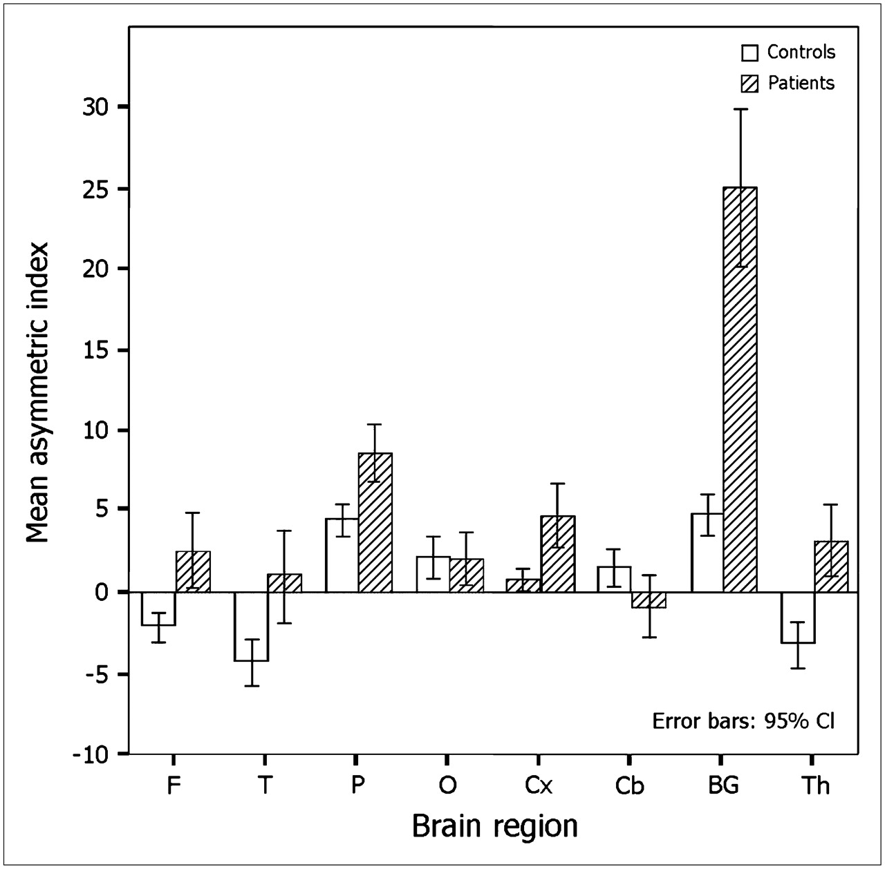
  • FIGURE 2. 
    • Download figure
    • Open in new tab
    • Download powerpoint
    FIGURE 2. 

    Comparisons of regional AIs between healthy control subjects and patients. F = frontal cortex; T = temporal cortex; P = parietal cortex; O = occipital cortex; Cx = whole cerebral cortex; Cb = cerebellar cortex; BG = basal ganglia; Th = thalamus; CI = confidence interval.
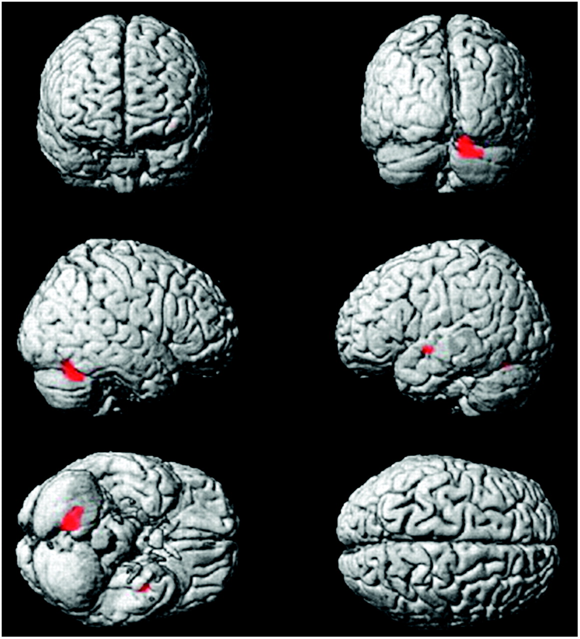
  • FIGURE 3. 
    • Download figure
    • Open in new tab
    • Download powerpoint
    FIGURE 3. 

    Voxel-based SPM analysis shows significant voxels on SPECT data to correlate with the AQ. Perfusion of left temporal cortex and right cerebellum was positively correlated with the AQ.

Tables

  • Figures
    • View popup
    TABLE 1

    Clinical Characteristics of Patients with Isolated SCI

    Patient no.Age (y)Aphasia typeAQ (points)Infarct size (mm)Regional asymmetric index on brain perfusion SPECT
    SexMRAFTPOCxCbBGTh
    180FGlobal6.838M1 occlusion3.422.9310.110.585.72−4.2327.219.63
    275MGlobal13.036M1 occlusion8.335.8911.532.668.61−8.4122.049.59
    367MTS53.437Normal2.783.467.345.265.40−4.2643.383.14
    464FMixed T54.028M1 occlusion3.344.8112.473.496.92−4.0328.794.22
    567FTS57.439M1 occlusion13.4314.2518.018.6814.932.6235.579.79
    677FMixed T66.033M1 occlusion4.865.789.51−1.256.29−2.2922.144.75
    768MMixed T67.544Normal4.213.067.71−1.264.67−3.8028.464.05
    849MTM79.230Normal−0.270.956.55−0.492.150.5234.003.92
    968FAnomic80.331Normal1.43−1.858.602.483.88−0.2616.090.25
    1067MTM81.628Normal2.10−4.105.20−2.681.460.8318.061.17
    1175FAnomic84.418M1 stenosis−3.84−5.797.554.330.733.7111.34−1.20
    1263FAnomic86.822Normal4.18−1.418.861.694.840.8028.08−1.39
    1346MAnomic87.640Normal0.211.237.082.973.63−2.0329.553.42
    1456MAnomic88.811M1 stenosis−2.88−4.567.44−1.020.651.6211.25−3.71
    1556FAnomic90.922Normal−1.07−1.925.735.332.531.0614.14−0.54
    1658MNormal95.127Normal1.64−4.554.452.082.134.6130.623.43
    • MRA = MR angiography; F = frontal cortex; T = temporal cortex; P = parietal cortex; O = occipital cortex; Cx = whole cerebral cortex; Cb = cerebellar cortex; BG = basal ganglia; Th = thalamus; M1 = horizontal segment of middle cerebral artery; TS = transcortical sensory; mixed T = mixed transcortical; TM = transcortical motor.

    • View popup
    TABLE 2

    Results of Correlation Analysis Between Regional AIs, AQ, Infarct Size, and Age

    Correlation with AQCorrelation with infarct sizeCorrelation with age
    AIMean ± SDRangeρP valueρP valueρP value
    Frontal cortex2.62 ± 4.19−3.84 ∼ 13.43−0.653<0.010.594<0.050.532<0.05
    Temporal cortex1.14 ± 5.21−5.79 ∼ 14.25−0.782<0.0010.7290.0010.379NS
    Parietal cortex8.63 ± 3.304.45 ∼ 18.01−0.694<0.0050.328NS0.597<0.05
    Occipital cortex2.05 ± 3.01−2.68 ∼ 8.68−0.053NS0.010NS0.321NS
    Whole cerebral cortex4.66 ± 3.570.65 ∼ 14.93−0.7680.0010.599<0.050.491NS
    Cerebellar cortex−0.85 ± 3.49−8.41 ∼ 4.610.7650.001−0.588<0.05−0.388NS
    Basal ganglia25.05 ± 9.1711.25 ∼ 43.38−0.312NS0.560<0.05−0.206NS
    Thalamus3.16 ± 4.04−3.71 ∼ 9.79−0.721<0.0050.713<0.0050.453NS
    Infarct size (mm)30 ± 911 ∼ 44−0.594<0.05NANA0.311NS
    Age (y)64.8 ± 9.646.4 ∼ 79.6−0.6650.0050.311NSNANA
    • NS = not significant; NA = not applicable.

PreviousNext
Back to top

In this issue

Journal of Nuclear Medicine: 48 (2)
Journal of Nuclear Medicine
Vol. 48, Issue 2
February 2007
  • Table of Contents
  • Table of Contents (PDF)
  • About the Cover
  • Index by author
Print
Download PDF
Article Alerts
Sign In to Email Alerts with your Email Address
Email Article

Thank you for your interest in spreading the word on Journal of Nuclear Medicine.

NOTE: We only request your email address so that the person you are recommending the page to knows that you wanted them to see it, and that it is not junk mail. We do not capture any email address.

Enter multiple addresses on separate lines or separate them with commas.
Subcortical Aphasia After Striatocapsular Infarction: Quantitative Analysis of Brain Perfusion SPECT Using Statistical Parametric Mapping and a Statistical Probabilistic Anatomic Map
(Your Name) has sent you a message from Journal of Nuclear Medicine
(Your Name) thought you would like to see the Journal of Nuclear Medicine web site.
Citation Tools
Subcortical Aphasia After Striatocapsular Infarction: Quantitative Analysis of Brain Perfusion SPECT Using Statistical Parametric Mapping and a Statistical Probabilistic Anatomic Map
Joon Young Choi, Kwang Ho Lee, Duk L. Na, Hong Sik Byun, Soo Joo Lee, Hyanghee Kim, Miseon Kwon, Kyung-Han Lee, Byung-Tae Kim
Journal of Nuclear Medicine Feb 2007, 48 (2) 194-200;

Citation Manager Formats

  • BibTeX
  • Bookends
  • EasyBib
  • EndNote (tagged)
  • EndNote 8 (xml)
  • Medlars
  • Mendeley
  • Papers
  • RefWorks Tagged
  • Ref Manager
  • RIS
  • Zotero
Share
Subcortical Aphasia After Striatocapsular Infarction: Quantitative Analysis of Brain Perfusion SPECT Using Statistical Parametric Mapping and a Statistical Probabilistic Anatomic Map
Joon Young Choi, Kwang Ho Lee, Duk L. Na, Hong Sik Byun, Soo Joo Lee, Hyanghee Kim, Miseon Kwon, Kyung-Han Lee, Byung-Tae Kim
Journal of Nuclear Medicine Feb 2007, 48 (2) 194-200;
Twitter logo Facebook logo LinkedIn logo Mendeley logo
  • Tweet Widget
  • Facebook Like
  • Google Plus One
Bookmark this article

Jump to section

  • Article
    • Abstract
    • MATERIALS AND METHODS
    • RESULTS
    • DISCUSSION
    • CONCLUSION
    • Acknowledgments
    • Footnotes
    • References
  • Figures & Data
  • Info & Metrics
  • PDF

Related Articles

  • This Month in JNM
  • PubMed
  • Google Scholar

Cited By...

  • The Importance of Premotor Cortex for Supporting Speech Production after Left Capsular-Putaminal Damage
  • Google Scholar

More in this TOC Section

  • Feasibility of Ultra-Low-Activity 18F-FDG PET/CT Imaging Using a Long–Axial-Field-of-View PET/CT System
  • Cardiac Presynaptic Sympathetic Nervous Function Evaluated by Cardiac PET in Patients with Chronotropic Incompetence Without Heart Failure
  • Validation and Evaluation of a Vendor-Provided Head Motion Correction Algorithm on the uMI Panorama PET/CT System
Show more Clinical Investigations

Similar Articles

SNMMI

© 2025 SNMMI

Powered by HighWire