


Abstract
18F-FDG PET/CT has rapidly become a widely used imaging modality for evaluating a variety of malignancies, including squamous cell carcinoma of the head and neck and thyroid cancer. Using both published data and the multidisciplinary experience at our institution, we provide a practical set of guidelines and algorithms for the use of 18F-FDG PET/CT in the evaluation and management of head and neck cancer and thyroid cancer.
Squamous cell carcinoma of the head and neck (HNSCC), which is newly diagnosed in nearly 40,000 patients annually in the United States, has a tremendous impact on basic functions, such as speech and swallowing, and its treatment can significantly affect patient appearance (1). The overall annual mortality rate for head and neck cancer in the United States is 23%, and the 5-y survival rate is 56% (1–3). Worldwide, HNSCC is an even larger issue. Nasopharyngeal cancer has an incidence of as high as 25 per 100,000 in southern China; in India, HNSCC accounts for 25% of all male carcinomas and 10% of all female carcinomas (4,5). Over 80% of early-stage tumors are cured, but nearly one half of patients have evidence of advanced local disease or lymph node metastases at the time of diagnosis. Therapy often requires extensive multidisciplinary collaboration among specialists in head and neck surgery, radiation oncology, medical oncology, prosthodontics, and speech therapy. Diagnostic imaging plays an important role in accurate staging, restaging, and treatment monitoring and is essential in both planning adequate treatment and minimizing treatment-related toxicity and functional impairment. MRI and CT remain the primary imaging modalities for the assessment of HNSCC, but 18F-FDG PET/CT has emerged as a vital adjunct when used in the appropriate clinical setting.
The use of 18F-FDG PET/CT for the evaluation of head and neck cancer has been most widely assessed for HNSCC, although it has also been studied for other malignancies, such as those of salivary gland and thyroid origins. PET/CT rarely adds additional useful information regarding the initial T stage of the primary tumor because the combination of clinical mucosal evaluation and MRI or CT better evaluates local soft-tissue and bony anatomy. PET/CT can be helpful, however, in several clinical scenarios: delineation of extent of regional lymph node involvement, detection of distant metastases, identification of an unknown primary tumor, detection of an occasional synchronous primary tumor, monitoring of the treatment response, and long-term surveillance for recurrence and metastases (Table 1).
Key Benefits of 18F-FDG PET/CT for HNSCC
Here we review the literature on the use of 18F-FDG PET and PET/CT for HNSCC, discuss how diagnostic imaging and PET/CT are used at our institution, and provide practical guidelines and algorithms for the use of 18F-FDG PET/CT for head and neck cancer. We also review the use of PET/CT for thyroid cancer. It should be noted that we do not routinely use intravenous contrast material for the CT component of PET/CT examinations at our institution (these noncontrast CT examinations are reviewed by a board-certified radiologist, and the results are integrated into the PET report). When we do, PET/CT is treated as 2 separate examinations, with a specialist in nuclear medicine interpreting the PET examination and a radiologist interpreting the neck CT and whole-body CT examinations. Our head and neck PET/CT protocol consists of a whole-body scan with the arms above the patient's head and a separate, dedicated acquisition of the head and neck with the arms down and a slightly longer acquisition time.
INITIAL STAGING OF HNSCC
Accurate delineation of the primary tumor and the extent of regional lymph node metastases is critical for staging of the tumor and for determination of the optimal initial therapeutic approach, including planning the extent of surgery and delineating targets for radiation therapy. Although numerous reports on initial staging have shown that PET is at least as sensitive as MRI or CT in detecting the primary tumor (6–8), PET and PET/CT (without contrast material) do not provide the anatomic definition that MRI and contrast-enhanced multislice CT can provide. Therefore, there is a limited role for PET in defining the T stage of the primary tumor. One exception is the use of 18F-FDG PET in identifying the primary tumor site in patients who present with cervical lymph node metastatic HNSCC from a clinically undetectable or unknown primary tumor. This use of 18F-FDG PET is discussed further in the next section.
For evaluating metastatic disease in cervical lymph nodes, previous studies showed that 18F-FDG PET/CT is comparable or superior to conventional imaging in detecting regional lymph node metastases during initial staging (9–12). In a review by Schoder and Yeung, an average sensitivity of 87%–90% and a specificity of 80%–93% were reported for 18F-FDG PET/CT (13); a sensitivity of 61%–97% and a specificity of 21%–100% were reported for MRI or CT. A more recent study suggested that MRI may be slightly more sensitive than PET for identifying regional lymph node metastases (93% for MRI vs. 85% for PET) (14); specificity was comparable between the 2 imaging modalities (95% for MRI vs. 98% for PET). However, a nonhybrid PET scanner without PET/CT fusion was used in that study; integrated PET/CT may be as good as or better than both MRI and PET used alone.
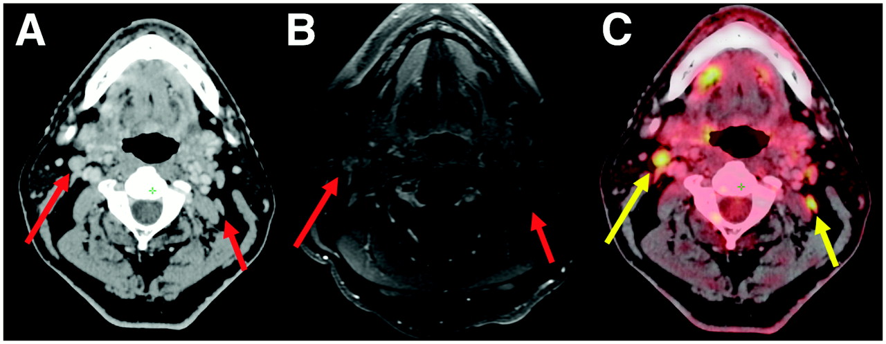
18F-FDG PET/CT can certainly play an important role in identifying disease in lymph nodes in unexpected locations (upper mediastinum and axilla) and detecting unsuspected distant metastatic disease (Fig. 1). In this setting, PET/CT has an advantage over conventional imaging because of its whole-body coverage and its sensitivity to lesions that may be missed by conventional imaging, such as subtle bone metastases that may not be detectable on a routine chest or abdominal CT scan. Several studies have demonstrated that PET may detect occult distant metastatic disease in as many as 10% of patients with advanced local–regional disease (8,9,11,13,15). Furthermore, patients with HNSCC have an elevated risk of having a synchronous malignancy, particularly in the upper aerodigestive tract. PET may be useful in detecting such tumors, although its sensitivity in this circumstance is not certain because of the relatively small number of reports addressing this topic (16–18).

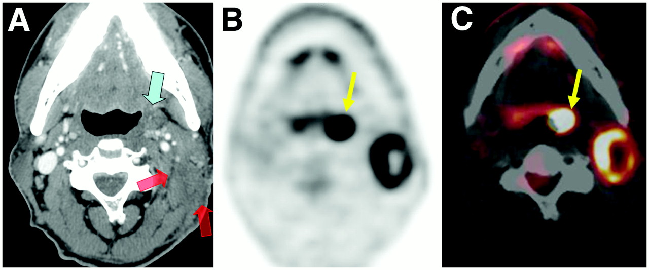
Detection of unexpected malignant involvement of lymph nodes at initial staging. Patient with locally advanced nasopharyngeal cancer was referred for initial staging evaluation. Contrast-enhanced CT (A) and MRI (B) revealed 2 lymph nodes that were within normal limits by size criteria and for which the presence of malignant disease was uncertain (red arrows). (C) These 2 nodes clearly had abnormal activity on 18F-FDG PET/CT (yellow arrows), and radiation treatment field was adjusted to accommodate these regions.
We have developed a schema for the role of 18F-FDG PET/CT in the staging of head and neck cancer (Fig. 2). MRI is typically our initial imaging study of choice because, compared with PET/CT without contrast material, it is capable of more accurately delineating the extent of tumor and evaluating perineural involvement and intracranial extent of disease and is nearly comparable in accuracy in detecting regional lymph node metastases (13,19). Contrast-enhanced CT is used only in cases of laryngeal cancer. However, the merits of MRI over contrast-enhanced CT are still deeply debated between institutions, and PET/CT performed with intravenous contrast material may be a reasonably accurate alternative for delineating the extent of disease at initial presentation (20).

Algorithm for initial staging of HNSCC. *Primarily to detect distant metastatic disease, additional regional lymph nodes with metastatic disease, and synchronous tumors. +Primarily to serve as baseline before therapy. Bx+ = biopsy positive; Bx– = biopsy negative; FNA = fine-needle aspiration.
At our institution, PET/CT is generally suggested when there is concern for distant metastases on the basis of the extent of local–regional disease. If a distant metastasis were identified, then surgery that would be associated with potential functional loss and comorbidity could be avoided. In addition, PET/CT may be used to further evaluate possibly abnormal incidental findings from another imaging examination (for example, mediastinal adenopathy detected by chest CT).
18F-FDG PET/CT does not perform as well in the staging of lymph node involvement as it does in the assessment of distant metastatic disease. In patients whose disease is clinically stage N0 after an initial evaluation (no evidence of lymph node involvement, as determined by physical examination and anatomic imaging), 2 small studies with sentinel lymph node biopsy as a gold standard showed that PET failed to detect lymph nodes with malignant involvement in 8 of 9 patients (from both studies collectively) (21,22). A larger and more recent prospective report of 31 patients with stage N0 disease showed that 18F-FDG PET/CT failed to detect minimally involved (≤3 mm) lymph nodes in 3 patients and yielded false-positive findings in 4 patients (23). The most likely causes of false-negative findings are the limited spatial resolution of PET and its poor performance in detecting lesions of less than ∼5 mm. Therefore, selective neck dissection or sentinel lymph node biopsy is more definitive.
Even in patients with clinically stage N0 disease, a PET/CT scan at the time of initial staging may still serve as a useful baseline examination for subsequent follow-up after therapy (see later discussion) and is included as an optional measure at our institution (Fig. 2). It is particularly useful to have a baseline PET/CT scan to help differentiate incidental physiologic 18F-FDG-avid foci from malignant foci on subsequent posttreatment scans. Normal variant 18F-FDG uptake can be seen in a variety of locations, including the pharyngeal muscles, salivary glands, and lymphoid tissue, and may pose a significant interpretive challenge when comparison images are not available.
IDENTIFICATION OF UNKNOWN PRIMARY TUMOR
Approximately 2%–9% of all HNSCCs will present with cervical lymph node metastases without clear evidence of a primary tumor site (24). Such HNSCCs present both a diagnostic challenge and a treatment quandary. Treatment consists of radiation therapy that is directed at the full extent of the pharyngeal mucosa, which may harbor the putative primary tumor site, and that is associated with significant morbidity, possibly for little gain. Identification of the primary tumor site is critical because it may identify a site for primary tumor surgical resection or define and limit the extent of radiotherapy.
The initial evaluation for patients with an HNSCC lymph node metastasis from an unknown primary tumor should include a thorough physical examination, in-office endoscopy, and anatomic imaging with MRI (or high-quality CT). MRI and CT scans may be negative if the primary tumor site is subtle or difficult to separate from adjacent normal structures (such as lingual tonsillar tissue), if the primary tumor site is superficial or very small, or if the scan is limited by motion or streak artifacts. Multiple studies have assessed the use of PET for detecting an occult primary tumor site, and success rates have generally been at least comparable to if not better than those of anatomic imaging (25). In a large review by Schoder and Yeung of 11 studies that included more than 300 patients, the sensitivity of PET ranged from 10% to 60%, a finding that was attributed to differences in inclusion criteria and clinical verification (13). A more recent metaanalysis concentrated on studies that specifically addressed the subpopulation of patients who had an initially negative physical examination and MRI results. For this group, 18F-FDG PET and PET/CT were able to detect the primary tumor in 40 of 150 patients (27%) (26).
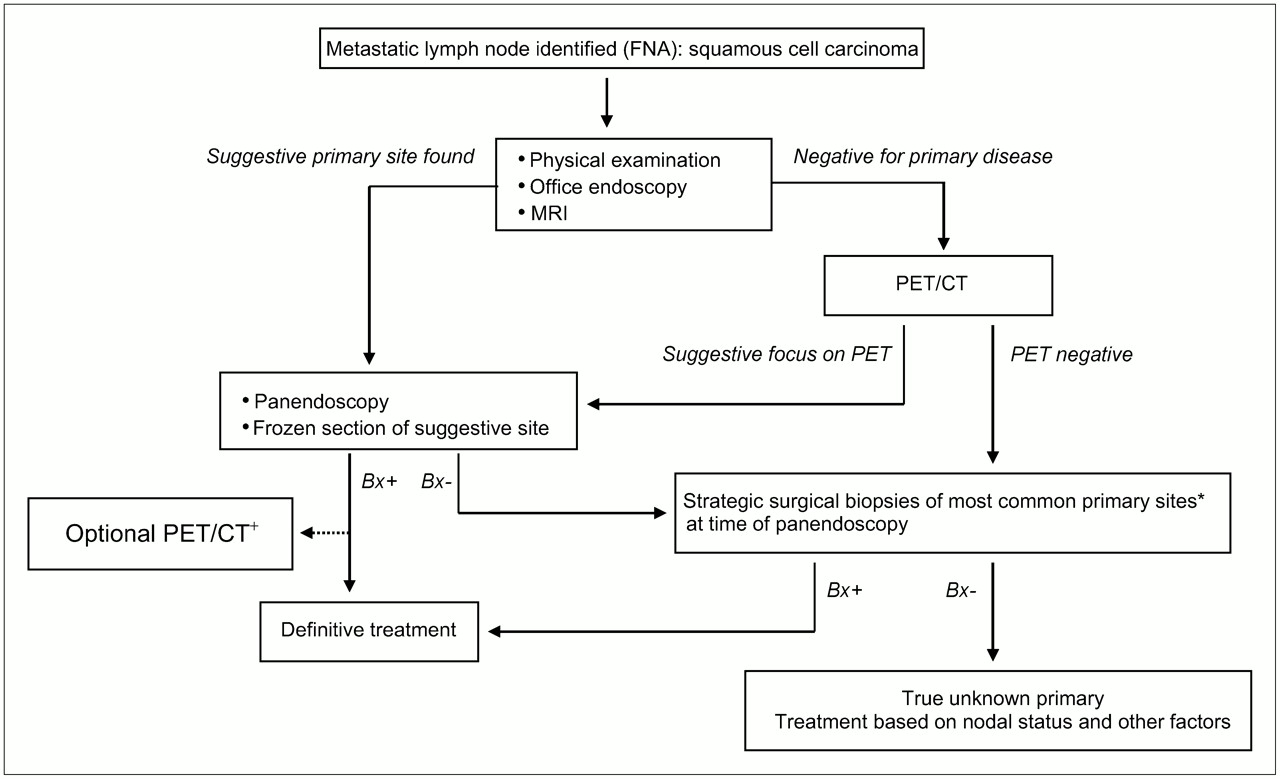
At our institution, when physical examination, in-office endoscopy, and MRI have been unrevealing, a PET/CT scan is obtained (Fig. 3). If there is a suspect focus on metabolic imaging, then the patient undergoes panendoscopy, during which a frozen-section biopsy of the suspect site found by PET/CT is obtained. If the frozen-section biopsy is negative, then further strategic biopsy specimens are obtained from the most common sites for primary tumors. These sites include the base of the tongue, the ipsilateral tonsillar fossa, and, in some cases, the pyriform sinus and contralateral tonsillar fossa and the nasopharynx. If the PET/CT scan does not show any evidence of the primary tumor, then the patient undergoes panendoscopy and subsequent strategic surgical biopsy specimens are obtained in the hope that permanent tissue sections will identify the occult primary tumor. If the primary tumor is discovered, then PET/CT may be performed before treatment to assess for local–regional disease, distant metastases, or a synchronous tumor (Fig. 4).

Algorithm for evaluation of unknown primary malignancy with cervical lymph node metastases. *Ipsilateral base of tongue, tonsillar fossa (TF), nasopharynx, and, in some cases, pyriform sinus and other TF. +PET/CT may be helpful in this setting for detecting distant metastatic disease, additional regional lymph nodes with metastatic disease, and synchronous tumors. Bx+ = biopsy positive; Bx– = biopsy negative; FNA = fine-needle aspiration.
HNSCC metastasis from unknown primary tumor. Patient had enlarging 3-cm left neck mass felt on physical examination. (A) Contrast-enhanced CT depicted space-occupying lesion (red arrows) in left neck. Biopsy of lesion was obtained by fine-needle aspiration, and lesion was proven to be lymph node metastatic squamous cell carcinoma. There was no clear evidence of primary tumor except for subtle asymmetry at left tonsillar fossa (blue arrow) that was of uncertain significance. 18F-FDG PET (B) and fusion PET/CT (C) clearly identified primary tumor at left tonsillar fossa (yellow arrows); finding was confirmed by endoscopic biopsy.
MONITORING TREATMENT RESPONSE AND SURVEILLANCE
Chemoradiation that includes cisplatin-based chemotherapy and intensity-modulated radiation therapy is frequently the primary treatment for HNSCC. This nonsurgical route may be chosen for either of 2 general reasons. First, an organ preservation protocol may be chosen for patients with laryngeal and tongue cancer in an effort to avoid surgical resection altogether. Second, chemoradiation is often chosen in clinical scenarios in which it has been demonstrated to achieve local–regional control similar to or better than that of protocols that include surgery. In both clinical settings, PET/CT is useful for monitoring the treatment response and for accurately identifying patients with residual disease for salvage surgery.
Assessment of the treatment response and surveillance for recurrence in HNSCC may pose a diagnostic challenge. Radiation and surgery can cause significant changes to the normal tissues of the head and neck (27). Such tissue distortions can obscure persistent or recurrent disease on physical examination, CT, and MRI until relatively late in the course of disease (27,28). For 18F-FDG PET, likewise, nonspecific increases in tracer uptake can occur in lymphoid tissue, salivary glands, muscles, and soft tissue, usually as a result of posttreatment inflammation (29). A number of studies, however, have shown that PET can detect residual tumor after chemoradiation more accurately than conventional imaging (30–34). For a series of 108 patients at our institution, we found that PET/CT detected local–regional persistent or recurrent HNSCC with a sensitivity of 82%, a specificity of 92%, a positive predictive value (PPV) of 64%, a negative predictive value (NPV) of 97%, and an overall accuracy of 90% (35). Similarly, in a recent study of 39 patients by Porceddu et al., PET had an NPV of 97% when it was performed at a median of 12 wk after the completion of therapy (36). For the detection of distant metastases, PET/CT had a sensitivity of 89%, a specificity of 97%, a PPV of 85%, an NPV of 98%, and an overall accuracy of 96% (35). A negative PET/CT result is highly reliable for all sites when PET/CT is performed at least 2–3 mo after the completion of therapy (Fig. 5) (36). A positive PET/CT result in the head and neck region must be correlated with information from the physical examination and cross-sectional imaging modalities, as nonspecific inflammation may often be 18F-FDG avid.

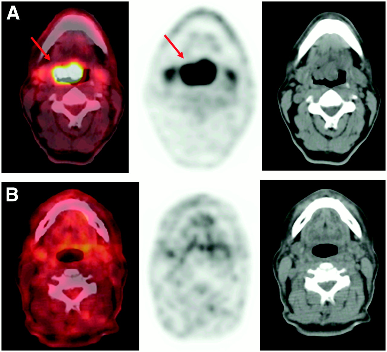
Monitoring chemoradiation. (A) Pretreatment transaxial 18F-FDG PET/CT revealed intensely hypermetabolic primary carcinoma at base of tongue (red arrows). (B) 18F-FDG PET/CT after chemoradiation revealed complete resolution of abnormal activity without evidence of residual disease in treated region.
Decisions about the optimal timing of PET/CT scans relative to radiation therapy, once greatly debated, are achieving greater consensus. PET/CT is more accurate when performed at 2–3 mo after the completion of radiation therapy than at earlier time points (32,36,37). This improvement is attributed to subsidence of the nonspecific inflammation over time. Like others, we found that PET/CT performed at 3 mo after the completion of radiation therapy had significantly higher sensitivity (P < 0.01) and NPV (P < 0.05) in the head and neck than PET/CT performed within 1 mo (35). Similarly, PET/CT had a higher specificity for detecting persistent tumor after radiation therapy than contrast CT alone, and the specificity of PET/CT was highest when it was performed after 8 wk of treatment (35). Other investigators described similar results at a range of 8–16 wk after the completion of radiotherapy (32,37,38).
In the setting of surveillance, numerous studies have demonstrated that 18F-FDG PET/CT has a relatively high sensitivity for detecting recurrent disease at the primary tumor site and regional lymph node metastases (39–45). For patients who underwent PET or PET/CT at least 12 wk after the completion of all therapy, the reported sensitivity ranged between 93% and 100% for the detection of recurrence. Because there may be nonspecific 18F-FDG uptake at sites other than sites of malignancy, such as in inflamed lymph nodes or musculature, the specificity of PET and PET/CT is between 63% and 94%, and additional confirmatory tests should be used when there are abnormal findings (Fig. 6).

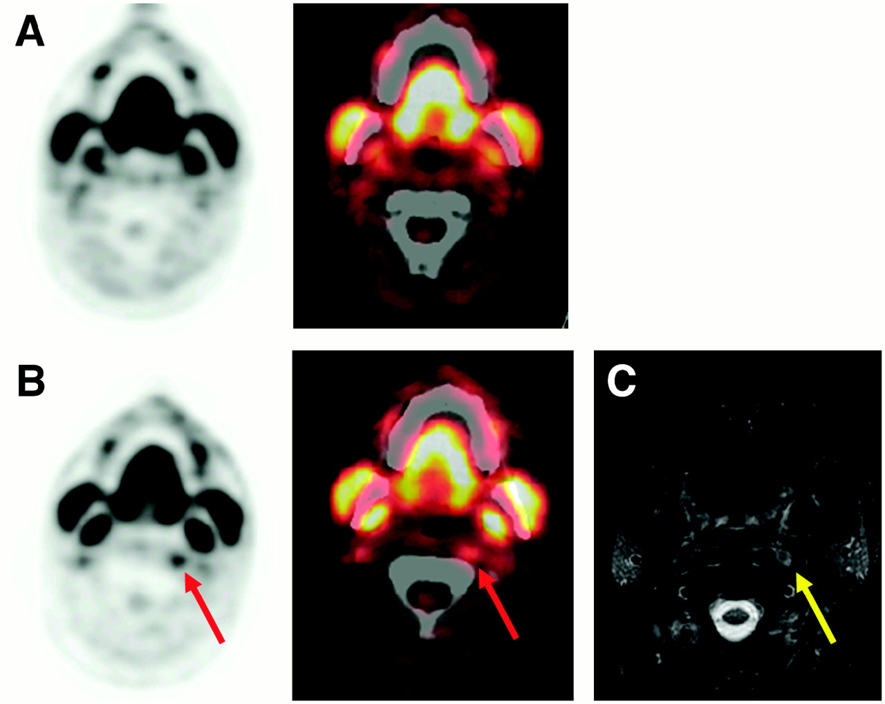
PET/CT for detection of recurrence. (A) Surveillance 18F-FDG PET/CT after therapy revealed prominent physiologic mandibular muscle uptake without evidence of recurrent disease. (B) Interval follow-up 18F-FDG PET/CT revealed new subtle hypermetabolic focus in left retropharyngeal region (red arrows) suspected of indicating recurrence. (C) T1-weighted contrast-enhanced MRI confirmed presence of suspect subcentimeter retropharyngeal lymph node (yellow arrow).
Researchers at our institution have reached a reasonable consensus opinion for assessing the treatment response and surveillance (Fig. 7). At approximately 1 mo after the completion of chemoradiation, patients with obvious residual disease or tumor progression are considered for immediate salvage surgery. If there is no clear evidence of residual disease, then patients are clinically evaluated 1 mo later (2 mo after the completion of chemoradiation). If a suspect site is found on examination, then biopsy specimens are obtained. Patients who appear to have had a complete therapeutic response undergo MRI (or CT, in cases of laryngeal cancer). Fine-needle aspiration biopsy is performed if there are suspect areas on cross-sectional imaging. If no evidence of disease is found on physical examination, in-office endoscopy, and MRI at 2 mo after the completion of chemoradiation, then patients are evaluated by PET/CT beginning at 3 mo after the completion of chemotherapy. The value of this examination may be increased if a comparison baseline PET scan had been obtained at initial staging. Subsequent surveillance PET/CT scans are scheduled either depending on clinical suspicion or at regular intervals, such as every 6 mo for the first 18 mo and annually thereafter. In the latter setting, frequent surveillance has a reasonable chance of detecting recurrence and may be of particular value when a potentially curative salvage modality is available (surgery or reirradiation).
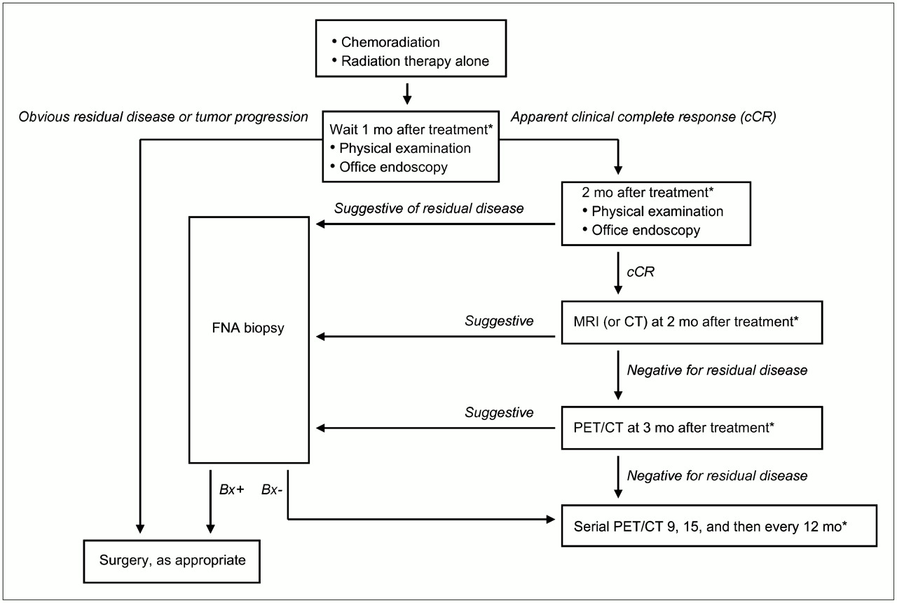
Algorithm for evaluation of treatment response. *Time after completion of radiotherapy. Bx+ = biopsy positive; Bx– = biopsy negative; FNA = fine-needle aspiration.
EMERGING ROLE OF PET/CT IN RADIOTHERAPY PLANNING
Integrated 18F-FDG PET/CT provides a bridge between anatomic imaging and functional imaging that appears to be ideally suited to radiotherapy planning in the era of highly conformal radiotherapy (46,47). Accurate spatial delineation of targets is especially important with treatment modalities that deliver highly conformal 3-dimensional dose distributions, such as intensity-modulated radiation therapy and stereotactic radiosurgery, both of which are used commonly for treating HNSCC at our institution. Immobilization devices can be used during the acquisition of the PET/CT scan to improve the registration of PET and CT data for treatment planning. Several publications have reported that target volumes may be modified in as many as 20% of cases with the use of 18F-FDG PET/CT versus CT alone (Fig. 8). Accordingly, PET/CT is commonly used as an adjunct examination for radiation treatment planning for head and neck cancers at our institution. We rely primarily on CT and MRI for target definition because of their high spatial resolution. In addition, we have found that PET frequently fails to identify hypermetabolism in areas of marrow space infiltration and perineural extension that are highly suspect on MRI. Therefore, we use PET/CT primarily to include normal-size, normal-appearance lymph nodes with increased metabolic activity as part of the high-dose target volume. In addition, PET/CT may be helpful for contouring primary tumors whose borders are difficult to distinguish by anatomic imaging alone, as is sometimes the case with tongue base tumors. At present, we do not reduce the target volumes to exclude PET-negative portions of tumors that appear abnormal on CT or MRI, such as areas of necrosis or enlarged lymph nodes with equivocal metabolic activity. Long-term outcome data identifying patterns of treatment failure in relationship to PET/CT-augmented target volumes are needed to define a standardized approach to the use of PET/CT in treatment planning before this can be recommended as routine practice.

Use of PET/CT to assist in radiation treatment planning. (A) MRI depicted asymmetric tissue in left nasopharynx. Radiotherapy contour line encircled this region (red line). (B) PET/CT revealed abnormal metabolic activity crossing midline from left nasopharynx to right nasopharynx, suggesting extent of tumor larger than that suggested by MRI. Radiotherapy contour line was adjusted accordingly (red line).
18F-FDG PET/CT IN THYROID CANCER
There are more than 30,000 new cases of thyroid cancer in the United States annually, and the incidence is increasing (48). The vast majority of thyroid cancers arise from follicular cells and are classified pathologically as papillary, follicular, and anaplastic. Papillary and follicular cancers and variants, such as the follicular variant of papillary cancer, are designated as differentiated because they retain the ability to trap iodine and produce thyroglobulin (Tg).
The normal thyroid traps little or no 18F-FDG. On PET scans obtained for the management of nonthyroid cancers, the uptake of 18F-FDG in the thyroid can occasionally be identified. When this uptake is diffuse and intense, it is usually attributable to autoimmune thyroiditis (49). In contrast, when the uptake is focal, there is a 30%–50% chance of primary thyroid cancer. These data were recently reviewed (50). With integrated PET/CT, the uptake can be shown to coincide with a solid nodule. This focal uptake can also be attributable to metastasis from primary nonthyroid cancer, including melanoma and kidney, lung, breast, and gastrointestinal tract cancers, but there is usually evidence of widespread metastases in these situations. In the case of focal uptake, fine-needle aspiration is advised to obtain a tissue diagnosis.
Suggested approaches to the management of thyroid cancer have been published (51,52). We have constructed an updated algorithm that includes integrated 18F-FDG PET/CT (Fig. 9). The fundamentals of the treatment of differentiated cancer are total thyroidectomy, thyroid hormone, and, in selected patients, radioiodine (131I). The prognosis worsens with each decade over the age of 45 y; with increasing size of the cancer; with invasion into surrounding structures, such as the trachea and esophagus; and with metastases to distant sites. The treatment should be least aggressive for patients with an excellent prognosis and progressively more aggressive when there are increasingly poor prognostic features. Thyroidectomy and thyroid hormone are adequate for the treatment of an intrathyroid differentiated cancer that measures less than 2 cm and is fully excised. When surgery is incomplete or the cancer is larger or invasive or there are local–regional or distant metastases, there is a role for 131I after total thyroidectomy. In preparation for 131I treatment, the level of thyrotropin (TSH) must be elevated (most authorities recommend a value of >30 mU/L) either by withdrawing thyroid hormone or by injecting recombinant human TSH, and the patient should adhere to a low-iodine diet for 2 wk. We prefer to obtain a diagnostic whole-body scan at 24 h after the administration of 148 MBq of 123I to define the extent of residual thyroid and metastases and to calculate the percentage of uptake in abnormal sites. Next, an appropriate therapeutic dose is administered on the same day, and a scan is obtained 5–8 d later. With 148 MBq of 123I, there is seldom a difference in the diagnostic and posttherapy scans; therefore, the extent of disease is defined before radioiodine therapy. After treatment, we conduct follow-up clinical evaluations at 2–3 mo, at intervals of 6 mo for 5 y, and annually thereafter. These visits include clinical examination and measurement of free thyroxine, TSH, and Tg. A follow-up whole-body scan is obtained after 1 and 5 y along with measurement of TSH-stimulated Tg; recombinant human TSH is generally used to produce an elevated TSH level. Some authorities use measurement of TSH-stimulated Tg without an accompanying scan (53,54). However, the follow-up scan can be compared with those obtained at the time of therapy and provides objective evidence in patients who have anti-Tg antibodies. Ultrasound of the neck is performed annually. When the results of a follow-up scan are positive, a second treatment with 131I is advised.
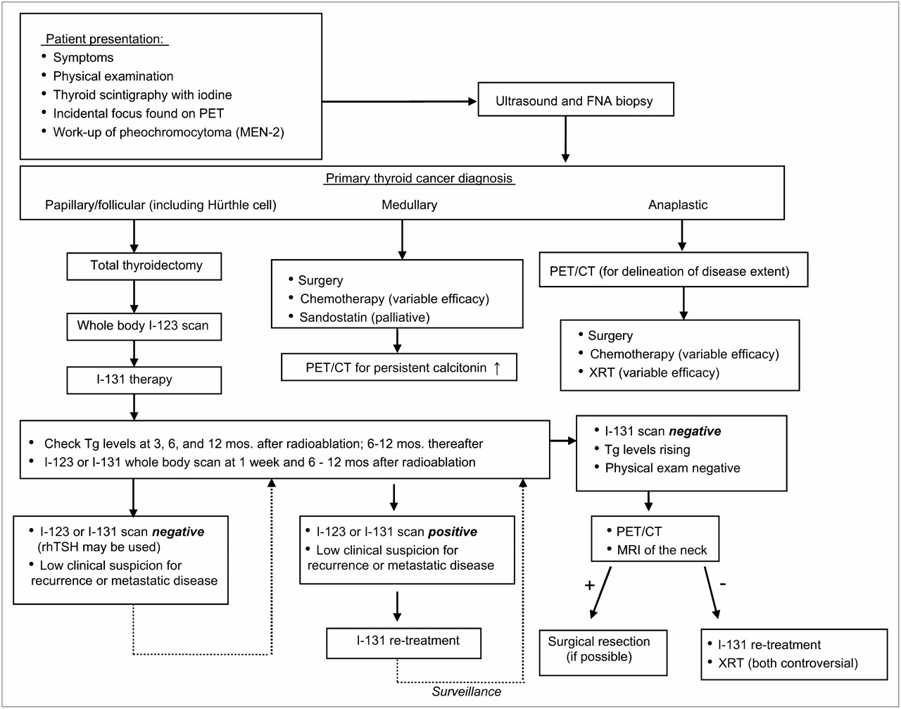
Algorithm for evaluation of thyroid carcinoma. FNA = fine-needle aspiration; rhTSH = recombinant human TSH; XRT = radiation therapy.
Usually the scintigraphic scan results and Tg levels match: both are negative or both are abnormal. One difficult problem is a disparity that usually involves measurable Tg but a negative radioiodine scan result. Most authorities consider a TSH-stimulated Tg level of 10 ng/mL or greater to merit action (51). Such action can include empirically determined therapy with 131I or imaging with PET or PET/CT to identify the source of Tg production (Fig. 10) (55–57). Reported sensitivities for PET and PET/CT range from 70% to 90%, and the specificity is also high. Our experience with 87 PET/CT studies revealed a sensitivity of 87% and a specificity of 80%. False-positive results, including 18F-FDG uptake in brown fat and muscles of speech, are correctly identified by fused PET/CT. Pathologic sites of 18F-FDG uptake can be cured by surgical excision. In contrast, we have been unimpressed with the success of empiric 131I treatment; frequently, there was no uptake of radioiodine on a posttherapy scan, and in none of our patients did the Tg level become undetectable after this treatment. However, an undetectable Tg level has been achieved with PET and ultrasound-guided surgery (58). There are reports that “blind” therapy with 131I can be hazardous (59).

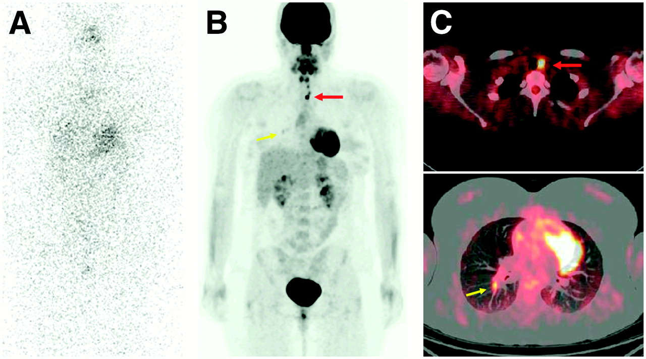
18F-FDG PET/CT for detecting scintigraphically occult metastatic thyroid cancer. Patient had persistently elevated Tg levels after thyroidectomy and 131I ablation. (A) Whole-body 131I scan appeared normal and did not reveal source of abnormal Tg production. (B and C) Follow-up 18F-FDG PET/CT detected several paratracheal lymph node metastases (red arrow) and lung metastasis (yellow arrow).
18F-FDG PET/CT also has a role in other thyroid cancers that do not trap iodine. These include anaplastic cancer, medullary cancer, and lymphoma. The roles of PET/CT in anaplastic cancer of the thyroid are to define the local extent of disease and to determine whether there are distant metastases (60). PET/CT is valuable in identifying the source of persistent measurable calcitonin in patients who have undergone total thyroidectomy. It is superior to anatomic imaging (61,62). The roles of PET/CT in primary lymphoma of the thyroid are to stage the disease and to monitor the response to therapy.
CONCLUSION
18F-FDG PET/CT has become a widely used imaging modality for a variety of common malignancies and is increasingly becoming a standard part of the management of HNSCC and thyroid cancer. For HNSCC, the primary benefits of this modality are 3-fold: initial staging, monitoring of the response to therapy, and long-term cancer surveillance. During initial management, PET/CT can delineate regional lymph node metastases, detect distant metastases, discover unknown primary tumors, and identify synchronous primary tumors. After chemoradiation, it is used for monitoring the response to therapy to accurately select patients for salvage surgery. PET can also be used for long-term surveillance, in which it has a very high sensitivity for recurrence and metastatic disease. The value of PET/CT for radiotherapy planning is under investigation; PET/CT is currently used only in an assistive role in such planning.
For well-differentiated thyroid cancers, PET/CT plays an important adjunct role in detecting sites of metastatic disease and recurrence that are scintigraphically occult on radioiodine imaging. PET plays a similar role for medullary tumors when there are persistently high calcitonin levels without a known source. For anaplastic tumors and thyroid lymphoma, PET may be used at initial staging to delineate the local extent of disease and to detect distant metastases.
Currently, no consensus national guidelines have been established for the use of PET/CT for HNSCC and thyroid cancer. Drawing on personal experience at our institution and on an analysis of the literature, we have outlined our specific strategy for its use.
Acknowledgments
We thank Aya Kamaya, MD, Department of Radiology, Stanford University, for editorial help and Andrei Iagaru, MD, Stanford University, for help in assembling figures.
Footnotes
-
COPYRIGHT © 2006 by the Society of Nuclear Medicine, Inc.
References
- Received for publication July 7, 2006.
- Accepted for publication October 3, 2006.


Jump to section
Related Articles
Cited By...
- CT Texture Analysis of Cervical Lymph Nodes on Contrast-Enhanced [18F] FDG-PET/CT Images to Differentiate Nodal Metastases from Reactive Lymphadenopathy in HIV-Positive Patients with Head and Neck Squamous Cell Carcinoma
- Effects of Intratumoral Inflammatory Process on 18F-FDG Uptake: Pathologic and Comparative Study with 18F-Fluoro-{alpha}-Methyltyrosine PET/CT in Oral Squamous Cell Carcinoma
- Usefulness of 3'-Deoxy-3'-18F-Fluorothymidine PET for Predicting Early Response to Chemoradiotherapy in Head and Neck Cancer
- 18F-FDG Metabolic Tumor Volume and Total Glycolytic Activity of Oral Cavity and Oropharyngeal Squamous Cell Cancer: Adding Value to Clinical Staging
- Fluorodeoxyglucose-Positron-Emission Tomography Imaging of Head and Neck Squamous Cell Cancer
- PET Monitoring of Therapy Response in Head and Neck Squamous Cell Carcinoma
- Tumor Volume Assessment by 18F-FDG PET/CT in Patients with Oral Cavity Cancer with Dental Artifacts on CT or MR Images
- Introduction