


Article Figures & Data
Figures
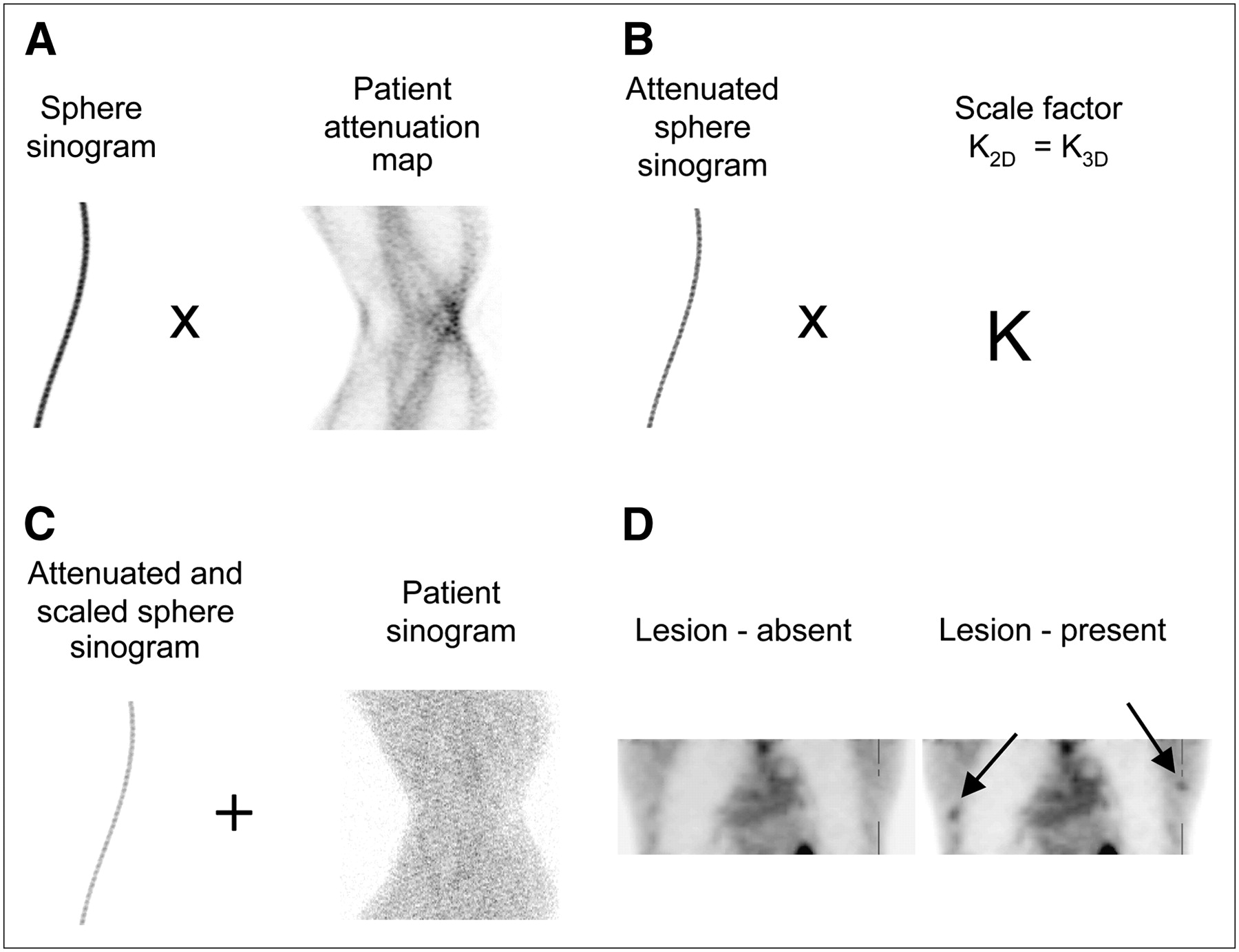
- FIGURE 1.
Generation of lesion-present studies. (A) Sphere sinograms were attenuated with the corresponding 2D or 3D attenuation map of the patient. (B) Attenuated sinograms of spheres were corrected for randoms and scaled to ensure marginal detectability, using the same scale factor for 2D and 3D modes. (C) These attenuated and scaled sphere sinograms were added to the patient's sinograms. (D) Reconstructed volumes of the patient's sinograms with and without the spheres added were, respectively, the signal-present and signal-absent data used in the observer study.
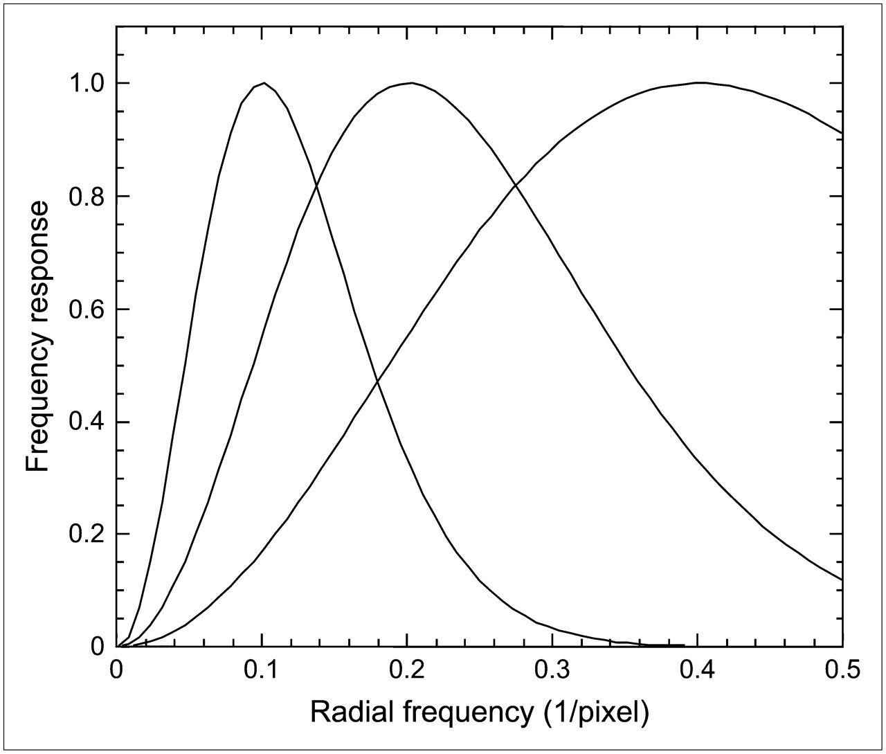
- FIGURE 2.
Three difference-of-gaussian channels used for the channelized Hotelling observer study.
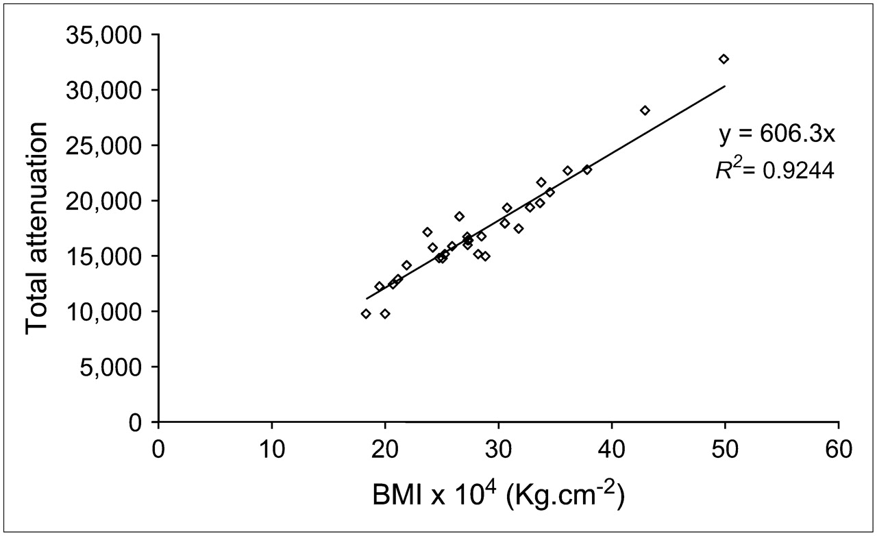
- FIGURE 3.
Correlation of BMI with weighted total attenuation volume in each bed position computed by summing the linear attenuation coefficients (in cm−1) over every voxel (cm) in the patient's reconstructed transmission image.
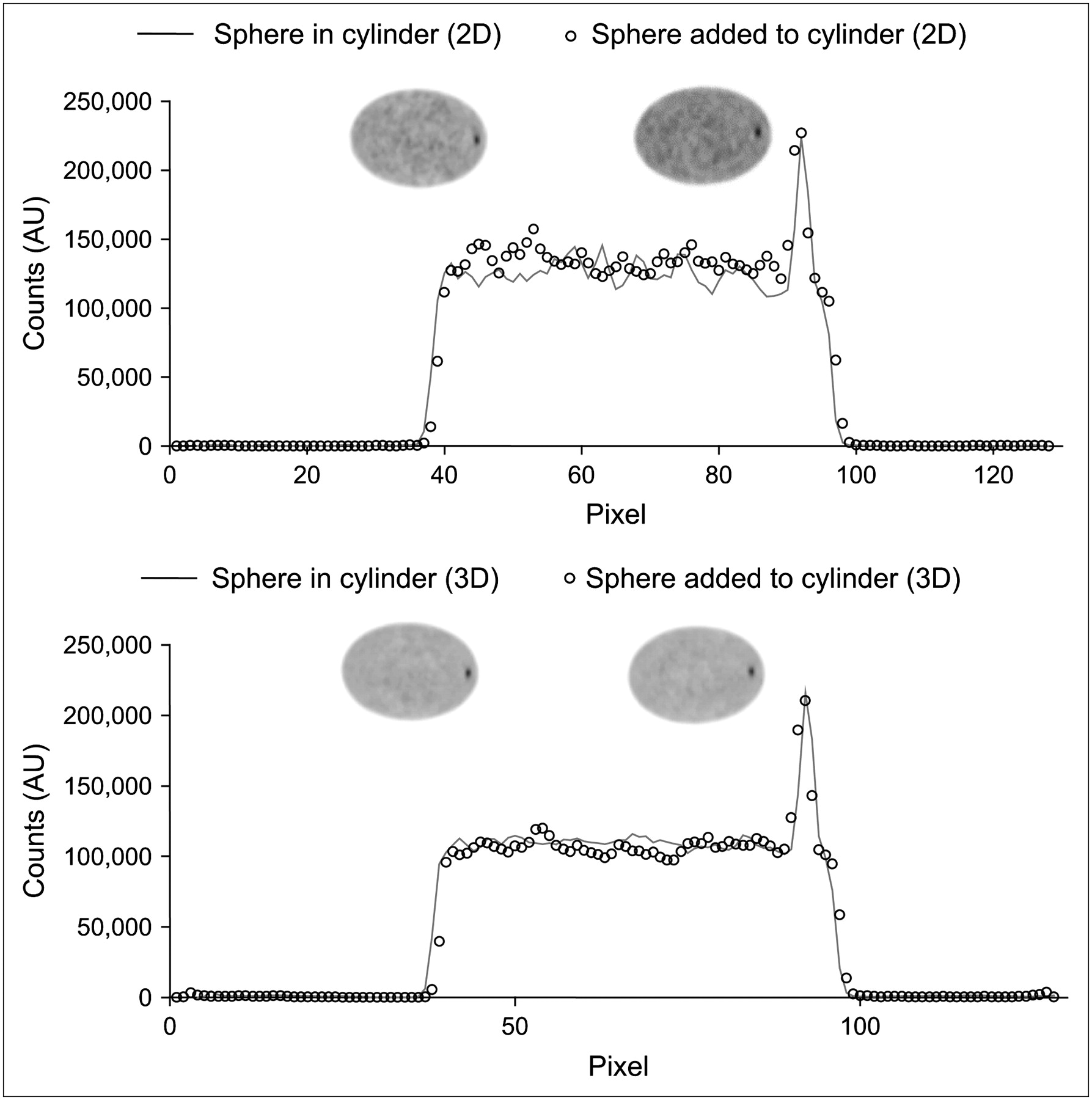
- FIGURE 4.
Transverse slices of 2D and 3D reconstructed images of the elliptic phantom acquired with the sphere inside the cylinder, as well as with the sphere synthetically added to the uniform cylinder without the sphere along with the corresponding profiles through the acquired and added spheres. AU = arbitrary units.
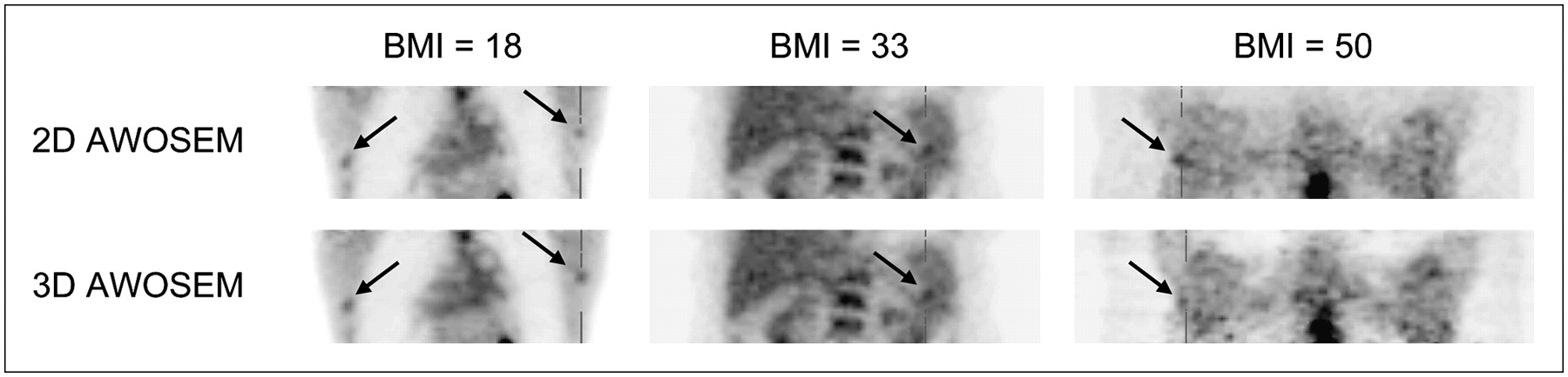
- FIGURE 5.
Lesion-present (arrow) coronal slices through normal-weight (BMI = 18, 2 lesions shown), obese (BMI = 33, 1 lesion shown), and extremely obese (BMI = 50, 1 lesion shown) patient studies in 2D and 3D modes reconstructed using the clinical protocol (AWOSEM).
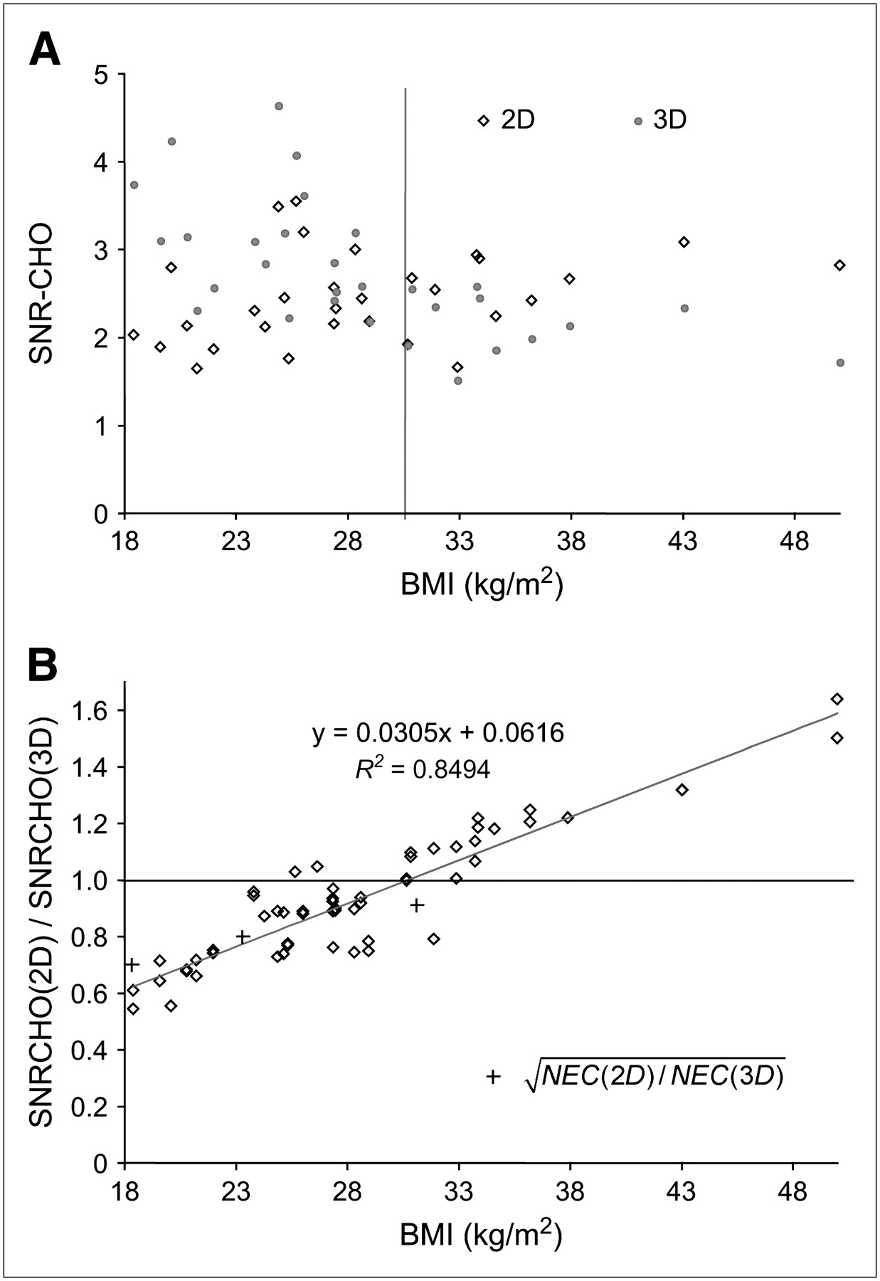
- FIGURE 6.
2D and 3D channelized Hotelling SNR computed over all lesion sizes and locations (30 conditions) as a function of BMI (A) as well as SNR ratio of 2D and 3D (B). √NEC (2D)/√NEC (3D) is plotted for 3 phantoms (20 × 70, 27 × 70, and 35 × 70 cm) corresponding to patients weighing 40, 60, and 95 kg, respectively.
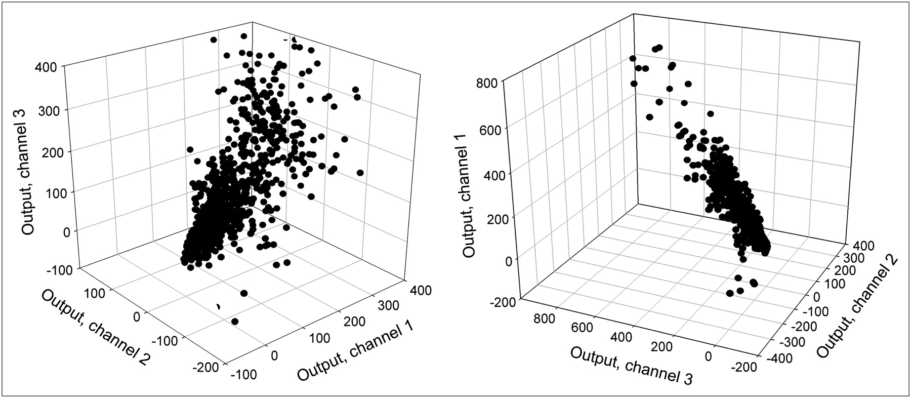
- FIGURE 7.
Two views of the 3D scatter plots of the individual CHO output values.
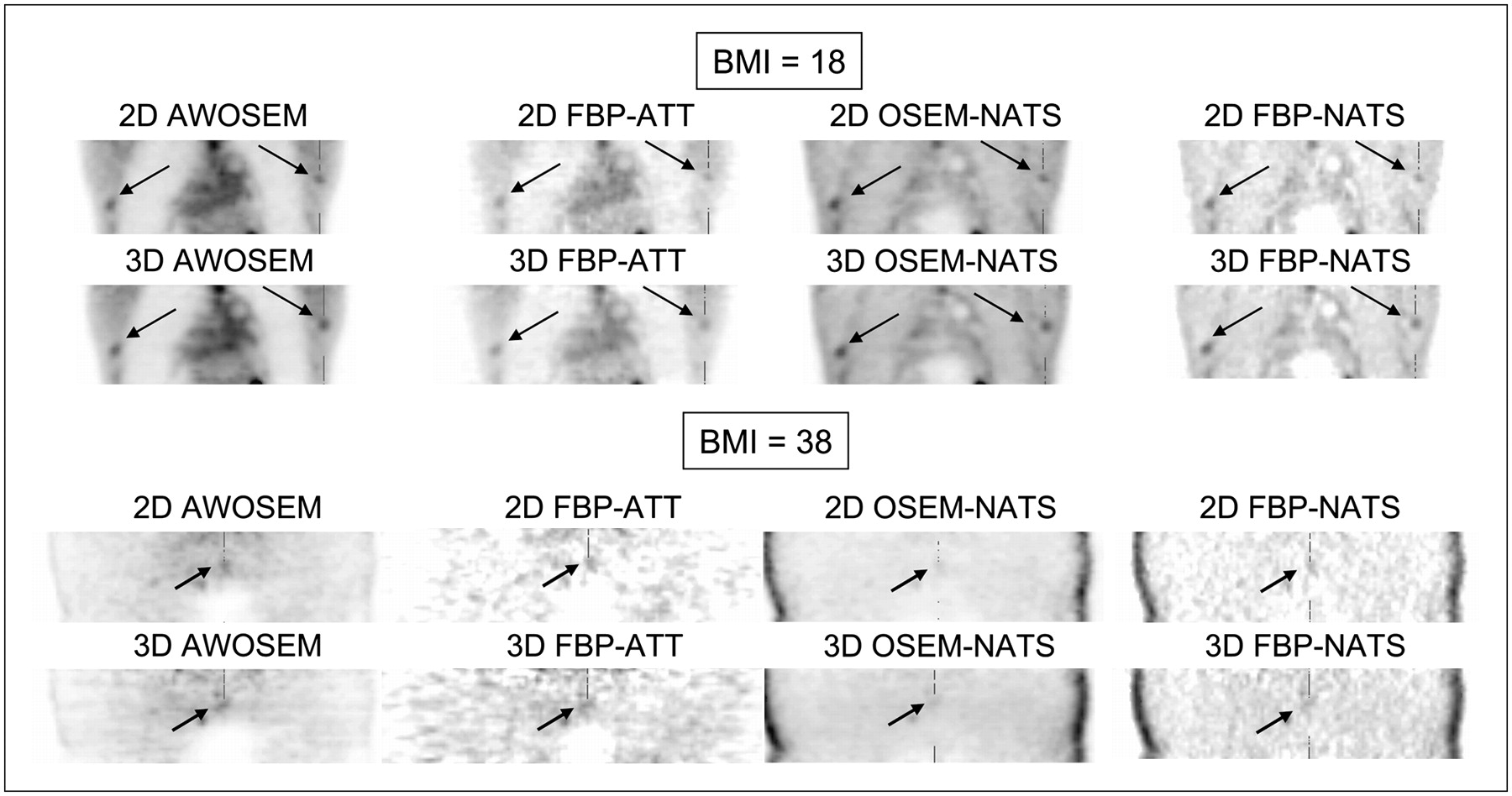
- FIGURE 8.
Coronal slices through normal-weight patient (BMI = 18) and obese patient (BMI = 38) in 2D and 3D corresponding to the following image processing protocols: FBP with (FBP-ATT) and without (FBP-NATS) attenuation and scatter corrections, OSEM without attenuation and scatter corrections (OSEM-NATS) and attenuation-weighted OSEM (AWOSEM). Arrows point to lesions added in mediastinum. Randoms were compensated for using the RVR technique.


Tables
- TABLE 1
Channelized Hotelling SNR Computed Over All 59 Bed Positions and 10 Locations (590 Conditions) as Function of Lesion Size

- TABLE 2
Channelized Hotelling SNR Computed Over All 59 Bed Positions and 10 Sphere Locations and 3 Sizes (1,770 Conditions) for the 8 Processing Schemes Evaluated in 2D and 3D*
Protocol 2D + RVR 3D + RVR 2D +DWS 3D + DWS AWOSEM 2.621 ± 0.305 2.759 ± 0.316 2.207 ± 0.312 2.279 ± 0.323 OSEM-NATS 1.451 ± 0.198 1.513 ± 0.258 1.394 ± 0.195 1.482 ± 0.242 FBP-ATT 2.307 ± 0.269 2.399 ± 0.293 2.014 ± 0.266 2.142 ± 0.285 FBP-NATS 2.304 ± 0.260 2.309 ± 0.279 2.004 ± 0.260 2.103 ± 0.283 ↵* FBP with (FBP-ATT) and without (FBP-NATS) attenuation and scatter corrections, OSEM without attenuation and scatter corrections (OSEM-NATS), and attenuation-weighted OSEM (AWOSEM) along with randoms variance reduction (RVR) or delayed window subtraction (DWS).


Jump to section
Related Articles
Cited By...
- Influence of Statistical Fluctuation on Reproducibility and Accuracy of SUVmax and SUVpeak: A Phantom Study
- Impact of Time-of-Flight PET on Whole-Body Oncologic Studies: A Human Observer Lesion Detection and Localization Study
- Improvement in Lesion Detection with Whole-Body Oncologic Time-of-Flight PET