


Article Figures & Data
Figures
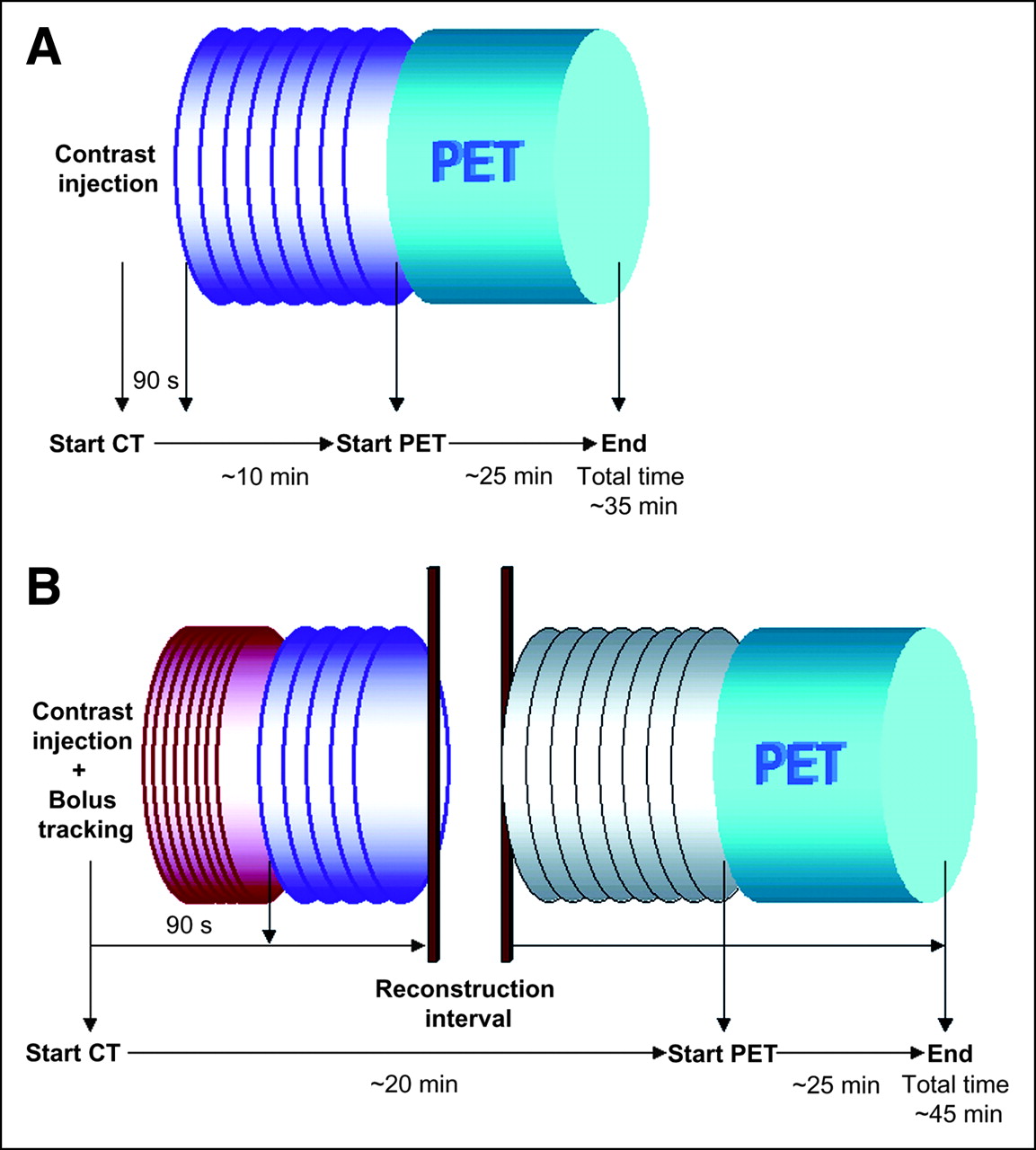
- FIGURE 1.
PET/CT protocols for single-phase (A) and multiphase (B) examinations. For protocol A, purple spiral represents whole body in portal-venous contrast enhancement. Scan range included base of skull to proximal thigh, as in PET (blue cylinder). For protocol B, 2 examination protocols had to be implemented because of technical standards of scanner. After injection and bolus tracking of contrast medium, first CT scan covered base of skull to lower borders of kidneys (red spiral). At 90 s after injection, CT ranging from base of lungs to proximal thighs was performed in portal-venous contrast enhancement phase (purple spiral). After short reconstruction interval, attenuation LD-CT (gray spiral) and PET were performed. Thickness of spiral lines indicates dose (thick line: 160 mAs; thin line: 30 mAs); width of spirals indicates collimation (narrow: 0.75; wide: 1.5).
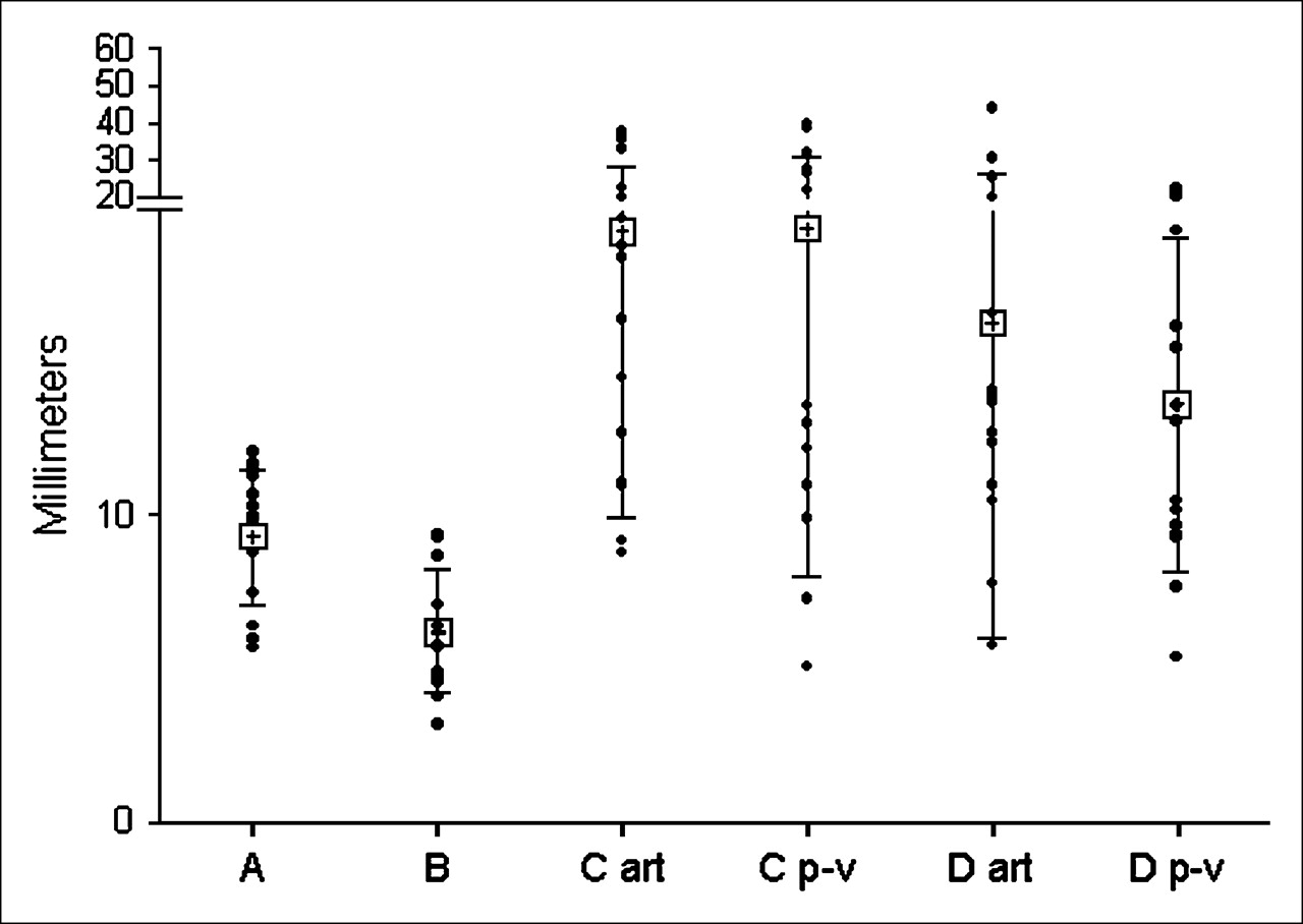
- FIGURE 2.
Comparison of misalignment of contrast-enhanced CT examinations for protocols A, B, C, and D. Protocol B (NormExp; single phase) had significantly lowest values for all abdominal organs. Significant differences between multiphase protocols (C and D) could not be assessed. art = arterial; p-v = portal-venous. • = individual values; + = mean values. Error bars indicate SDs.
- FIGURE 3.
Occurrence of image artifacts caused by breathing and severe mismatching caused by movements of patient in between CT and PET scans. Mismatching was distributed equally among protocols, whereas image quality correlated strongly with breathing protocols. NormExp protocols (B and D) had significantly lower occurrences of artifacts and, therefore, superior image quality. art = arterial; p-v = portal-venous.
- FIGURE 4.
Multiphase PET/CT. Comparison of misalignment in protocol C (A) and protocol D (B). LD-CT attenuation scans were performed during shallow breathing. However, with regard to misalignment in liver, values for LD-CT were significantly lower. For protocol D, contrast-enhanced CT scan during NormExp did not show superiority over LD-CT scan during shallow breathing. Instead, values for LD-CT were significantly lower than those for arterial phase for liver. LD = low dose; ART = arterial; P-V = portal-venous. • = individual values; + = mean values. Error bars indicate SDs.
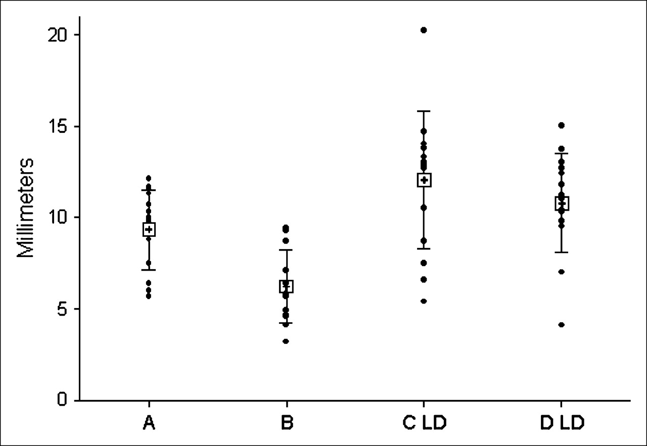
- FIGURE 5.
Comparison of CT attenuation scans. Protocol B showed lowest values for misalignment in all organs. Attenuation scans with protocol C (C LD) and protocol D (D LD) showed values similar to those with protocol A, as expected. • = individual values; + = mean values. Error bars indicate SDs.


Tables
Data for following protocol: Parameter A B C D Contrast phase Single Single Multi Multi Breathing protocol Shallow NormExp Shallow NormExp No. of patients 15 13* 15 15 Age (y)† 57.9 ± 13.1 53.5 ± 14.9 65.4 ± 9.2 55.3 ± 16.9 No. of men/women 9/6 8/5 11/4 9/6 Body mass index (kg/m2)† 27.2 ± 5.8 25.9 ± 3.2 24.8 ± 2.9 23.9 ± 3.7 Activity (MBq)† 432 ± 26 402 ± 34 429 ± 31 390 ± 59 Uptake time (min)† 72.5 ± 13.0 66.5 ± 11.1 80.9 ± 17.1 71.9 ± 10.3 No. of beds† 7.3 ± 0.8 7.0 ± 0.6 7.1 ± 0.7 7.5 ± 0.6 Bed time (min)† 3.1 ± 0.4 3 3.1 ± 0.3 3 No. in following protocol: Pathologic entities A B C D Genitourinary tumors 3 3 2 6 Tumors of bowel and intestines 1 2 2 1 Esophageal cancer 0 1 0 0 Stomach cancer 1 0 0 1 Breast cancer 1 1 2 0 Cancer of unknown primary source 2 2 0 1 Head and neck tumors 1 0 2 1 Lung cancer 1 2 2 1 Melanoma 0 0 1 1 Pancreatic tumor 0 0 0 1 Lymphoma 4 2 3 1 Inflammatory diseases 1 2 1 1


Jump to section
Related Articles
Cited By...
- MRI-Based Attenuation Correction for Whole-Body PET/MRI: Quantitative Evaluation of Segmentation- and Atlas-Based Methods
- Dual-Modality Imaging: Combining Anatomy and Function
- Limitations of CT During PET/CT
- Value of contrast-enhanced multiphase CT in combined PET/CT protocols for oncological imaging
- Evaluation of Image Registration in PET/CT of the Liver and Recommendations for Optimized Imaging
- Adequate Evaluation of Image Registration in Hybrid PET/CT
- Reply: Adequate Evaluation of Image Registration in Hybrid PET/CT.