
Main menu

  • Home
  • Content
    • Current
    • Ahead of print
    • Past Issues
    • JNM Supplement
    • SNMMI Annual Meeting Abstracts
    • Continuing Education
    • JNM Podcasts
  • Subscriptions
    • Subscribers
    • Institutional and Non-member
    • Rates
    • Journal Claims
    • Corporate & Special Sales
  • Authors
    • Submit to JNM
    • Information for Authors
    • Assignment of Copyright
    • AQARA requirements
  • Info
    • Reviewers
    • Permissions
    • Advertisers
  • About
    • About Us
    • Editorial Board
    • Contact Information
  • More
    • Alerts
    • Feedback
    • Help
    • SNMMI Journals
  • SNMMI
    • JNM
    • JNMT
    • SNMMI Journals
    • SNMMI

User menu

  • Subscribe
  • My alerts
  • Log in
  • My Cart

Search

  • Advanced search
Journal of Nuclear Medicine
  • SNMMI
    • JNM
    • JNMT
    • SNMMI Journals
    • SNMMI
  • Subscribe
  • My alerts
  • Log in
  • My Cart
Journal of Nuclear Medicine

Advanced Search

  • Home
  • Content
    • Current
    • Ahead of print
    • Past Issues
    • JNM Supplement
    • SNMMI Annual Meeting Abstracts
    • Continuing Education
    • JNM Podcasts
  • Subscriptions
    • Subscribers
    • Institutional and Non-member
    • Rates
    • Journal Claims
    • Corporate & Special Sales
  • Authors
    • Submit to JNM
    • Information for Authors
    • Assignment of Copyright
    • AQARA requirements
  • Info
    • Reviewers
    • Permissions
    • Advertisers
  • About
    • About Us
    • Editorial Board
    • Contact Information
  • More
    • Alerts
    • Feedback
    • Help
    • SNMMI Journals
  • View or Listen to JNM Podcast
  • Visit JNM on Facebook
  • Join JNM on LinkedIn
  • Follow JNM on Twitter
  • Subscribe to our RSS feeds
OtherBASIC SCIENCE INVESTIGATIONS

Evidence that Myocardial Na/I Symporter Gene Imaging Does Not Perturb Cardiac Function

Kyung-Han Lee, Jun-Sang Bae, Sang-Chul Lee, Jin-Young Paik, Takashi Matsui, Kyung-Ho Jung, Bong-Ho Ko and Byung-Tae Kim
Journal of Nuclear Medicine November 2006, 47 (11) 1851-1857;
Kyung-Han Lee
  • Find this author on Google Scholar
  • Find this author on PubMed
  • Search for this author on this site
Jun-Sang Bae
  • Find this author on Google Scholar
  • Find this author on PubMed
  • Search for this author on this site
Sang-Chul Lee
  • Find this author on Google Scholar
  • Find this author on PubMed
  • Search for this author on this site
Jin-Young Paik
  • Find this author on Google Scholar
  • Find this author on PubMed
  • Search for this author on this site
Takashi Matsui
  • Find this author on Google Scholar
  • Find this author on PubMed
  • Search for this author on this site
Kyung-Ho Jung
  • Find this author on Google Scholar
  • Find this author on PubMed
  • Search for this author on this site
Bong-Ho Ko
  • Find this author on Google Scholar
  • Find this author on PubMed
  • Search for this author on this site
Byung-Tae Kim
  • Find this author on Google Scholar
  • Find this author on PubMed
  • Search for this author on this site
  • Article
  • Figures & Data
  • Info & Metrics
  • PDF
Loading

Article Figures & Data

Figures

  • Tables
  • FIGURE 1. 
    • Download figure
    • Open in new tab
    • Download powerpoint
    FIGURE 1. 

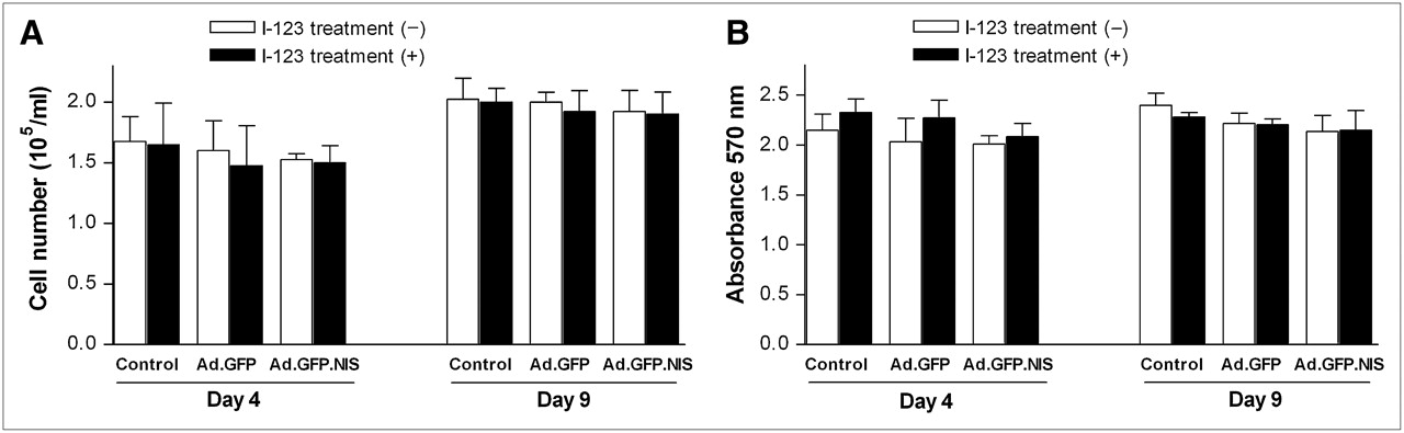
    Effect of adenoviral gene transfer and 123I treatment on cultured cardiac cells. (A) Number of viable cells at 4 and 9 d after infection with 10 MOIs of Ad.EGFP or Ad.EGFP.NIS and in uninfected controls. Cells were incubated with 0 or 185 kBq (0 or 0.5 μCi) 123I for 6 h at days 4 and 9. (B) MTT assay results of cells treated as in A. Cells are H9C2 myoblasts differentiated into cardiomyocytes by stimulation with retinoic acid. All results are mean ± SD of 4 samples obtained from a single experiment (representative of 2 experiments).

  • FIGURE 2. 
    • Download figure
    • Open in new tab
    • Download powerpoint
    FIGURE 2. 

    Time course of cardiac 123I uptake. (A) Serial scintigraphic rat images at 2, 4, and 9 d after myocardial injection with 3 × 10⁸ pfu of Ad.EGFP.NIS (n = 5). Arrow denotes focal cardiac 123I uptake consistent with site of adenovirus injection. (B) Semiquantitative C/M ratios expressed as mean ± SE of values obtained from 5 animals.

  • FIGURE 3. 
    • Download figure
    • Open in new tab
    • Download powerpoint
    FIGURE 3. 

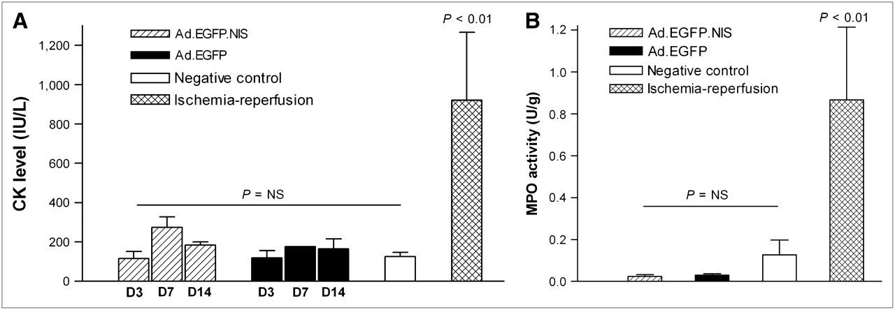
    Serum CK and myocardial MPO activity. (A) Total serum CK activity (mean ± SE; IU/L) at 3 (D3), 7 (D7), and 14 (D14) d after myocardial injection of Ad.EGFP.NIS (n = 3) or Ad.EGFP (n = 3), in negative controls (n = 3), and at 3 d of ischemia–reperfusion (n = 4). (B) Myocardial MPO activity (mean ± SE; U/g) in rats as in A (14 d after adenovirus injection). NS = not significant.

  • FIGURE 4. 
    • Download figure
    • Open in new tab
    • Download powerpoint
    FIGURE 4. 

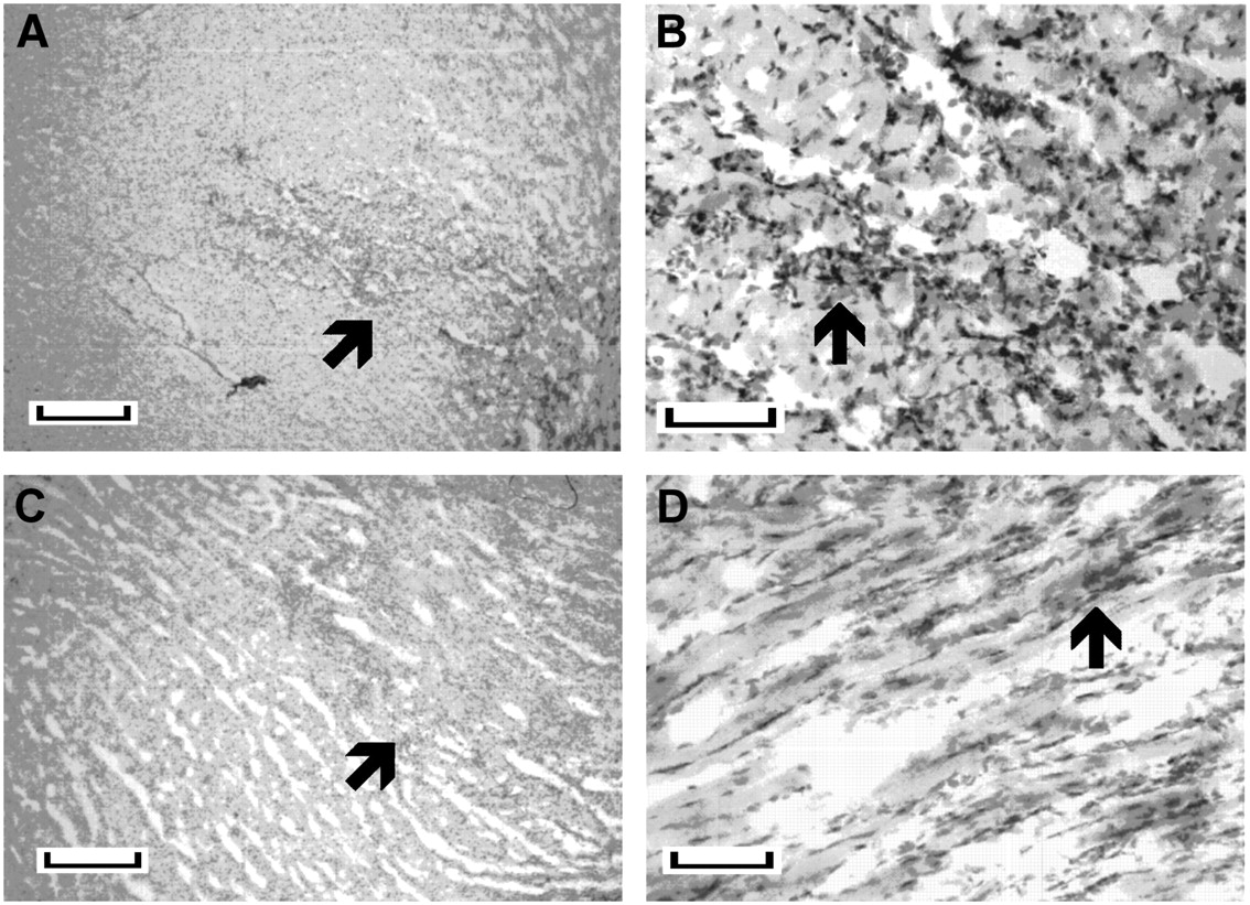
    Hematoxylin-stained myocardial microsections obtained 14 d after intramyocardial injection of Ad.EGFP.NIS (A and B) or Ad.EGFP (C and D). Arrows indicate areas of localized inflammatory infiltration, consistent with injection site. Length of bar indicates 400 μm for A and C and 100 μm for B and D.

Tables

  • Figures
    • View popup
    TABLE 1

    Echocardiographic Measurements Before and Serially After Intramyocardial Ad.EGFP or Ad.EGFP.NIS Injection

    Ad.EGFPAd.EGFP.NIS
    MeasurementBaselineDay 4Day 9BaselineDay 4Day 9
    Heart rate (bpm)341 ± 40328 ± 63306 ± 42300 ± 29292 ± 33318 ± 52
    LV dimension
     IVS thickness (mm)1.5 ± 0.41.6 ± 0.31.7 ± 0.31.6 ± 0.41.8 ± 0.61.8 ± 0.3
     PW thickness (mm)1.8 ± 0.11.8 ± 0.21.7 ± 0.21.7 ± 0.31.6 ± 0.21.8 ± 0.3
     Diastolic diameter (mm)6.8 ± 0.66.8 ± 0.97.5 ± 1.06.9 ± 0.86.8 ± 0.67.5 ± 0.9
     Systolic diameter (mm)3.1 ± 0.83.7 ± 1.04.2 ± 0.7*3.1 ± 0.93.3 ± 0.84.4 ± 0.8*
     IVS/LVPW ratio0.9 ± 0.10.9 ± 0.11.0 ± 0.21.0 ± 0.21.1 ± 0.31.0 ± 0.2
     Diastolic volume (mL)0.7 ± 0.20.7 ± 0.31.0 ± 0.3†0.7 ± 0.20.7 ± 0.21.0 ± 0.3†
     Systolic volume (mL)0.1 ± 0.00.2 ± 0.10.2 ± 0.1*0.1 ± 0.10.1 ± 0.10.2 ± 0.1†
     LV mass (g)0.7 ± 0.10.8 ± 0.20.9 ± 0.2†0.7 ± 0.10.8 ± 0.21.0 ± 0.2‡
    LV functional parameter
     LV FS (%)54.7 ± 10.046.7 ± 7.6†43.6 ± 4.9‡54.3 ± 9.148.6 ± 5.042.4 ± 4.7*
     LV EF (%)88.2 ± 6.482.3 ± 7.779.6 ± 5.0*88.0 ± 5.486.5 ± 5.778.7 ± 4.6*
    • ↵* P < 0.005 compared with baseline measurements.

    • ↵† P < 0.05 compared with baseline measurements.

    • ↵‡ P < 0.01 compared with baseline measurements.

    • IVS = interventricular septum; PW = posterior wall; LVPW = LV posterior wall; LVFS = LV fractional shortening.

    • Data are mean ± SD.

PreviousNext
Back to top

In this issue

Journal of Nuclear Medicine: 47 (11)
Journal of Nuclear Medicine
Vol. 47, Issue 11
November 2006
  • Table of Contents
  • Table of Contents (PDF)
  • About the Cover
  • Index by author
Print
Download PDF
Article Alerts
Sign In to Email Alerts with your Email Address
Email Article

Thank you for your interest in spreading the word on Journal of Nuclear Medicine.

NOTE: We only request your email address so that the person you are recommending the page to knows that you wanted them to see it, and that it is not junk mail. We do not capture any email address.

Enter multiple addresses on separate lines or separate them with commas.
Evidence that Myocardial Na/I Symporter Gene Imaging Does Not Perturb Cardiac Function
(Your Name) has sent you a message from Journal of Nuclear Medicine
(Your Name) thought you would like to see the Journal of Nuclear Medicine web site.
Citation Tools
Evidence that Myocardial Na/I Symporter Gene Imaging Does Not Perturb Cardiac Function
Kyung-Han Lee, Jun-Sang Bae, Sang-Chul Lee, Jin-Young Paik, Takashi Matsui, Kyung-Ho Jung, Bong-Ho Ko, Byung-Tae Kim
Journal of Nuclear Medicine Nov 2006, 47 (11) 1851-1857;

Citation Manager Formats

  • BibTeX
  • Bookends
  • EasyBib
  • EndNote (tagged)
  • EndNote 8 (xml)
  • Medlars
  • Mendeley
  • Papers
  • RefWorks Tagged
  • Ref Manager
  • RIS
  • Zotero
Share
Evidence that Myocardial Na/I Symporter Gene Imaging Does Not Perturb Cardiac Function
Kyung-Han Lee, Jun-Sang Bae, Sang-Chul Lee, Jin-Young Paik, Takashi Matsui, Kyung-Ho Jung, Bong-Ho Ko, Byung-Tae Kim
Journal of Nuclear Medicine Nov 2006, 47 (11) 1851-1857;
Twitter logo Facebook logo LinkedIn logo Mendeley logo
  • Tweet Widget
  • Facebook Like
  • Google Plus One
Bookmark this article

Jump to section

  • Article
    • Abstract
    • MATERIALS AND METHODS
    • RESULTS
    • DISCUSSION
    • CONCLUSION
    • Acknowledgments
    • Footnotes
    • References
  • Figures & Data
  • Info & Metrics
  • PDF

Related Articles

  • This Month in JNM
  • PubMed
  • Google Scholar

Cited By...

  • No citing articles found.
  • Google Scholar

More in this TOC Section

  • Design and Fabrication of Kidney Phantoms for Internal Radiation Dosimetry Using 3D Printing Technology
  • Synthesis and Biologic Evaluation of Novel 18F-Labeled Probes Targeting Prostate-Specific Membrane Antigen for PET of Prostate Cancer
  • Tumor-Specific Binding of Radiolabeled PEGylated GIRLRG Peptide: A Novel Agent for Targeting Cancers
Show more Basic Science Investigations

Similar Articles

SNMMI

© 2025 SNMMI

Powered by HighWire