


Article Figures & Data
Figures
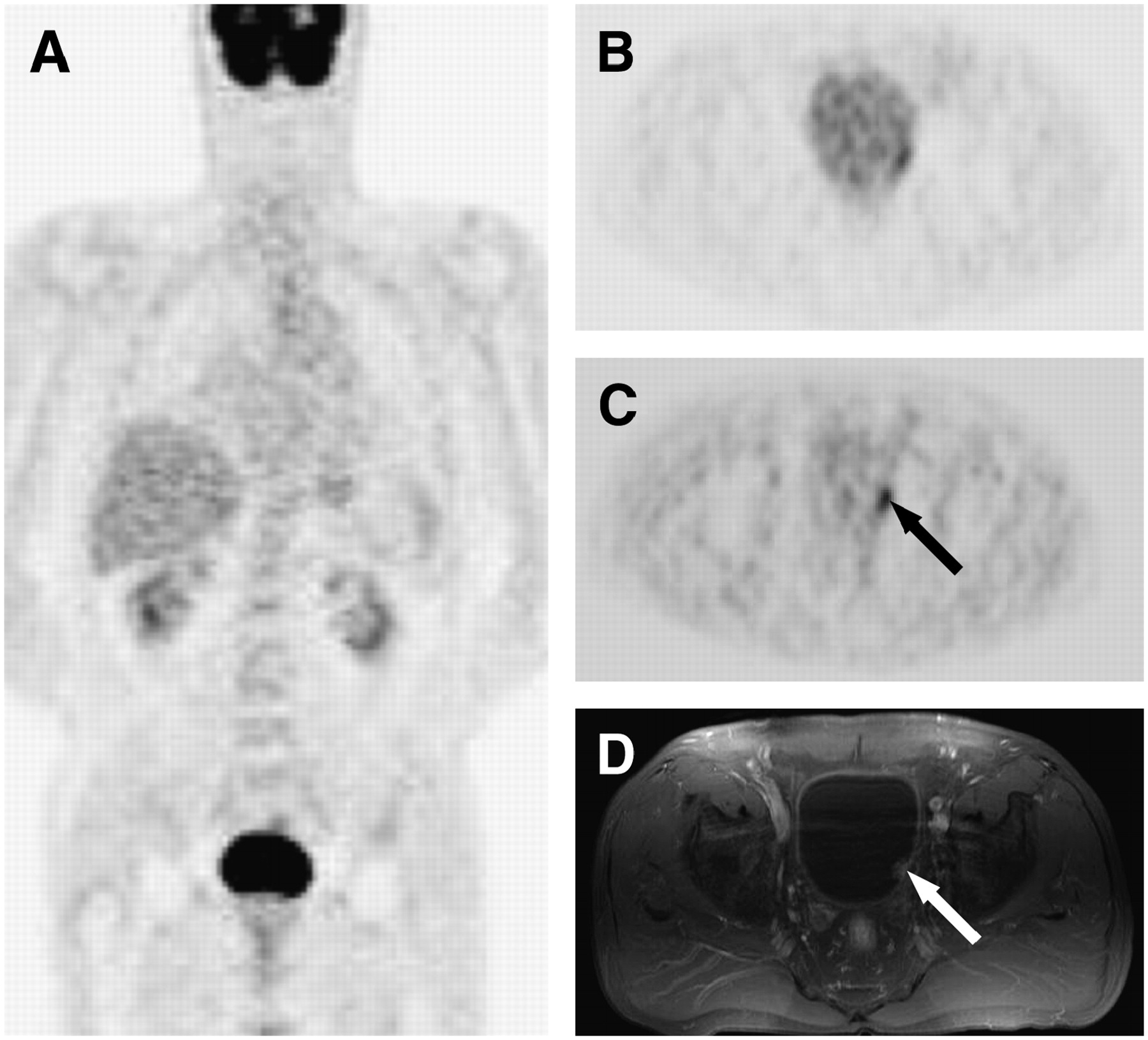
- FIGURE 1.
A 69-y-old man with papillary bladder cancer. (A) Coronal prediuretic 18F-FDG PET image shows complete masking of primary tumor by urinary activity. (B) Transaxial postdiuretic 18F-FDG PET image after 2 voidings of urinary bladder shows equivocal finding. (C and D) Transaxial postdiuretic 18F-FDG PET after 3 voidings (C) and correlative MRI (D) reveal T1 tumor in left lateral wall of bladder (arrows).
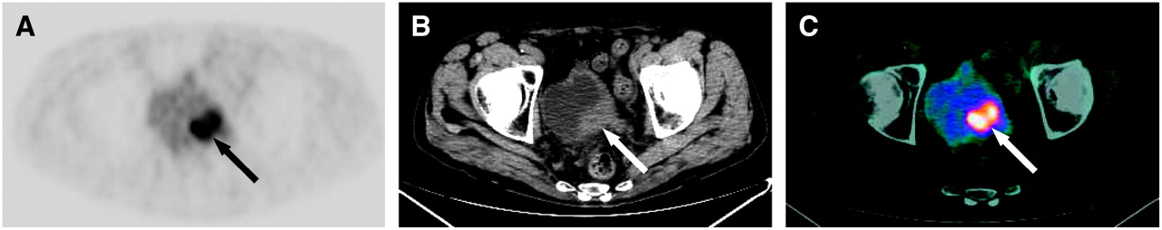
- FIGURE 2.
Transaxial postdiuretic 18F-FDG PET (A) and correlative transaxial CT (B) and PET/CT (C) images of 71-y-old man show 18F-FDG–avid T2 sarcomatoid carcinoma of bladder after residual urinary activity has been discarded (arrows).
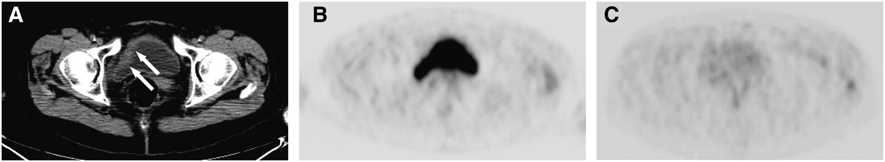
- FIGURE 3.
Sagittal 18F-FDG PET (A) and correlative PET/CT (B) images of 73-y-old man after furosemide challenge show poorly differentiated adenocarcinoma of prostate (arrowheads) that invades bladder base (large arrows) and metastasizes to sacrum (small arrows).
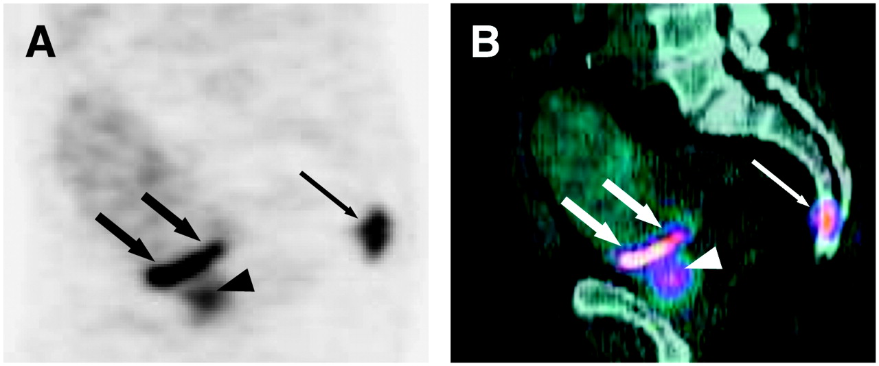
- FIGURE 4.
(A) Transaxial CT image of 68-y-old woman with bladder lymphoma shows residual wall thickening after chemotherapy (arrows). (B and C) Transaxial 18F-FDG PET images of bladder before (B) and after (C) furosemide challenge show significant residual urinary activity and complete, postdiuretic, washout with no evidence of active tumor manifestation. True-negative postdiuretic PET finding was confirmed by clinical and radiologic follow-up.
- FIGURE 5.
(A–C) Coronal and sagittal 18F-FDG PET (A and B) and sagittal PET/CT (C) images of 67-y-old man previously treated for rectosigmoid cancer show focal hot spot along sigmoid colon that mimics local recurrence (arrows). (D) Postdiuretic coronal PET image shows disappearance of this factitious hot spot, suggesting urinary origin. (E) Correlative intravenous urography reveals medially displaced left ureterovesical junction after recent reimplantation surgery of left ureter (arrowheads).
- FIGURE 6.
(A and B) Transaxial (A) and sagittal (B) prediuretic 18F-FDG PET images of 67-y-old woman previously treated for uterine adenocarcinoma show no convincing evidence of local disease recurrence. (C and D) Transaxial (C) and sagittal (D) postdiuretic 18F-FDG PET images display sharply delineated focal hot spot (arrows) that corresponds to disease recurrence in vaginal stump, as confirmed by histologic analysis.


Jump to section
Related Articles
Cited By...
- Elevated 18F-FDG Levels in Blood and Organs After Angiotensin II Receptor Blocker Administration: Experiment in Mice Administered Telmisartan
- Could Different Hydration Protocols Affect the Quality of 18F-FDG PET/CT Images?
- Impact of 18F-FDG PET/CT with Retrograde Filling of the Urinary Bladder in Patients with Suspected Pelvic Malignancies
- Preliminary Study of Detecting Urothelial Malignancy with FDG PET in Taiwanese ESRD Patients
- 18F-FDG PET/CT Delayed Images After Diuretic for Restaging Invasive Bladder Cancer