
Main menu

  • Home
  • Content
    • Current
    • Ahead of print
    • Past Issues
    • JNM Supplement
    • SNMMI Annual Meeting Abstracts
    • Continuing Education
    • JNM Podcasts
  • Subscriptions
    • Subscribers
    • Institutional and Non-member
    • Rates
    • Journal Claims
    • Corporate & Special Sales
  • Authors
    • Submit to JNM
    • Information for Authors
    • Assignment of Copyright
    • AQARA requirements
  • Info
    • Reviewers
    • Permissions
    • Advertisers
  • About
    • About Us
    • Editorial Board
    • Contact Information
  • More
    • Alerts
    • Feedback
    • Help
    • SNMMI Journals
  • SNMMI
    • JNM
    • JNMT
    • SNMMI Journals
    • SNMMI

User menu

  • Subscribe
  • My alerts
  • Log in
  • My Cart

Search

  • Advanced search
Journal of Nuclear Medicine
  • SNMMI
    • JNM
    • JNMT
    • SNMMI Journals
    • SNMMI
  • Subscribe
  • My alerts
  • Log in
  • My Cart
Journal of Nuclear Medicine

Advanced Search

  • Home
  • Content
    • Current
    • Ahead of print
    • Past Issues
    • JNM Supplement
    • SNMMI Annual Meeting Abstracts
    • Continuing Education
    • JNM Podcasts
  • Subscriptions
    • Subscribers
    • Institutional and Non-member
    • Rates
    • Journal Claims
    • Corporate & Special Sales
  • Authors
    • Submit to JNM
    • Information for Authors
    • Assignment of Copyright
    • AQARA requirements
  • Info
    • Reviewers
    • Permissions
    • Advertisers
  • About
    • About Us
    • Editorial Board
    • Contact Information
  • More
    • Alerts
    • Feedback
    • Help
    • SNMMI Journals
  • View or Listen to JNM Podcast
  • Visit JNM on Facebook
  • Join JNM on LinkedIn
  • Follow JNM on Twitter
  • Subscribe to our RSS feeds
OtherBasic Science Investigations

Monitoring Left Ventricular Dilation in Mice with PET

Lars Stegger, Klaus P. Schäfers, Ulrich Flögel, Lefteris Livieratos, Sven Hermann, Christoph Jacoby, Petra Keul, Edward M. Conway, Otmar Schober, Jürgen Schrader, Bodo Levkau and Michael Schäfers
Journal of Nuclear Medicine September 2005, 46 (9) 1516-1521;
Lars Stegger
  • Find this author on Google Scholar
  • Find this author on PubMed
  • Search for this author on this site
Klaus P. Schäfers
  • Find this author on Google Scholar
  • Find this author on PubMed
  • Search for this author on this site
Ulrich Flögel
  • Find this author on Google Scholar
  • Find this author on PubMed
  • Search for this author on this site
Lefteris Livieratos
  • Find this author on Google Scholar
  • Find this author on PubMed
  • Search for this author on this site
Sven Hermann
  • Find this author on Google Scholar
  • Find this author on PubMed
  • Search for this author on this site
Christoph Jacoby
  • Find this author on Google Scholar
  • Find this author on PubMed
  • Search for this author on this site
Petra Keul
  • Find this author on Google Scholar
  • Find this author on PubMed
  • Search for this author on this site
Edward M. Conway
  • Find this author on Google Scholar
  • Find this author on PubMed
  • Search for this author on this site
Otmar Schober
  • Find this author on Google Scholar
  • Find this author on PubMed
  • Search for this author on this site
Jürgen Schrader
  • Find this author on Google Scholar
  • Find this author on PubMed
  • Search for this author on this site
Bodo Levkau
  • Find this author on Google Scholar
  • Find this author on PubMed
  • Search for this author on this site
Michael Schäfers
  • Find this author on Google Scholar
  • Find this author on PubMed
  • Search for this author on this site
  • Article
  • Figures & Data
  • Info & Metrics
  • PDF
Loading

Article Figures & Data

Figures

  • Tables
  • FIGURE 1.
    • Download figure
    • Open in new tab
    • Download powerpoint
    FIGURE 1.

    PET and MR images depict a short-axis slice through the heart of a wild-type mouse (A) with normal heart size and a transgenic mouse (B) with dilated left ventricle. MR images shown were acquired in end-diastole.

  • FIGURE 2.
    • Download figure
    • Open in new tab
    • Download powerpoint
    FIGURE 2.

    Midmyocardial (solid line) and endo- or epicardial (dashed lines) contours superimposed on 18F-FDG PET short-axis image (A) and vertical long-axis image (B) of mouse heart as measured by PET. Only the midmyocardial contour is used in this study.
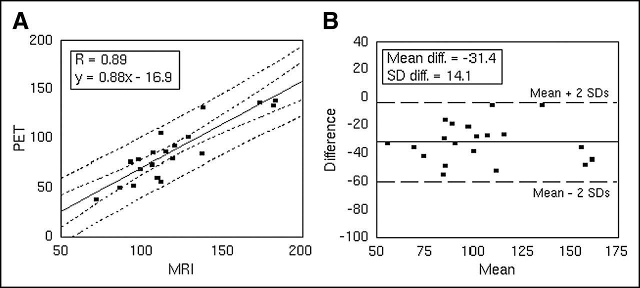
  • FIGURE 3.
    • Download figure
    • Open in new tab
    • Download powerpoint
    FIGURE 3.

    Comparison of LV volume indices LV-VIPET/MRI (in μL) measured by PET and MRI. Equal fractional contributions of end-diastolic or end-systolic volumes were used for LV-VIMRI (xED = xES = 0.5). (A) Scatter plot of LV volume indices measured by PET vs. MRI. Confidence curves for regression fit as well as confidence curves of individual values are also shown (dashed lines, α = 0.05). (B) Difference of PET and MRI measurements plotted against the mean of both measurements. Horizontal lines for mean (solid) and mean ± 2 SD (dashed) are included. Values for linear fit (intercept, 30.0; slope, 0.011) are not shown.

Tables

  • Figures
    • View popup
    TABLE 1

    Weight and Sex of Wild-Type and Transgenic Mice (Matched Pairs)

    AgeWild-type (g)Transgenic (g)Sex
    Young21.624.3F
    27.427.9M
    25.927.3M
    19.516.8F
    23.023.6F
    φ23.5φ24.0
    Old34.032.3M
    26.826.9F
    35.632.3M
    28.132.7M
    29.722.3M
    φ30.8φ29.3
    • ↵φ indicates mean weight.

    • View popup
    TABLE 2

    Choice of Fractional Contributions of End-Diastolic and End-Systolic Volumes in Calculation of LV-VIMRI and its Influence on Correlation Between PET- and MRI-Derived LV-VI

    Fraction xED of end-diast. volume in LV-VIMRIFraction xES of end-syst. volume in LV-VIMRICorrelation coefficient (R)Intercept of correlation lineSlope of correlation line
    0.01.00.8811.00.83
    0.20.80.89−0.260.85
    0.50.50.89−16.90.88
    0.80.20.89−32.60.89
    1.00.00.88−41.90.89
    • end-diast. = end-diastolic; end-syst. = end-systolic.

    • View popup
    TABLE 3

    Matrix of Group Results

    PETYoungOldP value
    Wild-type65.0 ± 16.982.6 ± 20.90.185 (NS)
    Transgenic73.8 ± 15.0129.3 ± 15.3<0.001 (S)
    P value0.413 (NS)0.004 (S)
    MRIYoung*†Old*†P value*†
    Wild-type94.7 ± 14.6106.2 ± 12.30.214 (NS)
    111.3 ± 16.0†122.8 ± 12.3†0.27† (NS)
    Transgenic114.7 ± 15.2160.5 ± 25.70.012 (S)
    132.3 ± 15.8†174.6 ± 25.5†0.014† (S)
    P value0.067 (NS)0.006 (S)
    0.071† (NS)0.004† (S)
    • ↵* First row of results for equal fractional contributions of end-diastolic or end-systolic volumes in definition of LV-VIMRI (xED = xES = 0.5).

    • ↵† Denotes values with unequal fractional contributions (xED = 0.8, xES = 0.2).

    • NS = nonsignificant results; S = significant results.

      Mean values of midmyocardial volumes (in μL) ± SDs for all 4 groups of animals (young—old, wild-type—transgenic) are presented together with t-test results (P values). Results are presented separately for PET and MRI measurement techniques.

PreviousNext
Back to top

In this issue

Journal of Nuclear Medicine: 46 (9)
Journal of Nuclear Medicine
Vol. 46, Issue 9
September 1, 2005
  • Table of Contents
  • About the Cover
  • Index by author
Print
Download PDF
Article Alerts
Sign In to Email Alerts with your Email Address
Email Article

Thank you for your interest in spreading the word on Journal of Nuclear Medicine.

NOTE: We only request your email address so that the person you are recommending the page to knows that you wanted them to see it, and that it is not junk mail. We do not capture any email address.

Enter multiple addresses on separate lines or separate them with commas.
Monitoring Left Ventricular Dilation in Mice with PET
(Your Name) has sent you a message from Journal of Nuclear Medicine
(Your Name) thought you would like to see the Journal of Nuclear Medicine web site.
Citation Tools
Monitoring Left Ventricular Dilation in Mice with PET
Lars Stegger, Klaus P. Schäfers, Ulrich Flögel, Lefteris Livieratos, Sven Hermann, Christoph Jacoby, Petra Keul, Edward M. Conway, Otmar Schober, Jürgen Schrader, Bodo Levkau, Michael Schäfers
Journal of Nuclear Medicine Sep 2005, 46 (9) 1516-1521;

Citation Manager Formats

  • BibTeX
  • Bookends
  • EasyBib
  • EndNote (tagged)
  • EndNote 8 (xml)
  • Medlars
  • Mendeley
  • Papers
  • RefWorks Tagged
  • Ref Manager
  • RIS
  • Zotero
Share
Monitoring Left Ventricular Dilation in Mice with PET
Lars Stegger, Klaus P. Schäfers, Ulrich Flögel, Lefteris Livieratos, Sven Hermann, Christoph Jacoby, Petra Keul, Edward M. Conway, Otmar Schober, Jürgen Schrader, Bodo Levkau, Michael Schäfers
Journal of Nuclear Medicine Sep 2005, 46 (9) 1516-1521;
Twitter logo Facebook logo LinkedIn logo Mendeley logo
  • Tweet Widget
  • Facebook Like
  • Google Plus One
Bookmark this article

Jump to section

  • Article
    • Abstract
    • MATERIALS AND METHODS
    • RESULTS
    • DISCUSSION
    • CONCLUSION
    • Acknowledgments
    • Footnotes
    • REFERENCES
  • Figures & Data
  • Info & Metrics
  • PDF

Related Articles

  • THIS MONTH IN JNM
  • PubMed
  • Google Scholar

Cited By...

  • Micro-Positron Emission Tomography in the Evaluation of Trypanosoma cruzi-Induced Heart Disease: Comparison with Other Modalities
  • Quantification of Left Ventricular Volumes and Ejection Fraction in Mice Using PET, Compared with MRI
  • Accurate Noninvasive Measurement of Infarct Size in Mice with High-Resolution PET
  • Google Scholar

More in this TOC Section

  • 11C-Methionine PET of Myocardial Inflammation in a Rat Model of Experimental Autoimmune Myocarditis
  • Counting Rate Characteristics and Image Distortion in Preclinical PET Imaging During Radiopharmaceutical Therapy
  • Design and Fabrication of Kidney Phantoms for Internal Radiation Dosimetry Using 3D Printing Technology
Show more Basic Science Investigations

Similar Articles

SNMMI

© 2025 SNMMI

Powered by HighWire