


Article Figures & Data
Figures
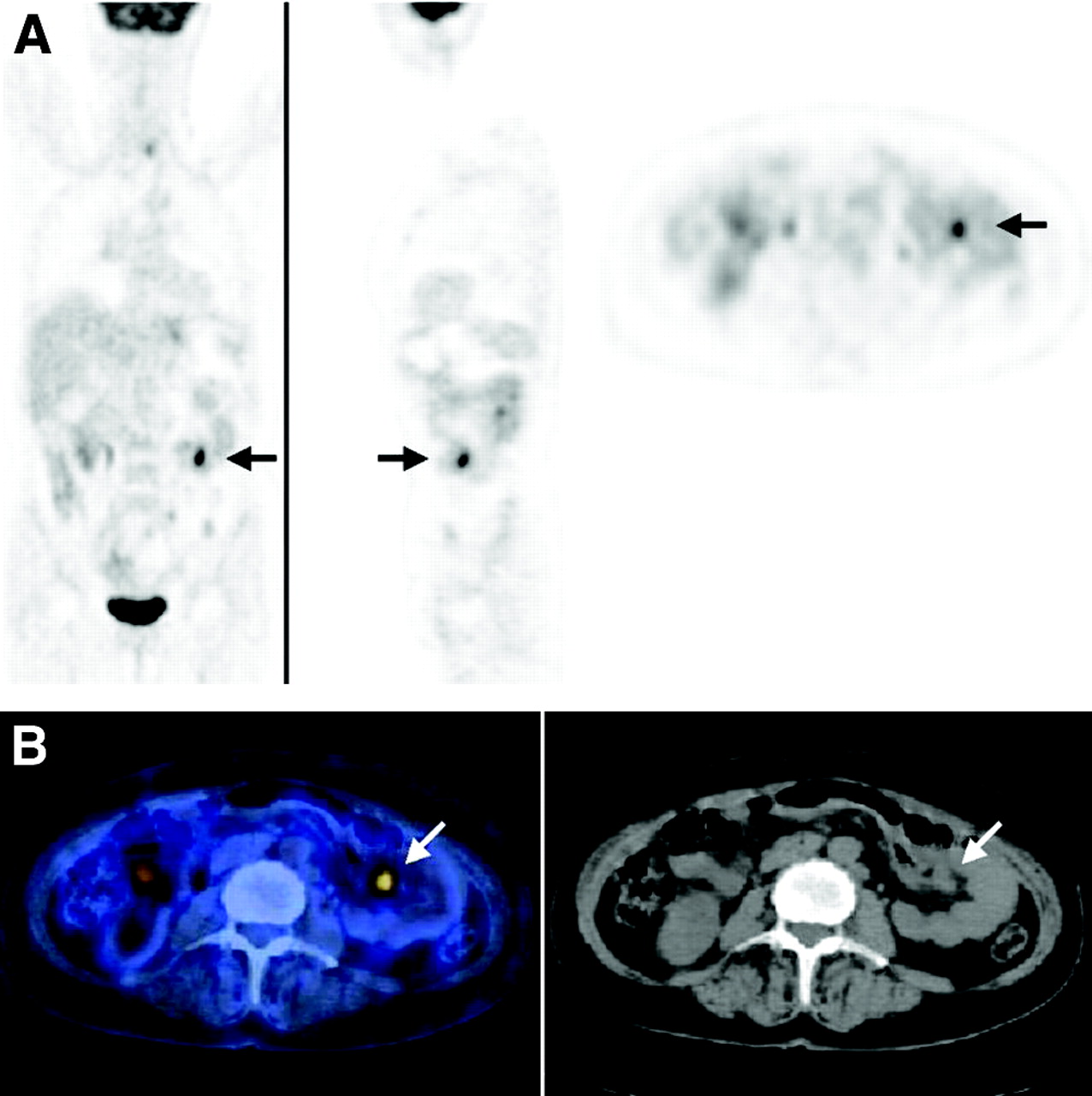
- FIGURE 1.
Focal 18F-FDG uptake in 57-y-old woman who had undergone total gastrectomy for stomach cancer and was being evaluated for fatigue, abdominal pain, frequent vomiting, equivocal endoscopic findings at level of anastomosis, and negative findings on whole-body CT. (A) From left to right, coronal, sagittal, and transaxial PET slices show focus of increased 18F-FDG uptake (arrows) in left lower abdomen. (B) Area of increased uptake (arrows) was localized by PET/CT (left panel) to small bowel, as seen on corresponding CT image (right panel). PET/CT-guided surgery revealed small-bowel metastasis originating from primary gastric cancer. No abnormal 18F-FDG uptake was seen in region of anastomosis, and there was no further evidence of disease in this area.
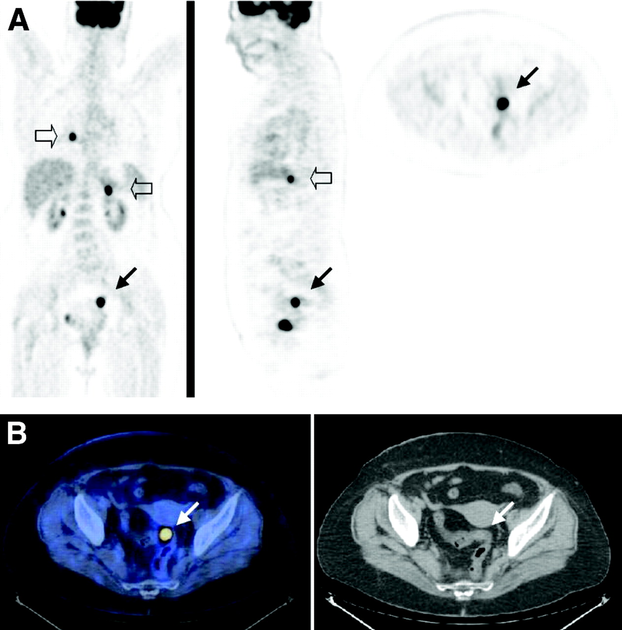
- FIGURE 2.
Focal 18F-FDG uptake in 64-y-old woman who was being evaluated for staging of aggressive non-Hodgkin’s lymphoma. (A) From left to right, coronal, sagittal, and transaxial PET slices show focus of increased 18F-FDG uptake (arrows) in left pelvis. Coronal and sagittal PET images show additional areas of abnormal 18F-FDG uptake in mediastinum and left upper abdomen (open arrows), consistent with sites of active lymphoma. (B) Pelvic area of increased uptake (arrows) was localized by PET/CT (left panel) to sigmoid, as seen on corresponding CT image (right panel). Villous adenoma was diagnosed from biopsy sample taken during colonoscopy.
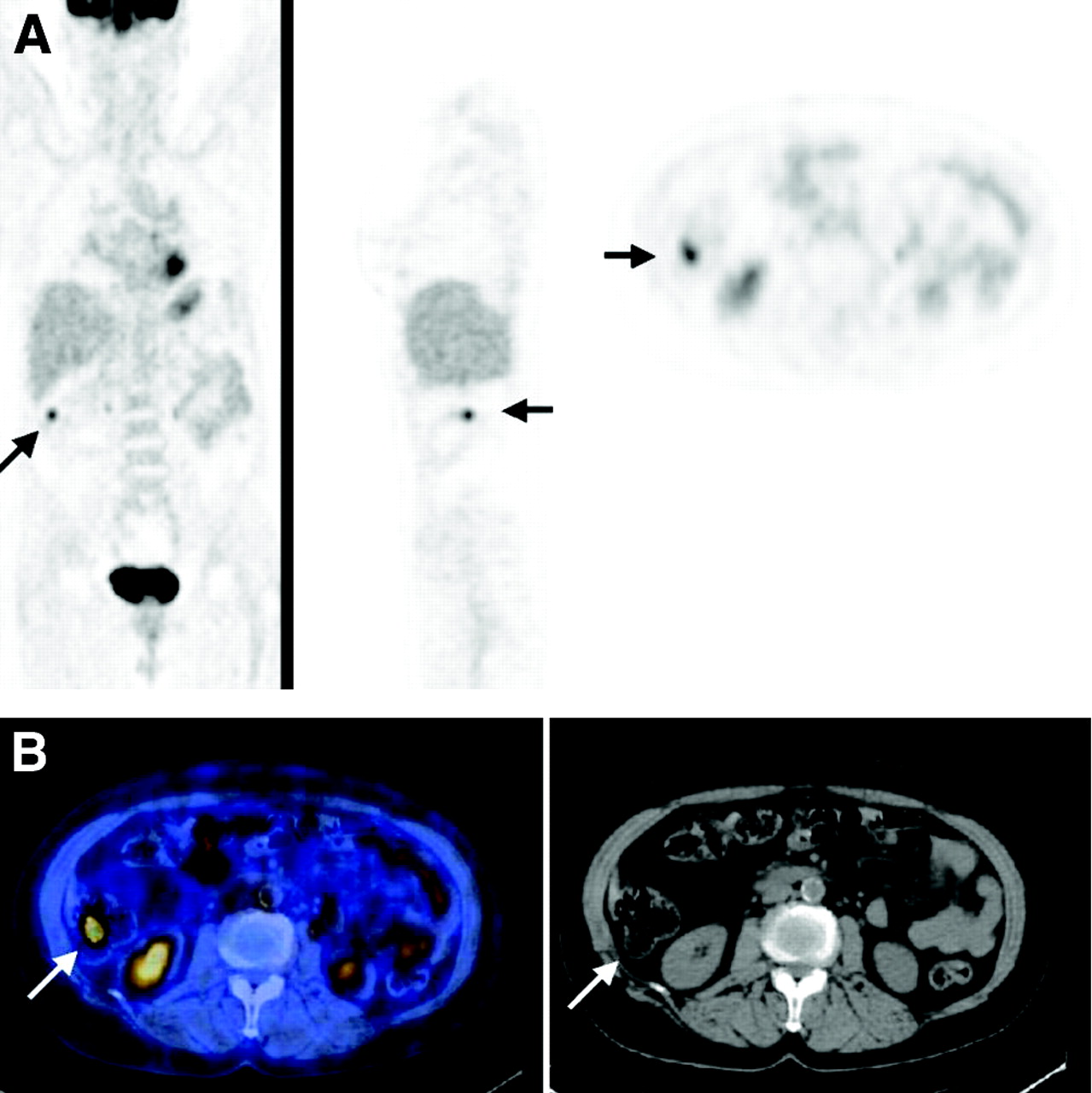
- FIGURE 3.
Focal 18F-FDG uptake in 70-y-old woman with low-grade non-Hodgkin’s lymphoma who was undergoing routine follow-up examination. (A) Coronal, sagittal, and transaxial PET slices show focus of increased 18F-FDG uptake (arrows) in right upper abdomen. (B) Area of increased uptake (arrows) was localized by PET/CT (left panel) to ascending colon, as seen on corresponding CT image (right panel). Colonoscopy had negative findings, and patient showed no evidence of disease after 16 mo of follow-up. Focus of increased 18F-FDG uptake was considered to represent physiologic bowel activity.

Tables
Parameter Malignant Premalignant Benign Physiologic Foci (n) 11 9 4 10 Anatomic site Stomach 3 — 1 — Small intestine 2 — — — Colon 6 9 3 10 SUVmax Mean ± SD 17.3 ± 10.2 14.0 ± 10.5 18.0 ± 12.1 11.1 ± 7.4 Range 8.1–40.3 4.5–40 8.7–35.6 5.7–30.8 PET/CT focus Diagnosis Location Patients (n) On referral to PET/CT After PET/CT Patients (n) Stomach 4 SPN Gastric cancer 2 Colon cancer Second primary gastric cancer 1 Colon cancer Active gastritis 1 Small bowel 2 Stomach cancer Metastasis 1 Colon cancer Metastasis 1 Colon 28 MCUO Colon cancer 1 SPN Colon cancer 1 Lung cancer Second primary colon cancer 2 Lung cancer Metastasis 1 Colon cancer Metastasis 1 Lymphoma Villous adenoma 2 Colon cancer Villous adenoma 2 Lung cancer Adenomatous polyp with low-grade dysplasia 2 Breast cancer Adenomatous polyp with low-grade dysplasia 1 Histiocytoma Villous adenoma 1 SPN Tubular adenoma 1 Lymphoma Benign polyps (hamartomatous and serrated) 2 Sarcoma Abscess of sigmoid 1 Lymphoma Physiologic uptake 3 Colon cancer Physiologic uptake 3 Esophagus cancer Physiologic uptake 1 Lung cancer Physiologic uptake 1 SPN Physiologic uptake 1 MCUO Physiologic uptake 1 SPN = single pulmonary nodule; MCUO = metastatic cancer of unknown origin.
In this issue


Jump to section
Related Articles
Cited By...
- Long-term Outcomes of Early-stage Non-stomach Gastrointestinal Mucosa-associated Lymphoid Tissue Lymphoma Treated With Radiation Therapy
- Correlation of BRAFV600E Mutation and Glucose Metabolism in Thyroid Cancer Patients: An 18F-FDG PET Study
- Assessment of incidental and clinically unsuspected fluorodeoxyglucose-avid foci detected on oncological positron emission tomography/CT
- The role of the breast radiologist in evaluation of breast incidentalomas detected on 18-fludeoxyglucose positron emission tomography/CT
- Incidental findings on positron emission tomography/CT scans performed in the investigation of lung cancer
- Amoebic Abscess Diagnosed on Fluorodeoxyglucose Positron Emission Tomography Scan in Patient With Recurrent Oropharyngeal Squamous Cell Carcinoma
- Incidental Focal 18F-FDG Uptake in the Pituitary Gland: Clinical Significance and Differential Diagnostic Criteria
- Total Abdominal 18F-FDG Uptake Reflects Intestinal Adenoma Burden in Apc Mutant Mice
- Nonlaxative PET/CT Colonography: Feasibility, Acceptability, and Pilot Performance in Patients at Higher Risk of Colonic Neoplasia
- Incidental findings in imaging diagnostic tests: a systematic review
- 18F-FDG PET and PET/CT in the Evaluation of Cancer Treatment Response
- PET/CT assessment of clinically unsuspected, incidental FDG-avid lesions in oncological patients
- 18F-FDG PET/CT in Evaluating Non-CNS Pediatric Malignancies
- Detection of extrapulmonary lesions with integrated PET/CT in the staging of lung cancer
- Screening for Cancer with PET and PET/CT: Potential and Limitations
- Improvements in Cancer Staging with PET/CT: Literature-Based Evidence as of September 2006
- Lung Cancer Presenting With a Solitary Colon Metastasis Detected on Positron Emission Tomography Scan