


Article Figures & Data
Figures
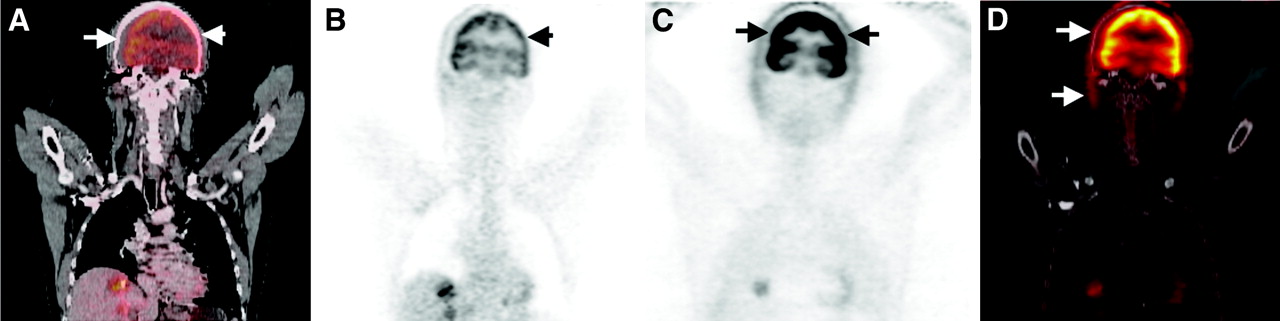
- FIGURE 1.
(A) Limited coronal view of WB FDG PET/CT study with severe misalignment (arrows) in area of head and neck. (B and C) PET image after CT-based attenuation correction (B) demonstrates biased FDG uptake (arrow), which appears normal on uncorrected PET image (C). (D) Fused CT and uncorrected PET illustrate magnitude of misalignment.
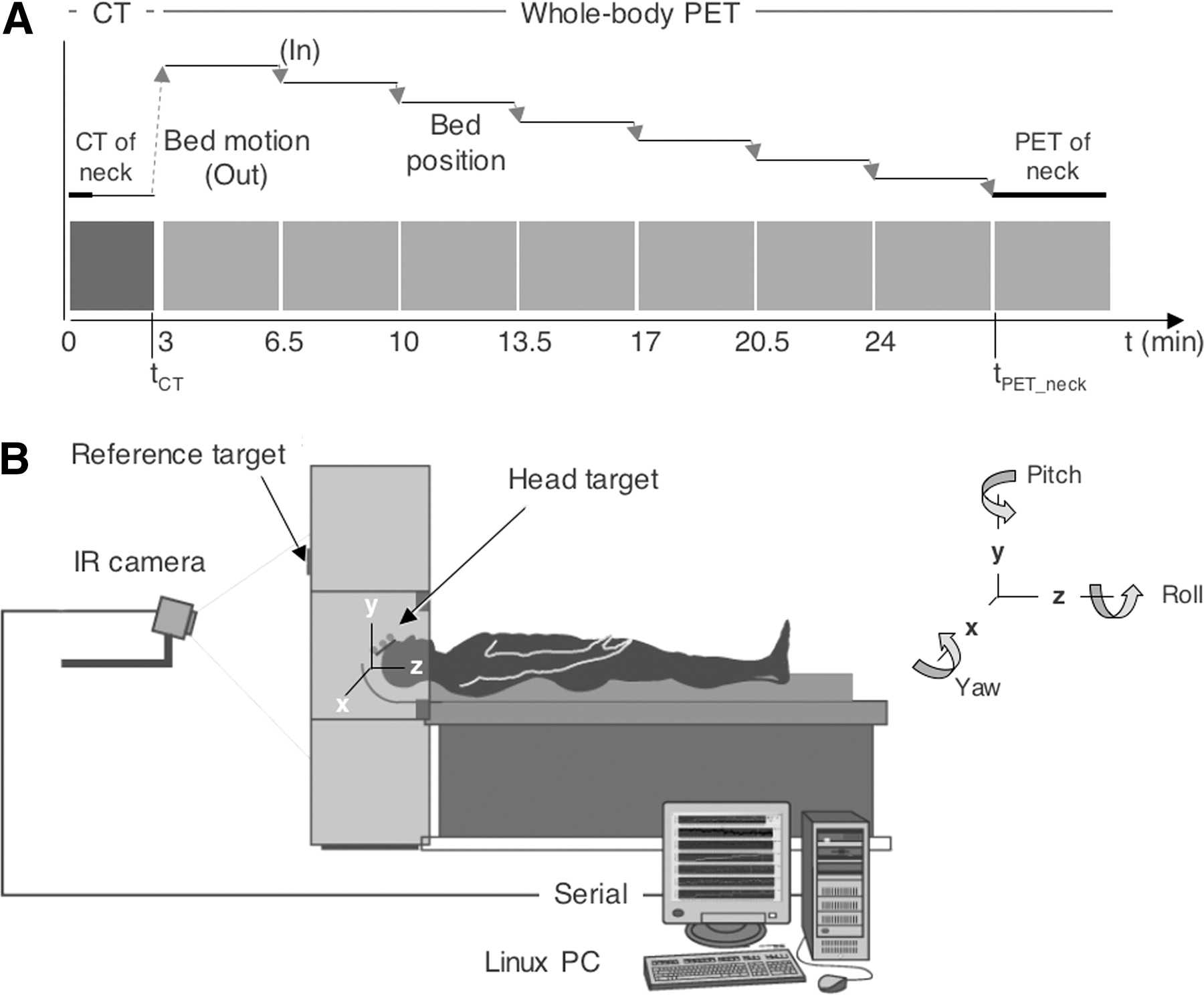
- FIGURE 2.
(A) Timeline of simulated WB PET/CT study and bed motion. Each plateau represents physically stationary bed position; connecting arrows indicate bed motion. (B) Drawing illustrates arrangement of motion-tracking device with coordinate system for monitoring misregistration of head and neck by means of 3 rotational and 3 translational parameters. IR = infrared.
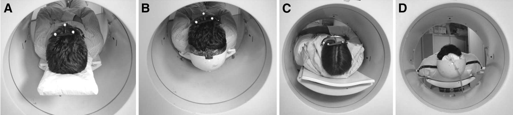
- FIGURE 3.
Supports for head and neck in simulated WB PET/CT studies: disposable cushion (A), head holder with disposable construction-foam insert (B), vacuum-lock bag (C), and smaller vacuum-lock bag (D) inside head holder shown in B. Photographs were taken from rear of PET tomograph used for simulations.
- FIGURE 4.
Boxcar plots for PET/CT misregistration in the 3 translational (Δx, Δy, Δz) and 3 rotational (pi, pitch; ya, yaw; ro, roll) parameters for subjects in setup A, head holder (A); setup B, head holder with construction foam (B); setup C, vacuum-lock bag (C); and setup D, head holder with vacuum-lock bag (D).
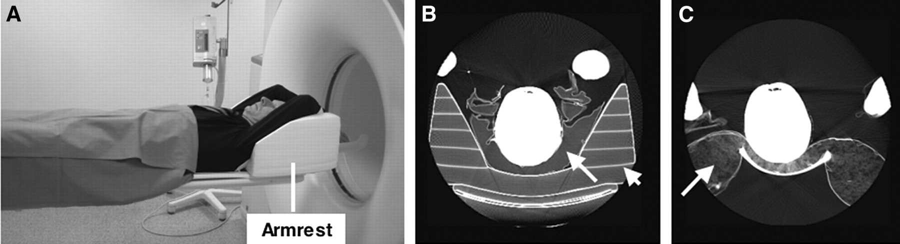
- FIGURE 5.
(A) Patient positioning in clinical PET/CT scanner with armrest to support arms above head during combined scanning. (B and C) Axial CT images (windowed) illustrate patient positioning with vacuum-lock bag (arrow) placed inside armrest (arrowhead) (B) and with head supported by cushion (arrow) placed in front of armrest (C). Measured CT attenuation values of cushion, armrest, and vacuum-lock bag were −970 HU, −900 HU, and −940 HU, respectively.
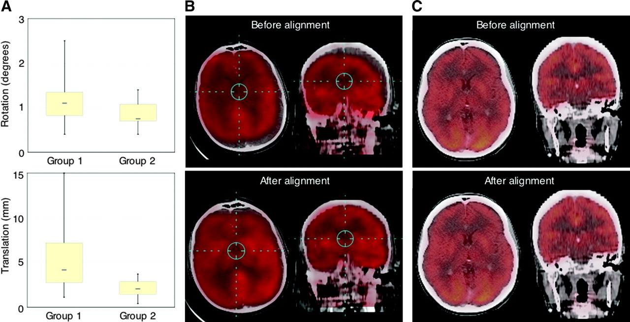
- FIGURE 6.
(A) Boxcar plots of center-to-center misalignment of head and neck in WB PET/CT studies of patients with standard neck support (group 1) and with vacuum-lock support (group 2). (B and C) Examples of axial (left) and coronal (right) fusion images are shown before and after linear realignment for group 1 (B) and group 2 (C). Effect of realignment is significant in patients without head restraint (group 1) and less noticeable in group 2 because of better intrinsic patient positioning.


Tables
- TABLE 1
Average (±SD) Absolute Displacement (Rotation [Pitch, Yaw, Roll] and Translation [Δx, Δy, Δz]) of Reference Markers for Subjects in Groups A–D
Parameter Group A B C D Rotation (degrees) Pitch 0.8 ± 0.5 0.3 ± 0.1 (0.6 ± 0.4) 0.3 ± 0.3 Yaw 1.1 ± 0.8 (0.6 ± 0.6) (1.2 ± 0.6) (0.5 ± 0.4) Roll 1.1 ± 0.9 0.3 ± 0.3 0.2 ± 0.2 0.2 ± 0.1 〈rot〉 1.0 ± 0.2 0.4 ± 0.1 (0.7 ± 0.5) 0.3 ± 0.2 Translation (mm) Δx 4 ± 2 1 ± 1 (4 ± 2) 0.4 ± 0.3 Δy 4 ± 3 1 ± 1 1.0 ± 0.8 0.6 ± 0.5 Δz 3 ± 1 (1 ± 1) (2 ± 1) 1.1 ± 0.5 〈Δr〉 7 ± 3 2 ± 2 (4 ± 2) 1.4 ± 0.5 Mean (±SD) of average absolute rotational displacement (<rot>) and mean (±SD) of individual translational displacements Δr (<Δr>) are listed also. Numbers in parentheses indicate no statistically significant difference with respect to group A for Student t test (2-tailed, P < 0.05).
In this issue


Jump to section
Related Articles
Cited By...
- Technical Considerations in Brain Amyloid PET Imaging with 18F-Florbetapir
- Value of a Lower-Limb Immobilization Device for Optimization of SPECT/CT Image Fusion
- Does Reducing CT Artifacts from Dental Implants Influence the PET Interpretation in PET/CT Studies of Oral Cancer and Head and Neck Cancer?
- Can PET/CT Replace Separate Diagnostic CT for Cancer Imaging? Optimizing CT Protocols for Imaging Cancers of the Chest and Abdomen
- Correction of Head Movement on PET Studies: Comparison of Methods
- Reply: Adequate Evaluation of Image Registration in Hybrid PET/CT.