
Main menu

  • Home
  • Content
    • Current
    • Ahead of print
    • Past Issues
    • JNM Supplement
    • SNMMI Annual Meeting Abstracts
    • Continuing Education
    • JNM Podcasts
  • Subscriptions
    • Subscribers
    • Institutional and Non-member
    • Rates
    • Journal Claims
    • Corporate & Special Sales
  • Authors
    • Submit to JNM
    • Information for Authors
    • Assignment of Copyright
    • AQARA requirements
  • Info
    • Reviewers
    • Permissions
    • Advertisers
  • About
    • About Us
    • Editorial Board
    • Contact Information
  • More
    • Alerts
    • Feedback
    • Help
    • SNMMI Journals
  • SNMMI
    • JNM
    • JNMT
    • SNMMI Journals
    • SNMMI

User menu

  • Subscribe
  • My alerts
  • Log in
  • My Cart

Search

  • Advanced search
Journal of Nuclear Medicine
  • SNMMI
    • JNM
    • JNMT
    • SNMMI Journals
    • SNMMI
  • Subscribe
  • My alerts
  • Log in
  • My Cart
Journal of Nuclear Medicine

Advanced Search

  • Home
  • Content
    • Current
    • Ahead of print
    • Past Issues
    • JNM Supplement
    • SNMMI Annual Meeting Abstracts
    • Continuing Education
    • JNM Podcasts
  • Subscriptions
    • Subscribers
    • Institutional and Non-member
    • Rates
    • Journal Claims
    • Corporate & Special Sales
  • Authors
    • Submit to JNM
    • Information for Authors
    • Assignment of Copyright
    • AQARA requirements
  • Info
    • Reviewers
    • Permissions
    • Advertisers
  • About
    • About Us
    • Editorial Board
    • Contact Information
  • More
    • Alerts
    • Feedback
    • Help
    • SNMMI Journals
  • View or Listen to JNM Podcast
  • Visit JNM on Facebook
  • Join JNM on LinkedIn
  • Follow JNM on Twitter
  • Subscribe to our RSS feeds
OtherClinical Investigations

Selective Neuronal Damage and Borderzone Infarction in Carotid Artery Occlusive Disease: A 11C-Flumazenil PET Study

Hiroshi Yamauchi, Takashi Kudoh, Yoshihiko Kishibe, Jinei Iwasaki and Shinya Kagawa
Journal of Nuclear Medicine December 2005, 46 (12) 1973-1979;
Hiroshi Yamauchi
  • Find this author on Google Scholar
  • Find this author on PubMed
  • Search for this author on this site
Takashi Kudoh
  • Find this author on Google Scholar
  • Find this author on PubMed
  • Search for this author on this site
Yoshihiko Kishibe
  • Find this author on Google Scholar
  • Find this author on PubMed
  • Search for this author on this site
Jinei Iwasaki
  • Find this author on Google Scholar
  • Find this author on PubMed
  • Search for this author on this site
Shinya Kagawa
  • Find this author on Google Scholar
  • Find this author on PubMed
  • Search for this author on this site
  • Article
  • Figures & Data
  • Info & Metrics
  • PDF
Loading

Article Figures & Data

Figures

  • Tables
  • FIGURE 1.
    • Download figure
    • Open in new tab
    • Download powerpoint
    FIGURE 1.

    Examples of PET images on 2 different levels show parallel decrease of FMZ-BP, CBF, CMRo2, and an increase of OEF in patient with right ICA occlusion who showed internal borderzone infarction (red arrow) with cortical extension (green arrow) on corresponding MR images. In addition to markedly reduced FMZ-BP in region with small cortical infarcts, a decrease of FMZ-BP was found in normal-appearing cerebral cortex beyond borderzone infarcts. Mean hemispheric values of ipsilateral/contralateral hemisphere: FMZ-BP ratio, 1.00/1.16; CBF, 20.4/26.1 mL/100 g/min; CMRo2, 2.37/2.87 mL/100 g/min; OEF, 61.9%/58.3%.

  • FIGURE 2.
    • Download figure
    • Open in new tab
    • Download powerpoint
    FIGURE 2.

    Examples of PET images show distribution of decrease of FMZ-BP in patient with right ICA occlusion (patient in Fig. 1). Although decreases of FMZ-BP predominated in frontoparietal region near borderzone infarction, an extensive decrease of FMZ-BP was found in MCA distribution.
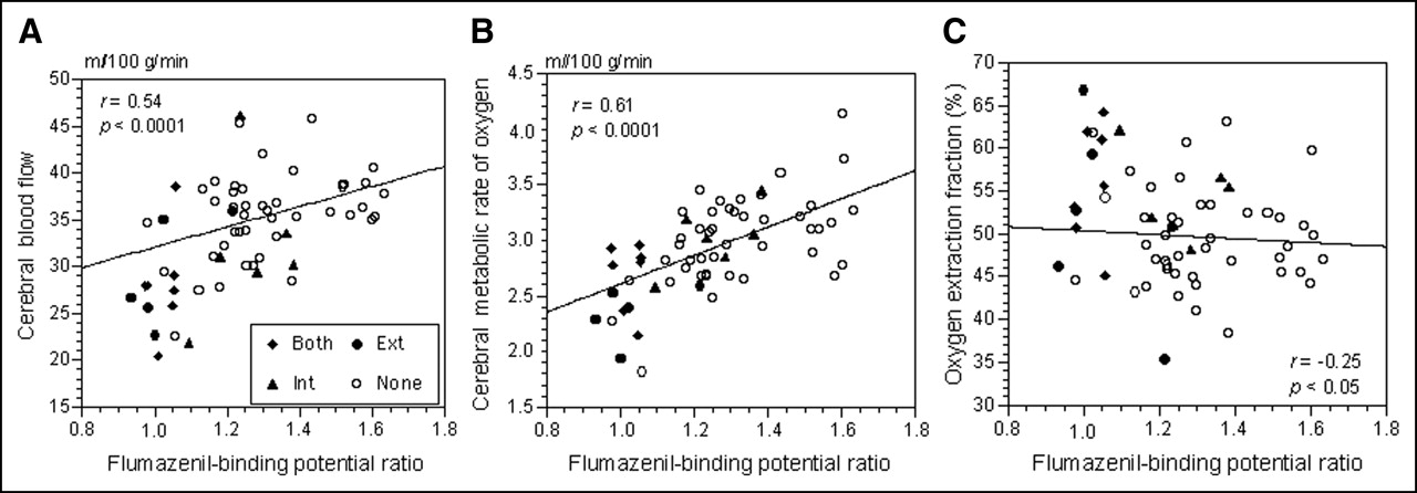
  • FIGURE 3.
    • Download figure
    • Open in new tab
    • Download powerpoint
    FIGURE 3.

    Scatter diagrams plot CBF (A), CMRo2 (B), and OEF (C) against cerebral/cerebellar FMZ-BP ratio in 62 hemispheres with carotid artery disease. The lines were generated by simple linear regression. Ext = solitary external borderzone infarct; Int = solitary internal borderzone infarct; Both = both borderzone infarct; None = no borderzone infarct.

Tables

  • Figures
    • View popup
    TABLE 1

    Characteristics of Patients

    CharacteristicAsymptomaticTIAStroke
    No. of patients211229
    ICA occlusion/stenosis8/135/716/13
        Asymptomatic contralateral carotid stenosis >50% (n)226
        Asymptomatic vertebral artery stenosis >50% (n)421
    Type of lesion (n)
        External borderzone infarcts0012
        Territorial infarcts1010
        Internal borderzone infarcts0112
        Other white matter infarcts447
        Striatocapsular infarcts005
        Lacunar infarcts8212
    • TIA = transient ischemic attack.

    • View popup
    TABLE 2

    Ischemic Lesions and Cerebral/Cerebellar FMZ BP Ratio in Hemisphere with Carotid Artery Disease

    Type of lesionYesNo
    External borderzone infarcts (n = 12)1.02 ± 0.07*1.31 ± 0.16
    Territorial infarcts (n = 11)1.22 ± 0.121.26 ± 0.20
    Internal borderzone infarcts (n = 13)1.13 ± 0.14*1.29 ± 0.19
    Other white matter infarcts (n = 15)1.29 ± 0.171.24 ± 0.19
    Striatocapsular infarcts (n = 5)1.20 ± 0.161.26 ± 0.19
    Lacunar infarcts (n = 22)1.22 ± 0.171.27 ± 0.20
    • ↵* P < 0.008 (0.05/6) vs. corresponding value in No group (Student t test).

    • View popup
    TABLE 3

    Multiple Linear Regression Analysis with Cerebral/Cerebellar FMZ BP Ratio in Hemisphere with Carotid Artery Disease as Dependent Variable

    ParameterCoefficientSEtP value
    External borderzone infarcts (no = 0, yes = 1)−0.2850.056−5.11<0.0001
    Age (y)−0.0030.003−1.060.292
    Territorial infarcts (no = 0, yes = 1)−0.0760.053−1.430.157
    Internal borderzone infarcts (no = 0, yes = 1)−0.0580.054−1.060.293
    Other white matter infarcts (no = 0, yes = 1)0.0110.0460.240.809
    Striatocapsular infarcts (no = 0, yes = 1)−0.1370.072−1.900.062
    Lacunar infarcts (no = 0, yes = 1)−0.0450.043−1.040.299
PreviousNext
Back to top

In this issue

Journal of Nuclear Medicine: 46 (12)
Journal of Nuclear Medicine
Vol. 46, Issue 12
December 1, 2005
  • Table of Contents
  • About the Cover
  • Index by author
Print
Download PDF
Article Alerts
Sign In to Email Alerts with your Email Address
Email Article

Thank you for your interest in spreading the word on Journal of Nuclear Medicine.

NOTE: We only request your email address so that the person you are recommending the page to knows that you wanted them to see it, and that it is not junk mail. We do not capture any email address.

Enter multiple addresses on separate lines or separate them with commas.
Selective Neuronal Damage and Borderzone Infarction in Carotid Artery Occlusive Disease: A 11C-Flumazenil PET Study
(Your Name) has sent you a message from Journal of Nuclear Medicine
(Your Name) thought you would like to see the Journal of Nuclear Medicine web site.
Citation Tools
Selective Neuronal Damage and Borderzone Infarction in Carotid Artery Occlusive Disease: A 11C-Flumazenil PET Study
Hiroshi Yamauchi, Takashi Kudoh, Yoshihiko Kishibe, Jinei Iwasaki, Shinya Kagawa
Journal of Nuclear Medicine Dec 2005, 46 (12) 1973-1979;

Citation Manager Formats

  • BibTeX
  • Bookends
  • EasyBib
  • EndNote (tagged)
  • EndNote 8 (xml)
  • Medlars
  • Mendeley
  • Papers
  • RefWorks Tagged
  • Ref Manager
  • RIS
  • Zotero
Share
Selective Neuronal Damage and Borderzone Infarction in Carotid Artery Occlusive Disease: A 11C-Flumazenil PET Study
Hiroshi Yamauchi, Takashi Kudoh, Yoshihiko Kishibe, Jinei Iwasaki, Shinya Kagawa
Journal of Nuclear Medicine Dec 2005, 46 (12) 1973-1979;
Twitter logo Facebook logo LinkedIn logo Mendeley logo
  • Tweet Widget
  • Facebook Like
  • Google Plus One
Bookmark this article

Jump to section

  • Article
    • Abstract
    • MATERIALS AND METHODS
    • RESULTS
    • DISCUSSION
    • CONCLUSION
    • Acknowledgments
    • Footnotes
    • REFERENCES
  • Figures & Data
  • Info & Metrics
  • PDF

Related Articles

  • THIS MONTH IN JNM
  • PubMed
  • Google Scholar

Cited By...

  • Selective neuronal damage and blood pressure in atherosclerotic major cerebral artery disease
  • Progressive Cortical Neuronal Damage and Chronic Hemodynamic Impairment in Atherosclerotic Major Cerebral Artery Disease
  • History of the Letzte Wiese/Last Meadow Concept of Brain Ischemia
  • Central Benzodiazepine Receptor Binding Potential and CBF Images on SPECT Correlate with Oxygen Extraction Fraction Images on PET in the Cerebral Cortex with Unilateral Major Cerebral Artery Occlusive Disease
  • Selective neuronal damage and Wisconsin Card Sorting Test performance in atherosclerotic occlusive disease of the major cerebral artery
  • Steal physiology is spatially associated with cortical thinning
  • Decreased Chronic-Stage Cortical 11C-Flumazenil Binding After Focal Ischemia-Reperfusion in Baboons: A Marker of Selective Neuronal Loss?
  • Advances in Imaging 2006
  • Google Scholar

More in this TOC Section

  • Feasibility of Ultra-Low-Activity 18F-FDG PET/CT Imaging Using a Long–Axial-Field-of-View PET/CT System
  • Cardiac Presynaptic Sympathetic Nervous Function Evaluated by Cardiac PET in Patients with Chronotropic Incompetence Without Heart Failure
  • Validation and Evaluation of a Vendor-Provided Head Motion Correction Algorithm on the uMI Panorama PET/CT System
Show more Clinical Investigations

Similar Articles

SNMMI

© 2025 SNMMI

Powered by HighWire