


Article Figures & Data
Figures
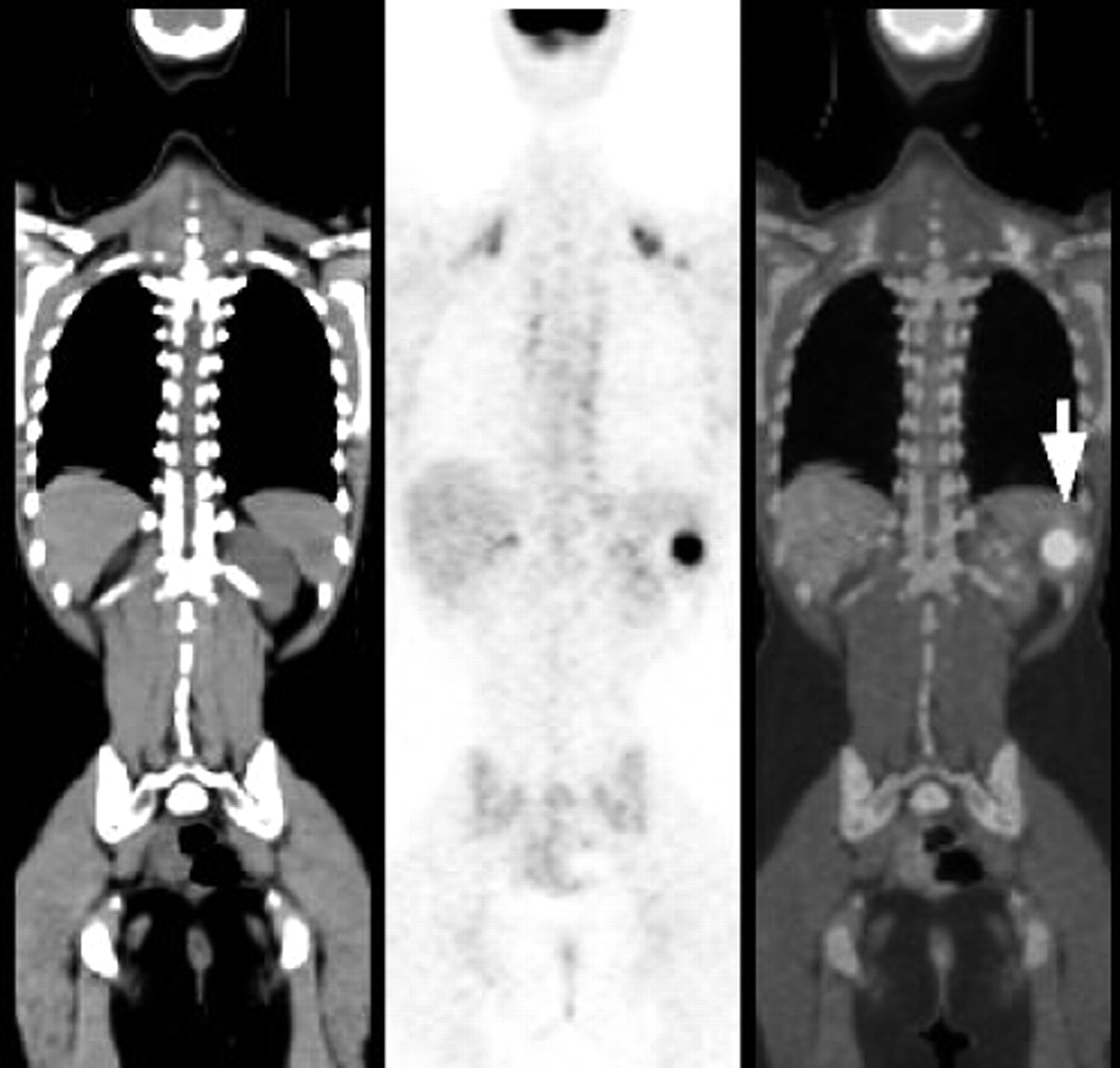
- FIGURE 1.
Solitary splenic metastasis in presence of disseminated metastatic disease: 62-y-old man with lung cancer. (A) Axial PET/CT images show 18F-FDG–avid metastatic deposit in anterior aspect of spleen (arrow). Follow-up contrast-enhanced CT showed progression of splenic mass (not shown). (B) Maximum-intensity-projection PET image shows disseminated metastatic disease in bone, liver, lymph nodes, and spleen.
- FIGURE 2.
Primary splenic lymphoma: 66-y-old man without previously diagnosed malignancy. Coronal PET/CT images show multiple 18F-FDG–avid splenic masses, proven to be non-Hodgkin’s lymphoma on biopsy, in absence of other 18F-FDG–avid disease.
- FIGURE 3.
Presumably benign solid splenic mass: 62-y-old woman without previously diagnosed malignancy. Axial PET/CT images show no increased uptake of 18F-FDG within hypodense mass on CT (arrows). Mass was stable on sonography for >1 year.
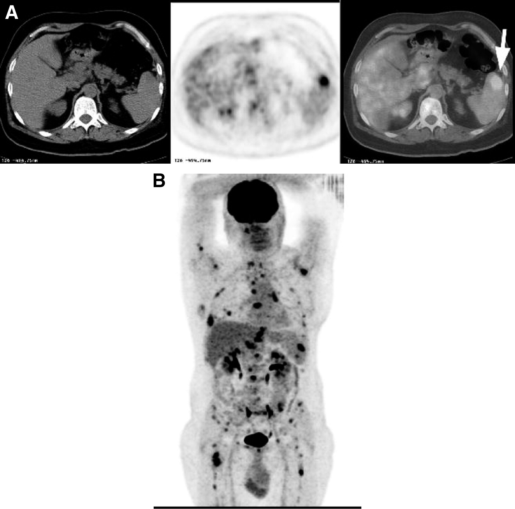
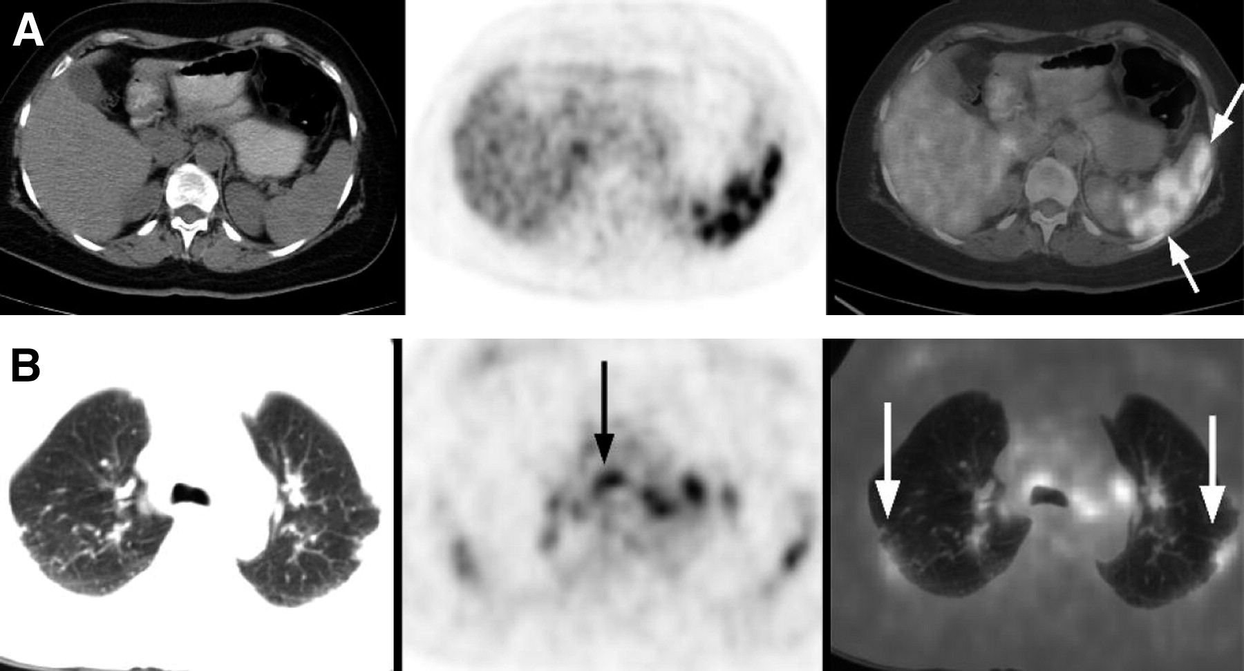
- FIGURE 4.
False-positive 18F-FDG PET/CT in sarcoidosis: 55-y-old woman without known malignancy. Multiple hypodense splenic nodules discovered on CT performed for abdominal pain. (A) Axial PET/CT images show innumerable 18F-FDG–avid nodules within spleen. (B) Axial PET/CT images (lung windows) show abnormal uptake of 18F-FDG in mediastinal lymph nodes (black arrow) as well as increased 18F-FDG uptake in subpleural lung densities (white arrows) proven to be sarcoidosis on lung biopsy.
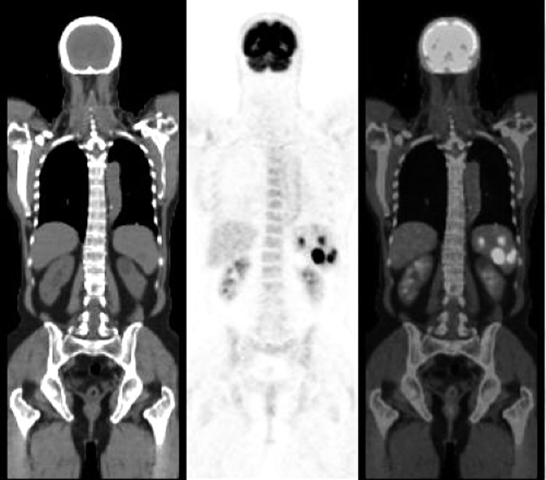
- FIGURE 5.
False-positive 18F-FDG PET/CT in splenic granulomas due to Brucella melitensis: 18-y-old woman with incidentally discovered solid splenic masses (arrow) on sonography performed because of epigastric pain. Coronal PET/CT images show abnormal uptake of 18F-FDG in splenic nodule, proven to be granuloma due to Brucella on splenectomy.
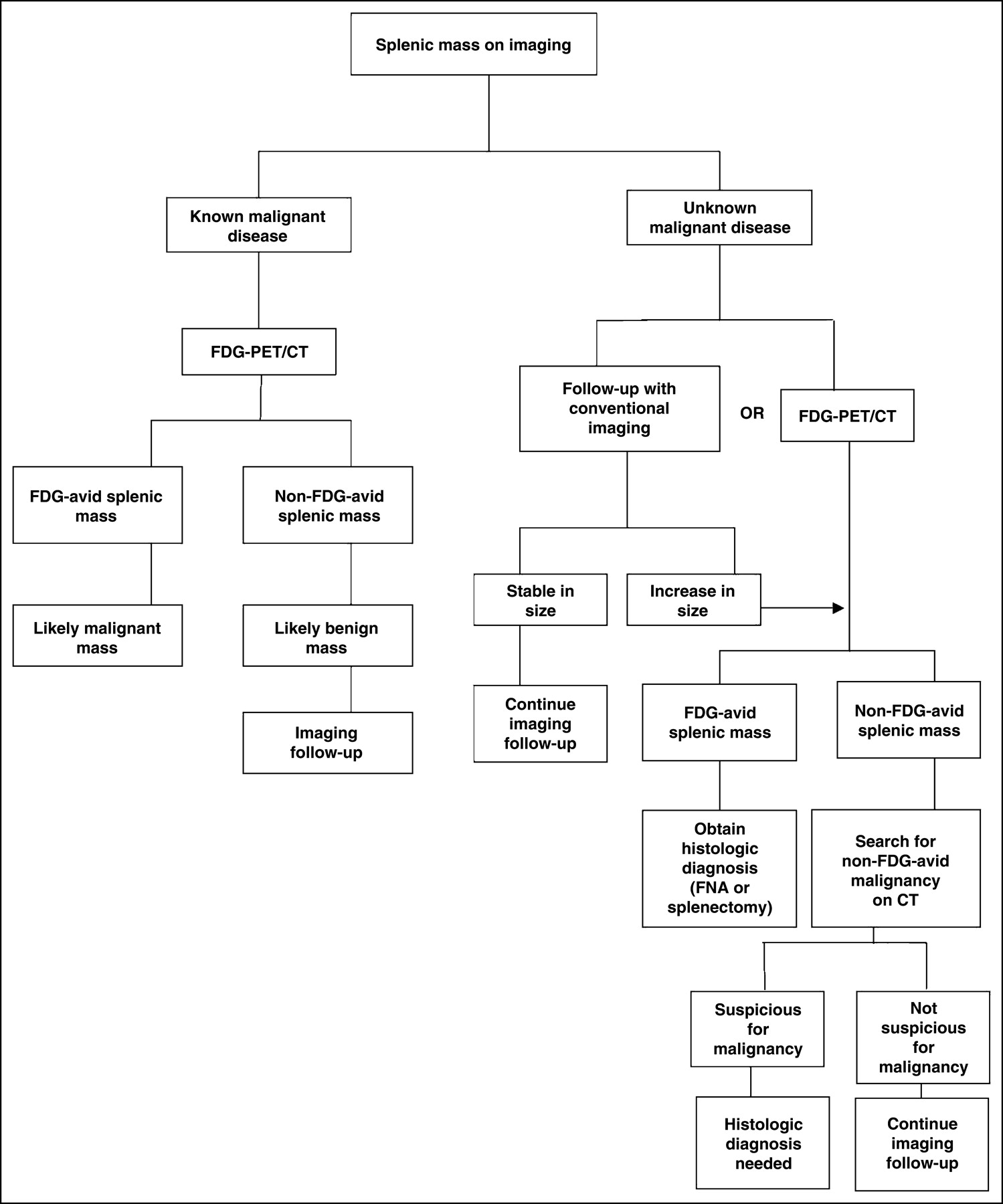
- FIGURE 6.
Suggested scheme for interpretation of a splenic lesion on PET/CT. FNA = fine-needle aspiration.


Tables
- TABLE 1
Primary Malignancies in Oncologic Patients (Group A) in Whom Splenic Lesions Were Suggested on PET/CT
Primary malignancy No. of patients with 18F-FDG–avid splenic lesion* (n = 60) No. of patients with non-18F-FDG–avid splenic lesion† (n = 8) Lymphoma 34* 3 Melanoma 6 1 Lung cancer 6 2 Colon cancer 5* Ovarian cancer 4 Squamous cell carcinoma 2 1 Breast cancer 1 Esophageal cancer 1 Adenocarcinoma of unknown primary 1 Papillary thyroid cancer 1 Seminoma 1 - TABLE 2
Clinical Data, 18F-FDG Avidity, and Final Diagnosis of Splenic Lesions in Patients Without Known Malignancy (Group B)
Patient no. Age (y) Sex Clinical indication 18F-FDG avidity Method of diagnosis Final diagnosis 1 89 M Abdominal pain; CT lesion + SPL FNA Gastric cancer 2 18 F Abdominal pain; CT lesion + Surgery Brucellosis 3 52 M Abdominal pain; CT lesions + LN Bx Lymphoma 4 66 M Incidental US finding + SPL FNA Lymphoma 5 55 F Incidental CT finding + Lung Bx Sarcoidosis 6 65 M Incidental CT finding + SPL FNA Lymphoma 7 69 F Incidental CT finding; previous malaria + Surgery Lymphoma 8 40 F Incidental US finding + Surgery Lymphoma 9 66 M Incidental CT finding + SPL FNA Lymphoma 10 59 M Abdominal pain; CT lesions + Surgery Lymphoma 11 62 F Incidental CT finding − C and I F/U NED 12 33 M Incidental US finding − C and I F/U NED 13 53 M Incidental US finding − C and I F/U NED 14 55 M Incidental CT finding − C and I F/U NED 15 48 F Incidental US finding − C and I F/U NED 16 58 M Incidental US finding − C and I F/U NED 17 48 M Incidental CT finding − C and I F/U NED 18 59 F Incidental US finding − C and I F/U NED 19 62 M Incidental CT finding − C and I F/U NED 20 65 M Incidental US finding − C and I F/U NED SPL = spleen; FNA = fine-needle aspiration; LN = lymph node; Bx = biopsy; US = ultrasound; C and I F/U = clinical and imaging follow-up; NED = no evidence of malignant disease on imaging work-up and stability of lesion on follow-up imaging.
- TABLE 3
Distribution of Splenic Lesions Based on Number of Lesions in Spleen and Type of Lesion in Both Groups of Patients
Splenic lesions Malignant Benign Group A Group B Group A Group B Single 42 4 7 4 Multiple 18 4 1 4


