
Main menu

  • Home
  • Content
    • Current
    • Ahead of print
    • Past Issues
    • JNM Supplement
    • SNMMI Annual Meeting Abstracts
    • Continuing Education
    • JNM Podcasts
  • Subscriptions
    • Subscribers
    • Institutional and Non-member
    • Rates
    • Journal Claims
    • Corporate & Special Sales
  • Authors
    • Submit to JNM
    • Information for Authors
    • Assignment of Copyright
    • AQARA requirements
  • Info
    • Reviewers
    • Permissions
    • Advertisers
  • About
    • About Us
    • Editorial Board
    • Contact Information
  • More
    • Alerts
    • Feedback
    • Help
    • SNMMI Journals
  • SNMMI
    • JNM
    • JNMT
    • SNMMI Journals
    • SNMMI

User menu

  • Subscribe
  • My alerts
  • Log in
  • My Cart

Search

  • Advanced search
Journal of Nuclear Medicine
  • SNMMI
    • JNM
    • JNMT
    • SNMMI Journals
    • SNMMI
  • Subscribe
  • My alerts
  • Log in
  • My Cart
Journal of Nuclear Medicine

Advanced Search

  • Home
  • Content
    • Current
    • Ahead of print
    • Past Issues
    • JNM Supplement
    • SNMMI Annual Meeting Abstracts
    • Continuing Education
    • JNM Podcasts
  • Subscriptions
    • Subscribers
    • Institutional and Non-member
    • Rates
    • Journal Claims
    • Corporate & Special Sales
  • Authors
    • Submit to JNM
    • Information for Authors
    • Assignment of Copyright
    • AQARA requirements
  • Info
    • Reviewers
    • Permissions
    • Advertisers
  • About
    • About Us
    • Editorial Board
    • Contact Information
  • More
    • Alerts
    • Feedback
    • Help
    • SNMMI Journals
  • View or Listen to JNM Podcast
  • Visit JNM on Facebook
  • Join JNM on LinkedIn
  • Follow JNM on Twitter
  • Subscribe to our RSS feeds
OtherClinical Investigations

Prospective Feasibility Trial of Radiotherapy Target Definition for Head and Neck Cancer Using 3-Dimensional PET and CT Imaging

Christopher Scarfone, William C. Lavely, Anthony J. Cmelak, Dominique Delbeke, William H. Martin, Dean Billheimer and Dennis E. Hallahan
Journal of Nuclear Medicine April 2004, 45 (4) 543-552;
Christopher Scarfone
  • Find this author on Google Scholar
  • Find this author on PubMed
  • Search for this author on this site
William C. Lavely
  • Find this author on Google Scholar
  • Find this author on PubMed
  • Search for this author on this site
Anthony J. Cmelak
  • Find this author on Google Scholar
  • Find this author on PubMed
  • Search for this author on this site
Dominique Delbeke
  • Find this author on Google Scholar
  • Find this author on PubMed
  • Search for this author on this site
William H. Martin
  • Find this author on Google Scholar
  • Find this author on PubMed
  • Search for this author on this site
Dean Billheimer
  • Find this author on Google Scholar
  • Find this author on PubMed
  • Search for this author on this site
Dennis E. Hallahan
  • Find this author on Google Scholar
  • Find this author on PubMed
  • Search for this author on this site
  • Article
  • Figures & Data
  • Info & Metrics
  • PDF
Loading

Article Figures & Data

Figures

  • Tables
  • FIGURE 1.
    • Download figure
    • Open in new tab
    • Download powerpoint
    FIGURE 1.

    Typical landmarks demonstrate correlation of images for coregistration (patient 6). High-resolution CT anatomic data (A) and corresponding color-enhanced 18F-FDG PET emission image (B). Image pair shows correspondence between alignment points used for registration of the 2 datasets.

  • FIGURE 2.
    • Download figure
    • Open in new tab
    • Download powerpoint
    FIGURE 2.

    PET contour contained within corresponding CT contour (patient 5). CT (A), corresponding color-enhanced 18F-FDG PET (B), and coregistered images (C). Extent of CT GTV is indicated by blue contour (A and B). Corresponding PET contour is in yellow (A and B). This example demonstrates an 18F-FDG–avid region within CT-defined GTV.

  • FIGURE 3.
    • Download figure
    • Open in new tab
    • Download powerpoint
    FIGURE 3.

    Example variations in agreement between PET and CT contours (patient 6). Transverse CT (A) and corresponding 18F-FDG PET image (B). In this example there is good agreement between CT (orange) and PET (yellow) contours of right lymph node. PET definition of primary target contour (red), however, includes more tissue laterally when compared with CT contour of this same area (green).

  • FIGURE 4.
    • Download figure
    • Open in new tab
    • Download powerpoint
    FIGURE 4.

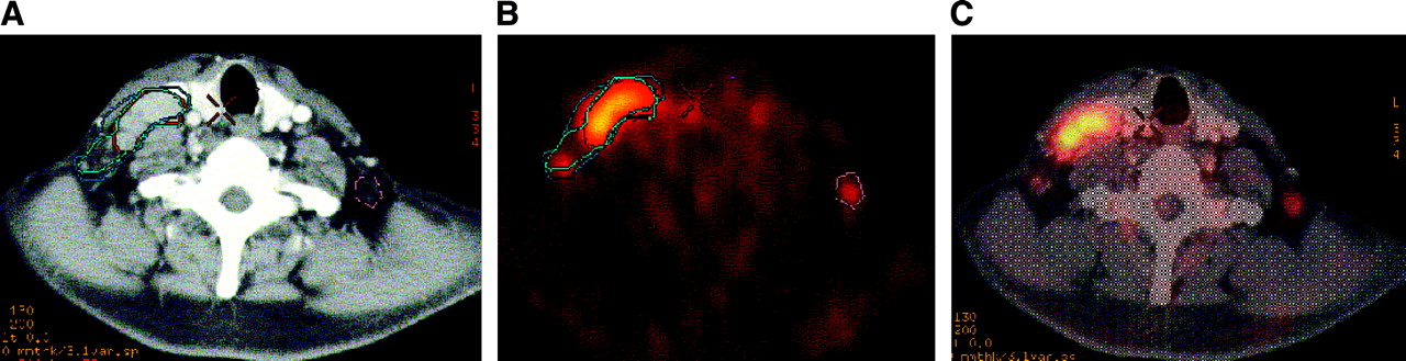
    Primary CT and PET contour overlap (patient 4). CT (A) and corresponding 18F-FDG PET image (B) of right side hypermetabolic lymph node. This slice level shows CT (yellow) and PET (green) contours nearly overlapping. Taking into account slight differences between contours on other slice levels, original CT volume was increased by 3.4 cm3 to include portions of PET-avid regions.

  • FIGURE 5.
    • Download figure
    • Open in new tab
    • Download powerpoint
    FIGURE 5.

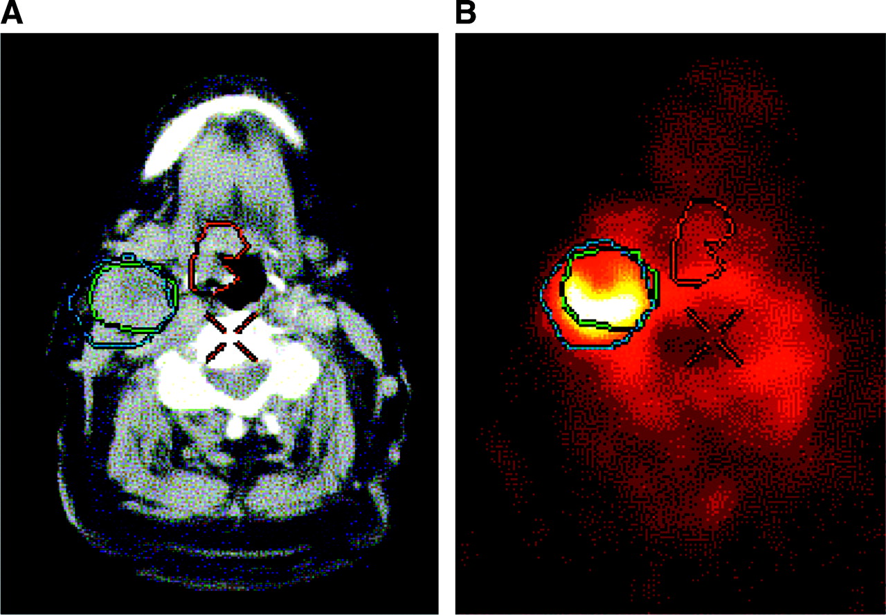
    PET contour extending outside CT-defined contour (patient 2). CT (A) and corresponding 18F-FDG PET image (B). PET definition of right side node (blue contour) extends beyond CT-defined node (green contour) to include other soft tissue. CT-defined GTV (central red contour) shows no corresponding PET uptake in this region at this slice level.

  • FIGURE 6.
    • Download figure
    • Open in new tab
    • Download powerpoint
    FIGURE 6.

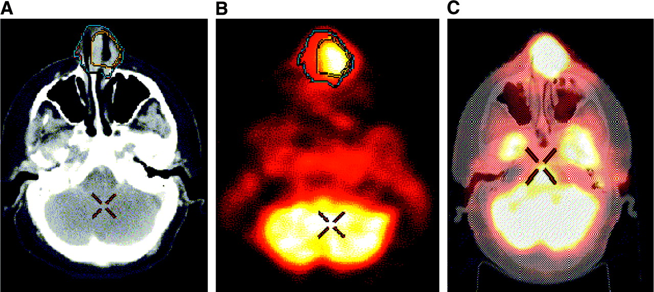
    Visualization of 18F-FDG PET–avid lymph nodes (patient 6). CT (A), 18F-FDG PET (B), and blended view (C). CT contour of right side lymph node is in aqua, and 18F-FDG PET contour of this same node is in red. 18F-FDG PET image (B) indicates 18F-FDG–avid node on left side not visualized on CT.
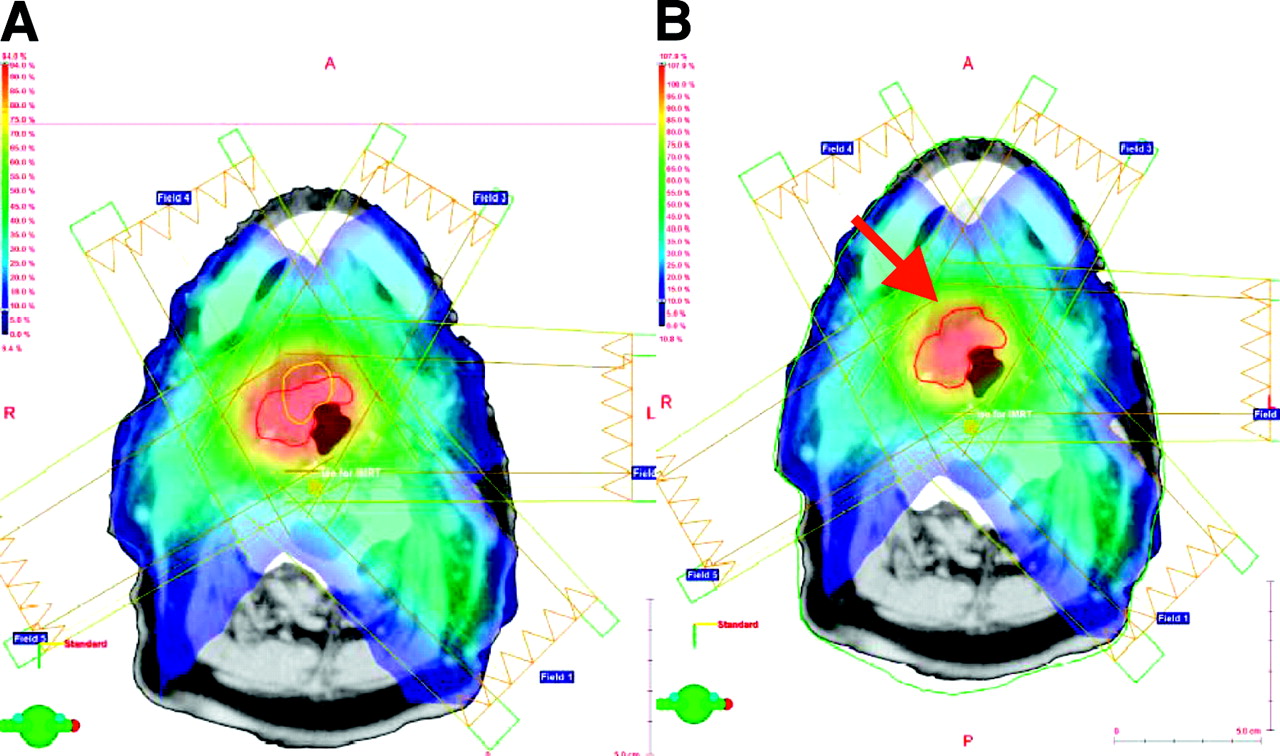
  • FIGURE 7.
    • Download figure
    • Open in new tab
    • Download powerpoint
    FIGURE 7.

    IMRT dose distribution (patient 2). Five-field IMRT plan is optimized to CT GTV (red contour in A). Corresponding PET GTV (yellow contour) is also shown in (A). IMRT plan is optimized to combined PET/CT GTV (red contour in B).

Tables

  • Figures
    • View popup
    TABLE 1

    Patient Demographics, Chemotherapy History, and Total Treatment Dose

    Patient no.Age (y)SexChemoTotal dose (Gy)
    141M+*66
    283F+70
    354M+70
    462F+70
    554F+50.4†
    663M+70
    • ↵* Before PET.

    • ↵† Small cell.

    • Chemo = chemotherapy.

    • View popup
    TABLE 2

    Target Volume Summary

    Patient no.CT primaryPET primaryCT lymph nodePET lymph nodeFinal primaryFinal lymph node
    123.5n/an/an/an/an/a
    3.5n/an/an/a
    2.3n/an/an/a
    1.8n/an/an/a
    1.6n/an/an/a
    1.5n/an/an/a
    211.86.626.028.314.728.3
    335.942.116.43.441.521.7
    1.316.5
    426.819.8n/an/a30.2n/a
    528.817.31.1n/a31.11.1
    66.613.818.718.28.718.7
    44.955.153.5
    n/a2.37.0
    n/a60.4n/a
    • n/a = not applicable.

    • All volumes are in cm3.

PreviousNext
Back to top

In this issue

Journal of Nuclear Medicine: 45 (4)
Journal of Nuclear Medicine
Vol. 45, Issue 4
April 1, 2004
  • Table of Contents
  • Index by author
Print
Download PDF
Article Alerts
Sign In to Email Alerts with your Email Address
Email Article

Thank you for your interest in spreading the word on Journal of Nuclear Medicine.

NOTE: We only request your email address so that the person you are recommending the page to knows that you wanted them to see it, and that it is not junk mail. We do not capture any email address.

Enter multiple addresses on separate lines or separate them with commas.
Prospective Feasibility Trial of Radiotherapy Target Definition for Head and Neck Cancer Using 3-Dimensional PET and CT Imaging
(Your Name) has sent you a message from Journal of Nuclear Medicine
(Your Name) thought you would like to see the Journal of Nuclear Medicine web site.
Citation Tools
Prospective Feasibility Trial of Radiotherapy Target Definition for Head and Neck Cancer Using 3-Dimensional PET and CT Imaging
Christopher Scarfone, William C. Lavely, Anthony J. Cmelak, Dominique Delbeke, William H. Martin, Dean Billheimer, Dennis E. Hallahan
Journal of Nuclear Medicine Apr 2004, 45 (4) 543-552;

Citation Manager Formats

  • BibTeX
  • Bookends
  • EasyBib
  • EndNote (tagged)
  • EndNote 8 (xml)
  • Medlars
  • Mendeley
  • Papers
  • RefWorks Tagged
  • Ref Manager
  • RIS
  • Zotero
Share
Prospective Feasibility Trial of Radiotherapy Target Definition for Head and Neck Cancer Using 3-Dimensional PET and CT Imaging
Christopher Scarfone, William C. Lavely, Anthony J. Cmelak, Dominique Delbeke, William H. Martin, Dean Billheimer, Dennis E. Hallahan
Journal of Nuclear Medicine Apr 2004, 45 (4) 543-552;
Twitter logo Facebook logo LinkedIn logo Mendeley logo
  • Tweet Widget
  • Facebook Like
  • Google Plus One
Bookmark this article

Jump to section

  • Article
    • Abstract
    • MATERIALS AND METHODS
    • RESULTS
    • DISCUSSION
    • CONCLUSION
    • Acknowledgments
    • Footnotes
    • REFERENCES
  • Figures & Data
  • Info & Metrics
  • PDF

Related Articles

  • No related articles found.
  • PubMed
  • Google Scholar

Cited By...

  • 18F-Fdg-PET-guided Planning and Re-Planning (Adaptive) Radiotherapy in Head and Neck Cancer: Current State of Art
  • Workflow and Radiation Safety Implications of 18F-FDG PET/CT Scans for Radiotherapy Planning
  • Positron emission tomography imaging approaches for external beam radiation therapies: current status and future developments
  • Autocontouring and Manual Contouring: Which Is the Better Method for Target Delineation Using 18F-FDG PET/CT in Non-Small Cell Lung Cancer?
  • 18F-FDG PET/CT for Image-Guided and Intensity-Modulated Radiotherapy
  • Metabolic Tumor Volume of [18F]-Fluorodeoxyglucose Positron Emission Tomography/Computed Tomography Predicts Short-Term Outcome to Radiotherapy With or Without Chemotherapy in Pharyngeal Cancer
  • PET Changes Management and Improves Prognostic Stratification in Patients with Head and Neck Cancer: Results of a Multicenter Prospective Study
  • Treatment Monitoring by 18F-FDG PET/CT in Patients with Sarcomas: Interobserver Variability of Quantitative Parameters in Treatment-Induced Changes in Histopathologically Responding and Nonresponding Tumors
  • The role of PET/CT scanning in radiotherapy planning
  • Advanced imaging applied to radiotherapy planning in head and neck cancer: a clinical review.
  • Expanding Role of Positron Emission Tomography in Cancer of the Uterine Cervix
  • The contribution of PET/CT to improved patient management
  • Comparison of Different Methods for Delineation of 18F-FDG PET-Positive Tissue for Target Volume Definition in Radiotherapy of Patients with Non-Small Cell Lung Cancer
  • Staging of Untreated Squamous Cell Carcinoma of Buccal Mucosa with 18F-FDG PET: Comparison with Head and Neck CT/MRI and Histopathology
  • Google Scholar

More in this TOC Section

  • Feasibility of Ultra-Low-Activity 18F-FDG PET/CT Imaging Using a Long–Axial-Field-of-View PET/CT System
  • Cardiac Presynaptic Sympathetic Nervous Function Evaluated by Cardiac PET in Patients with Chronotropic Incompetence Without Heart Failure
  • Validation and Evaluation of a Vendor-Provided Head Motion Correction Algorithm on the uMI Panorama PET/CT System
Show more Clinical Investigations

Similar Articles

SNMMI

© 2025 SNMMI

Powered by HighWire