
Main menu

  • Home
  • Content
    • Current
    • Ahead of print
    • Past Issues
    • JNM Supplement
    • SNMMI Annual Meeting Abstracts
    • Continuing Education
    • JNM Podcasts
  • Subscriptions
    • Subscribers
    • Institutional and Non-member
    • Rates
    • Journal Claims
    • Corporate & Special Sales
  • Authors
    • Submit to JNM
    • Information for Authors
    • Assignment of Copyright
    • AQARA requirements
  • Info
    • Reviewers
    • Permissions
    • Advertisers
  • About
    • About Us
    • Editorial Board
    • Contact Information
  • More
    • Alerts
    • Feedback
    • Help
    • SNMMI Journals
  • SNMMI
    • JNM
    • JNMT
    • SNMMI Journals
    • SNMMI

User menu

  • Subscribe
  • My alerts
  • Log in
  • Log out
  • My Cart

Search

  • Advanced search
Journal of Nuclear Medicine
  • SNMMI
    • JNM
    • JNMT
    • SNMMI Journals
    • SNMMI
  • Subscribe
  • My alerts
  • Log in
  • Log out
  • My Cart
Journal of Nuclear Medicine

Advanced Search

  • Home
  • Content
    • Current
    • Ahead of print
    • Past Issues
    • JNM Supplement
    • SNMMI Annual Meeting Abstracts
    • Continuing Education
    • JNM Podcasts
  • Subscriptions
    • Subscribers
    • Institutional and Non-member
    • Rates
    • Journal Claims
    • Corporate & Special Sales
  • Authors
    • Submit to JNM
    • Information for Authors
    • Assignment of Copyright
    • AQARA requirements
  • Info
    • Reviewers
    • Permissions
    • Advertisers
  • About
    • About Us
    • Editorial Board
    • Contact Information
  • More
    • Alerts
    • Feedback
    • Help
    • SNMMI Journals
  • View or Listen to JNM Podcast
  • Visit JNM on Facebook
  • Join JNM on LinkedIn
  • Follow JNM on Twitter
  • Subscribe to our RSS feeds
OtherBasic Science Investigations

In Vitro and In Vivo Evaluation of 111In-DTPAGlu-G-CCK8 for Cholecystokinin-B Receptor Imaging

Luigi Aloj, Corradina Caracò, Mariarosaria Panico, Antonella Zannetti, Silvana Del Vecchio, Diego Tesauro, Stefania De Luca, Claudio Arra, Carlo Pedone, Giancarlo Morelli and Marco Salvatore
Journal of Nuclear Medicine March 2004, 45 (3) 485-494;
Luigi Aloj
  • Find this author on Google Scholar
  • Find this author on PubMed
  • Search for this author on this site
Corradina Caracò
  • Find this author on Google Scholar
  • Find this author on PubMed
  • Search for this author on this site
Mariarosaria Panico
  • Find this author on Google Scholar
  • Find this author on PubMed
  • Search for this author on this site
Antonella Zannetti
  • Find this author on Google Scholar
  • Find this author on PubMed
  • Search for this author on this site
Silvana Del Vecchio
  • Find this author on Google Scholar
  • Find this author on PubMed
  • Search for this author on this site
Diego Tesauro
  • Find this author on Google Scholar
  • Find this author on PubMed
  • Search for this author on this site
Stefania De Luca
  • Find this author on Google Scholar
  • Find this author on PubMed
  • Search for this author on this site
Claudio Arra
  • Find this author on Google Scholar
  • Find this author on PubMed
  • Search for this author on this site
Carlo Pedone
  • Find this author on Google Scholar
  • Find this author on PubMed
  • Search for this author on this site
Giancarlo Morelli
  • Find this author on Google Scholar
  • Find this author on PubMed
  • Search for this author on this site
Marco Salvatore
  • Find this author on Google Scholar
  • Find this author on PubMed
  • Search for this author on this site
  • Article
  • Figures & Data
  • Info & Metrics
  • PDF
Loading

Article Figures & Data

Figures

  • FIGURE 1.
    • Download figure
    • Open in new tab
    • Download powerpoint
    FIGURE 1.

    Schematic representation of coupling of the DTPAGlu moiety to the resin-bound G-CCK8 peptide. Subsequent deprotection yields the final compound, which is ready for radiolabeling.
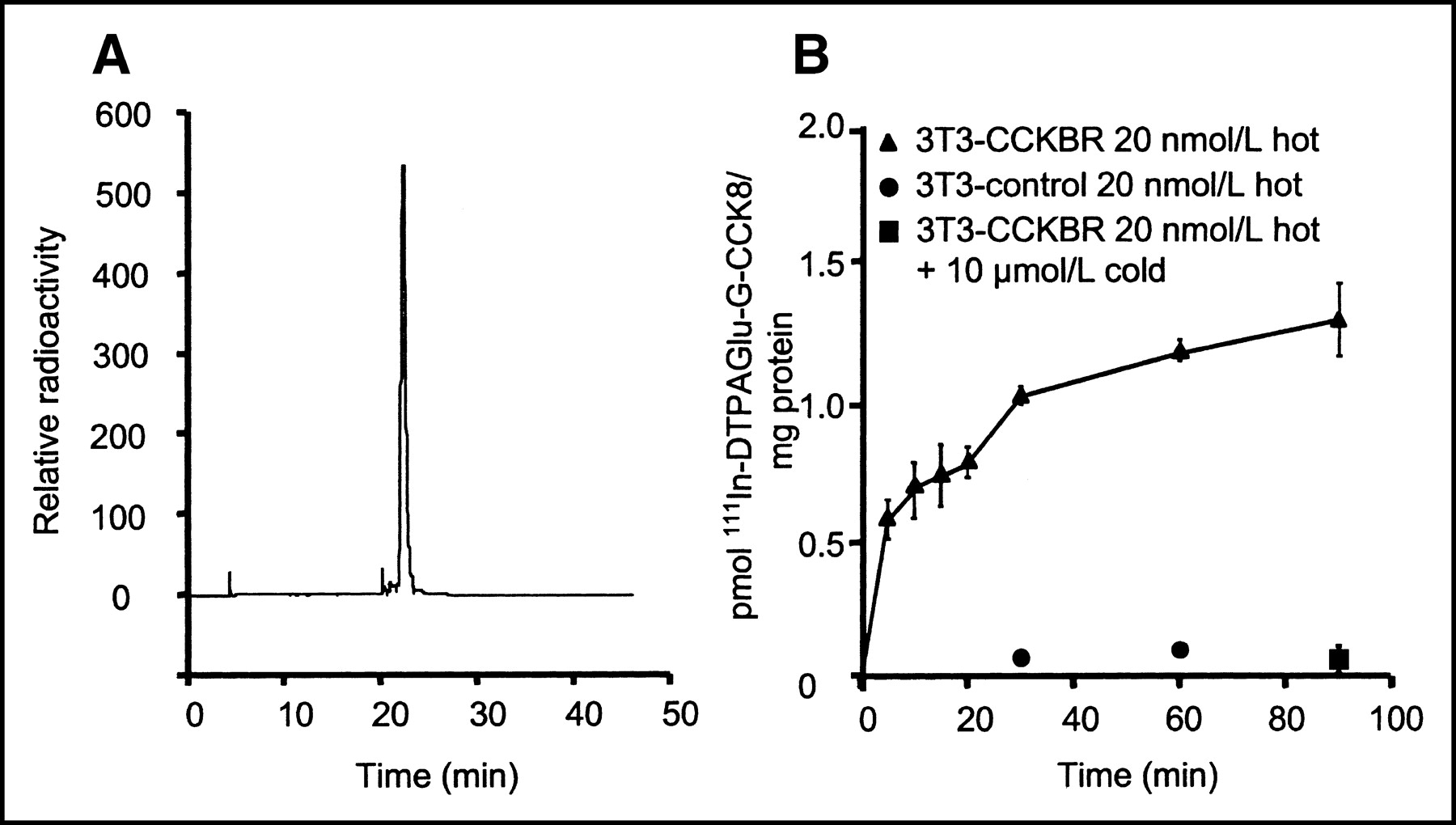
  • FIGURE 2.
    • Download figure
    • Open in new tab
    • Download powerpoint
    FIGURE 2.

    (A) Radioactive trace of RP HPLC analysis after labeling for 60 min at room temperature. 111In-DTPAGlu-G-CCK8 shows a retention time of approximately 22 min on the CH3CN:H2O gradient. Unincorporated 111In, which has a retention time of approximately 3 min, is barely detectable. (B) Specificity of interactions of 111In-DTPAGlu-G-CCK8 with NIH-3T3-CCKBR cells and control cells. There is a progressive increase of cell-associated activity in receptor-positive cells incubated with the radioactive peptide alone (▴). Receptor-negative cells (•) and receptor-positive cells incubated with excess unlabeled peptide (▪) show very little interaction with the radiolabeled peptide. There were 3 samples per time point; error bars indicate SDs.

  • FIGURE 3.
    • Download figure
    • Open in new tab
    • Download powerpoint
    FIGURE 3.

    Binding of 111In-DTPAGlu-G-CCK8 on NIH-3T3-CCKBR cells (A) and A431-CCKBR cells (B) at 4°C. Both cell lines showed saturable binding of the peptide, with equivalent Kds (22 ± 11 nmol/L [mean ± SD] for NIH-3T3-CCKBR cells; 23 ± 4 nmol/L for A431-CCKBR cells). A431-CCKBR cells had higher Bmax values (1.6 × 10⁶ ± 0.4 × 10⁶ sites per cell [mean ± SD] for NIH-3T3-CCKBR cells; 4.7 × 10⁶ ± 0.4 × 10⁶ sites per cell for A431-CCKBR cells). There were 2 samples per concentration; error bars indicate SDs.

  • FIGURE 4.
    • Download figure
    • Open in new tab
    • Download powerpoint
    FIGURE 4.

    Cellular internalization and release of 111In-DTPAGlu-G-CCK8 for A431-CCKBR cells. (A) Internalization. Cells were incubated at 4°C (□) and 37°C (▪) for 60 and 120 min with radiolabeled peptide (20 nmol/L). Excess unlabeled peptide was added during the 2-h incubation (third set of bars from left) or after the 2-h incubation and after washes in PBS to displace label on the cell surface (fourth set of bars from left). There is a progressive increase of cell-bound activity at 37°C, whereas a constant level is observed at 4°C. Coincubation with cold ligand produced a very low level of binding under both conditions. The addition of cold ligand after the 2-h incubation displaced most of the radioactivity from cells incubated at 4°C, consistent with the ligand being on the cell surface, whereas very little displacement was observed for cells incubated at 37°C, indicating that the label was in an intracellular compartment in these cells. (B) Release. Cells incubated at 37°C for 2 h were rinsed with PBS. Fresh medium was added, and cell-associated radioactivity was determined at the indicated times. There were 3 samples per time point; error bars indicate SDs.
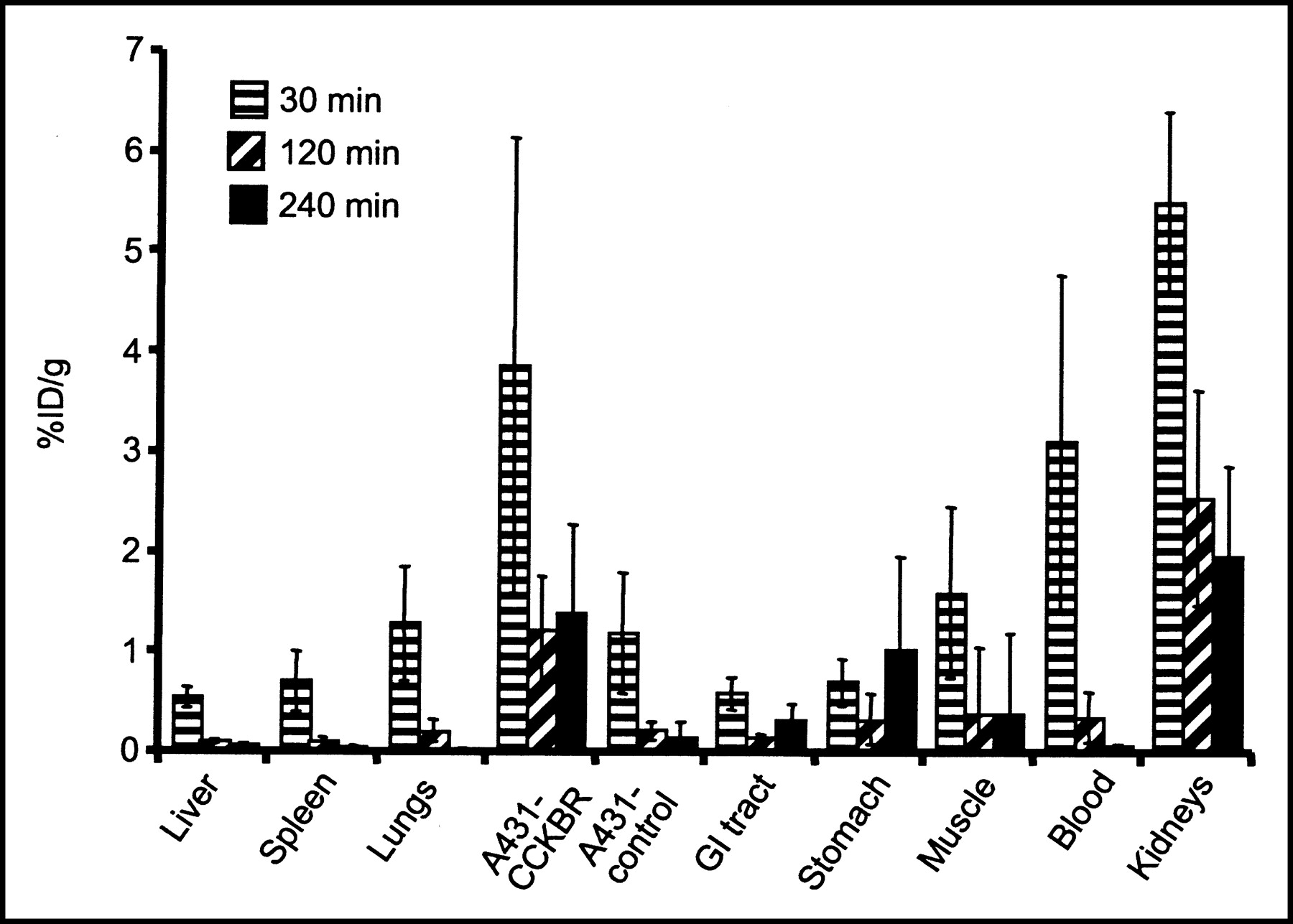
  • FIGURE 5.
    • Download figure
    • Open in new tab
    • Download powerpoint
    FIGURE 5.

    Biodistribution of 111In-DTPAGlu-G-CCK8 after intravenous injection. Organ-associated radioactivity is expressed as the %ID per gram of tissue normalized for a 20-g mouse. There were at least 5 samples per time point; error bars indicate SDs. GI = gastrointestinal.
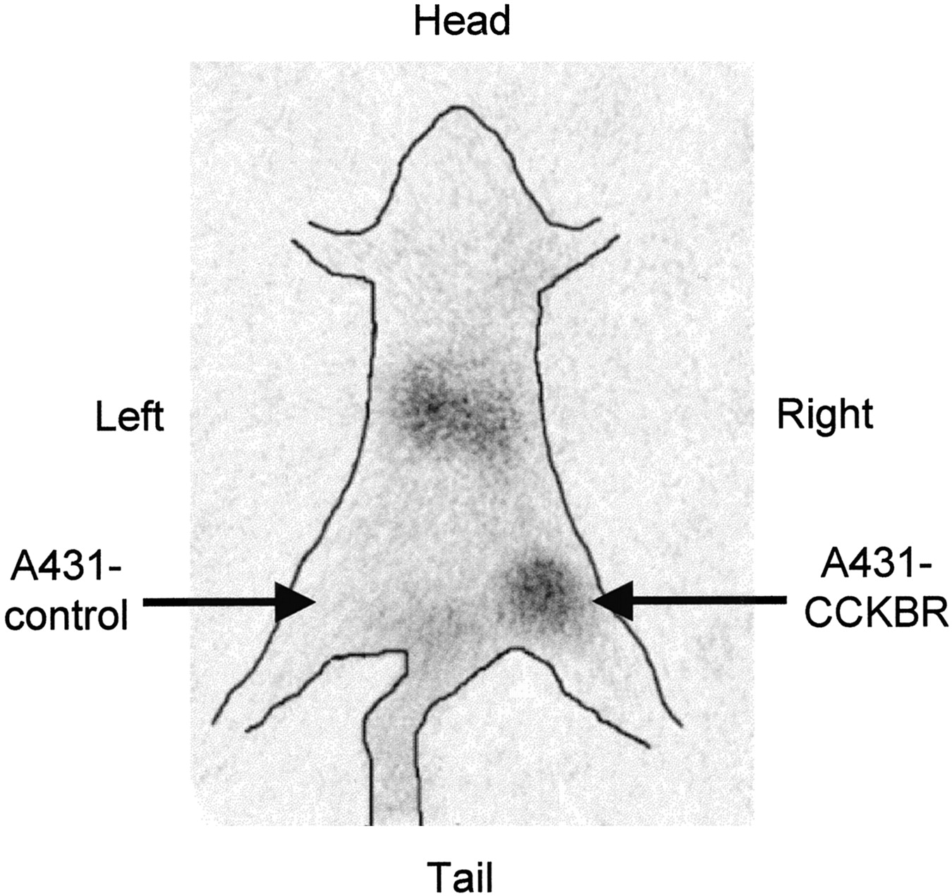
  • FIGURE 6.
    • Download figure
    • Open in new tab
    • Download powerpoint
    FIGURE 6.

    Pinhole γ-camera image obtained at 2 h after injection of 111In-DTPAGlu-G-CCK8. Avid accumulation of the peptide was seen in a CCKBR-positive xenograft (right thigh) but not in a control tumor (left thigh). Hot spots in the abdomen are consistent with kidney accumulation.
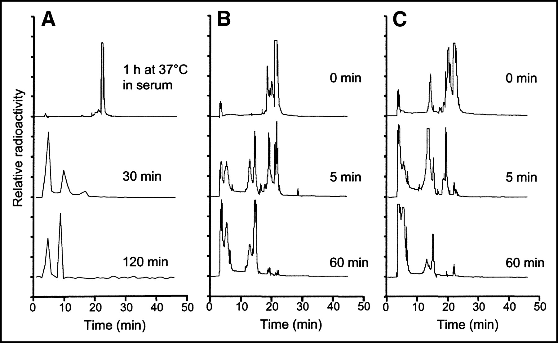
  • FIGURE 7.
    • Download figure
    • Open in new tab
    • Download powerpoint
    FIGURE 7.

    (A) RP HPLC analysis of 111In-DTPAGlu-G-CCK8 incubated for 1 h in serum (top trace) and radioactivity recovered from blood samples taken at different times after injection into a nude mouse (bottom 2 traces). The compound is very stable in serum but appears to be rapidly metabolized after injection to more hydrophilic compounds. The small amount of radioactivity in the blood samples taken after injection required the use of a fraction collector and γ-counting. (B and C) RP HPLC analysis of 111In-DTPAGlu-G-CCK8 incubated with tissue homogenates of liver (B) and kidneys (C). Homogenates were freshly prepared and incubated on ice. Radiolabeled peptide was added to the homogenates and either immediately extracted with CH3CN (0 min) or incubated at 37°C for the indicated times. A breakdown of the peptide to more hydrophilic forms appears to be more rapid and prominent in the kidneys.

PreviousNext
Back to top

In this issue

Journal of Nuclear Medicine: 45 (3)
Journal of Nuclear Medicine
Vol. 45, Issue 3
March 1, 2004
  • Table of Contents
  • About the Cover
  • Index by author
Print
Download PDF
Article Alerts
Sign In to Email Alerts with your Email Address
Email Article

Thank you for your interest in spreading the word on Journal of Nuclear Medicine.

NOTE: We only request your email address so that the person you are recommending the page to knows that you wanted them to see it, and that it is not junk mail. We do not capture any email address.

Enter multiple addresses on separate lines or separate them with commas.
In Vitro and In Vivo Evaluation of 111In-DTPAGlu-G-CCK8 for Cholecystokinin-B Receptor Imaging
(Your Name) has sent you a message from Journal of Nuclear Medicine
(Your Name) thought you would like to see the Journal of Nuclear Medicine web site.
Citation Tools
In Vitro and In Vivo Evaluation of 111In-DTPAGlu-G-CCK8 for Cholecystokinin-B Receptor Imaging
Luigi Aloj, Corradina Caracò, Mariarosaria Panico, Antonella Zannetti, Silvana Del Vecchio, Diego Tesauro, Stefania De Luca, Claudio Arra, Carlo Pedone, Giancarlo Morelli, Marco Salvatore
Journal of Nuclear Medicine Mar 2004, 45 (3) 485-494;

Citation Manager Formats

  • BibTeX
  • Bookends
  • EasyBib
  • EndNote (tagged)
  • EndNote 8 (xml)
  • Medlars
  • Mendeley
  • Papers
  • RefWorks Tagged
  • Ref Manager
  • RIS
  • Zotero
Share
In Vitro and In Vivo Evaluation of 111In-DTPAGlu-G-CCK8 for Cholecystokinin-B Receptor Imaging
Luigi Aloj, Corradina Caracò, Mariarosaria Panico, Antonella Zannetti, Silvana Del Vecchio, Diego Tesauro, Stefania De Luca, Claudio Arra, Carlo Pedone, Giancarlo Morelli, Marco Salvatore
Journal of Nuclear Medicine Mar 2004, 45 (3) 485-494;
Twitter logo Facebook logo LinkedIn logo Mendeley logo
  • Tweet Widget
  • Facebook Like
  • Google Plus One
Bookmark this article

Jump to section

  • Article
    • Abstract
    • MATERIALS AND METHODS
    • RESULTS
    • DISCUSSION
    • CONCLUSION
    • Acknowledgments
    • Footnotes
    • REFERENCES
  • Figures & Data
  • Info & Metrics
  • PDF

Related Articles

  • No related articles found.
  • PubMed
  • Google Scholar

Cited By...

  • Signaling Network Response to {alpha}-Particle-Targeted Therapy with the 225Ac-Labeled Minigastrin Analog 225Ac-PP-F11N Reveals the Radiosensitizing Potential of Histone Deacetylase Inhibitors
  • DOTA-MGS5, a New Cholecystokinin-2 Receptor-Targeting Peptide Analog with an Optimized Targeting Profile for Theranostic Use
  • "To Serve and Protect": Enzyme Inhibitors as Radiopeptide Escorts Promote Tumor Targeting
  • Evaluation of an 111In-Radiolabeled Peptide as a Targeting and Imaging Agent for ErbB-2 Receptor Expressing Breast Carcinomas
  • Selection of Radiolabeled Gastrin Analogs for Peptide Receptor-Targeted Radionuclide Therapy
  • CCK-2/Gastrin Receptor-Targeted Tumor Imaging with 99mTc-Labeled Minigastrin Analogs
  • Google Scholar

More in this TOC Section

  • 11C-Methionine PET of Myocardial Inflammation in a Rat Model of Experimental Autoimmune Myocarditis
  • Counting Rate Characteristics and Image Distortion in Preclinical PET Imaging During Radiopharmaceutical Therapy
  • Design and Fabrication of Kidney Phantoms for Internal Radiation Dosimetry Using 3D Printing Technology
Show more Basic Science Investigations

Similar Articles

SNMMI

© 2025 SNMMI

Powered by HighWire