
Main menu

  • Home
  • Content
    • Current
    • Ahead of print
    • Past Issues
    • JNM Supplement
    • SNMMI Annual Meeting Abstracts
    • Continuing Education
    • JNM Podcasts
  • Subscriptions
    • Subscribers
    • Institutional and Non-member
    • Rates
    • Journal Claims
    • Corporate & Special Sales
  • Authors
    • Submit to JNM
    • Information for Authors
    • Assignment of Copyright
    • AQARA requirements
  • Info
    • Reviewers
    • Permissions
    • Advertisers
  • About
    • About Us
    • Editorial Board
    • Contact Information
  • More
    • Alerts
    • Feedback
    • Help
    • SNMMI Journals
  • SNMMI
    • JNM
    • JNMT
    • SNMMI Journals
    • SNMMI

User menu

  • Subscribe
  • My alerts
  • Log in
  • My Cart

Search

  • Advanced search
Journal of Nuclear Medicine
  • SNMMI
    • JNM
    • JNMT
    • SNMMI Journals
    • SNMMI
  • Subscribe
  • My alerts
  • Log in
  • My Cart
Journal of Nuclear Medicine

Advanced Search

  • Home
  • Content
    • Current
    • Ahead of print
    • Past Issues
    • JNM Supplement
    • SNMMI Annual Meeting Abstracts
    • Continuing Education
    • JNM Podcasts
  • Subscriptions
    • Subscribers
    • Institutional and Non-member
    • Rates
    • Journal Claims
    • Corporate & Special Sales
  • Authors
    • Submit to JNM
    • Information for Authors
    • Assignment of Copyright
    • AQARA requirements
  • Info
    • Reviewers
    • Permissions
    • Advertisers
  • About
    • About Us
    • Editorial Board
    • Contact Information
  • More
    • Alerts
    • Feedback
    • Help
    • SNMMI Journals
  • View or Listen to JNM Podcast
  • Visit JNM on Facebook
  • Join JNM on LinkedIn
  • Follow JNM on Twitter
  • Subscribe to our RSS feeds
OtherClinical Investigations

Impact of Radioimmunoscintigraphy on Definition of Clinical Target Volume for Radiotherapy After Prostatectomy

Ashesh B. Jani, Danny Spelbring, Russell Hamilton, Michael J. Blend, Charles Pelizzari, Charles Brendler, Lani Krauz, Srinivasan Vijayakumar, Bipin Sapra and Ralph R. Weichselbaum
Journal of Nuclear Medicine February 2004, 45 (2) 238-246;
Ashesh B. Jani
  • Find this author on Google Scholar
  • Find this author on PubMed
  • Search for this author on this site
Danny Spelbring
  • Find this author on Google Scholar
  • Find this author on PubMed
  • Search for this author on this site
Russell Hamilton
  • Find this author on Google Scholar
  • Find this author on PubMed
  • Search for this author on this site
Michael J. Blend
  • Find this author on Google Scholar
  • Find this author on PubMed
  • Search for this author on this site
Charles Pelizzari
  • Find this author on Google Scholar
  • Find this author on PubMed
  • Search for this author on this site
Charles Brendler
  • Find this author on Google Scholar
  • Find this author on PubMed
  • Search for this author on this site
Lani Krauz
  • Find this author on Google Scholar
  • Find this author on PubMed
  • Search for this author on this site
Srinivasan Vijayakumar
  • Find this author on Google Scholar
  • Find this author on PubMed
  • Search for this author on this site
Bipin Sapra
  • Find this author on Google Scholar
  • Find this author on PubMed
  • Search for this author on this site
Ralph R. Weichselbaum
  • Find this author on Google Scholar
  • Find this author on PubMed
  • Search for this author on this site
  • Article
  • Figures & Data
  • Info & Metrics
  • PDF
Loading

Article Figures & Data

Figures

  • Tables
  • FIGURE 1.
    • Download figure
    • Open in new tab
    • Download powerpoint
    FIGURE 1.

    Flow diagram of image data, showing how planning CT was registered with simultaneously acquired dual-isotope 99mTc-labeled RBC SPECT and 111In-mAb RIS scans, enabling the projection of CTVRIS into the planning CT scan for assistance in modifying CTVpre to define CTVpost.
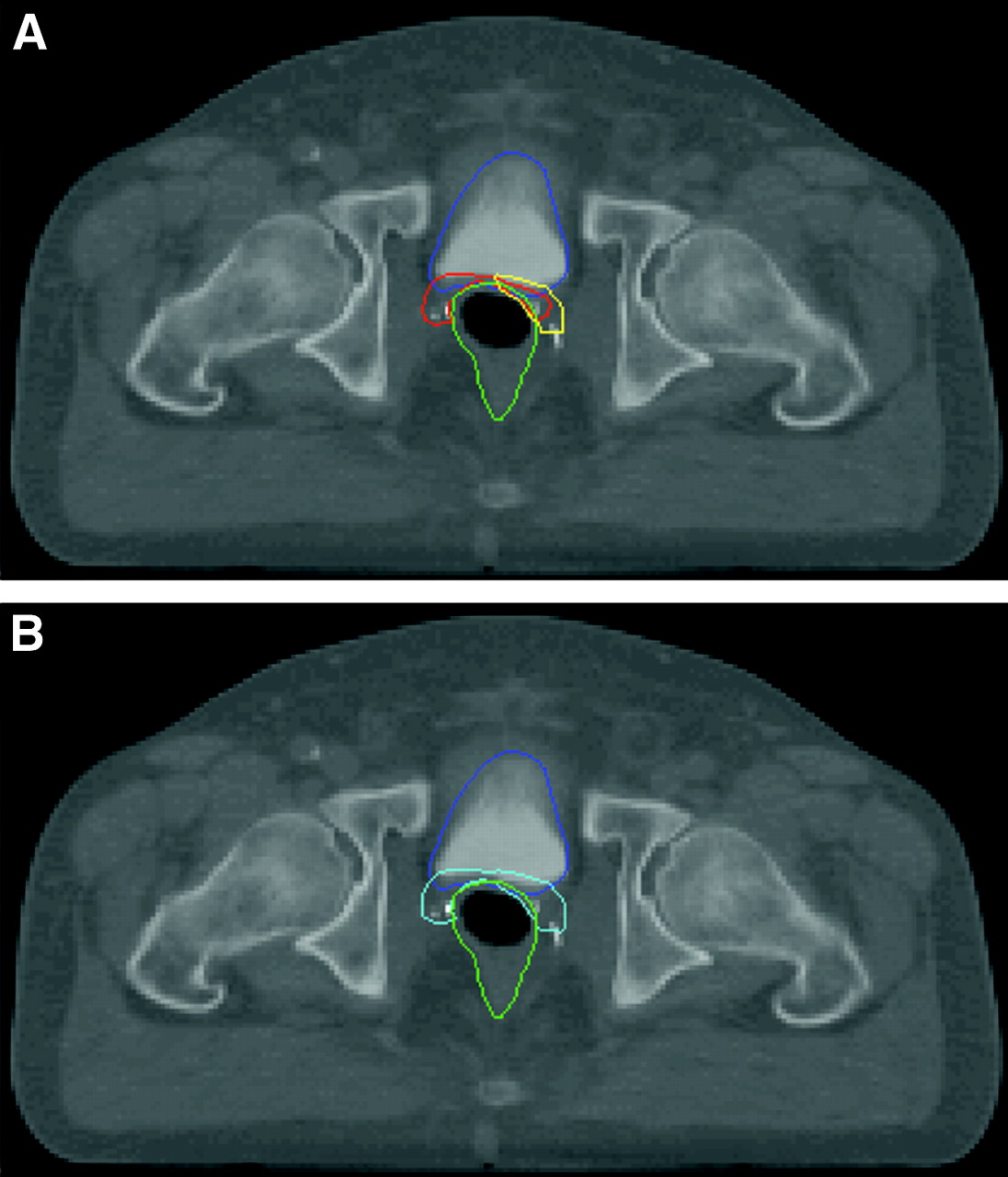
  • FIGURE 2.
    • Download figure
    • Open in new tab
    • Download powerpoint
    FIGURE 2.

    Axial images of planning CT showing the use of RIS in modifying the prostate-bed CTV. Normal structures: bladder (blue), and rectum (green). (A) The CTVpre (red) was entered before the RIS/CT fusion. CTVRIS (yellow) is the projection of the delineated uptake on the RIS scan into the planning CT. (B) CTVRIS was used to modify CTVpre to define CTVpost (aqua). In this case, CTVpost is the union of CTVpre and CTVRIS in A.
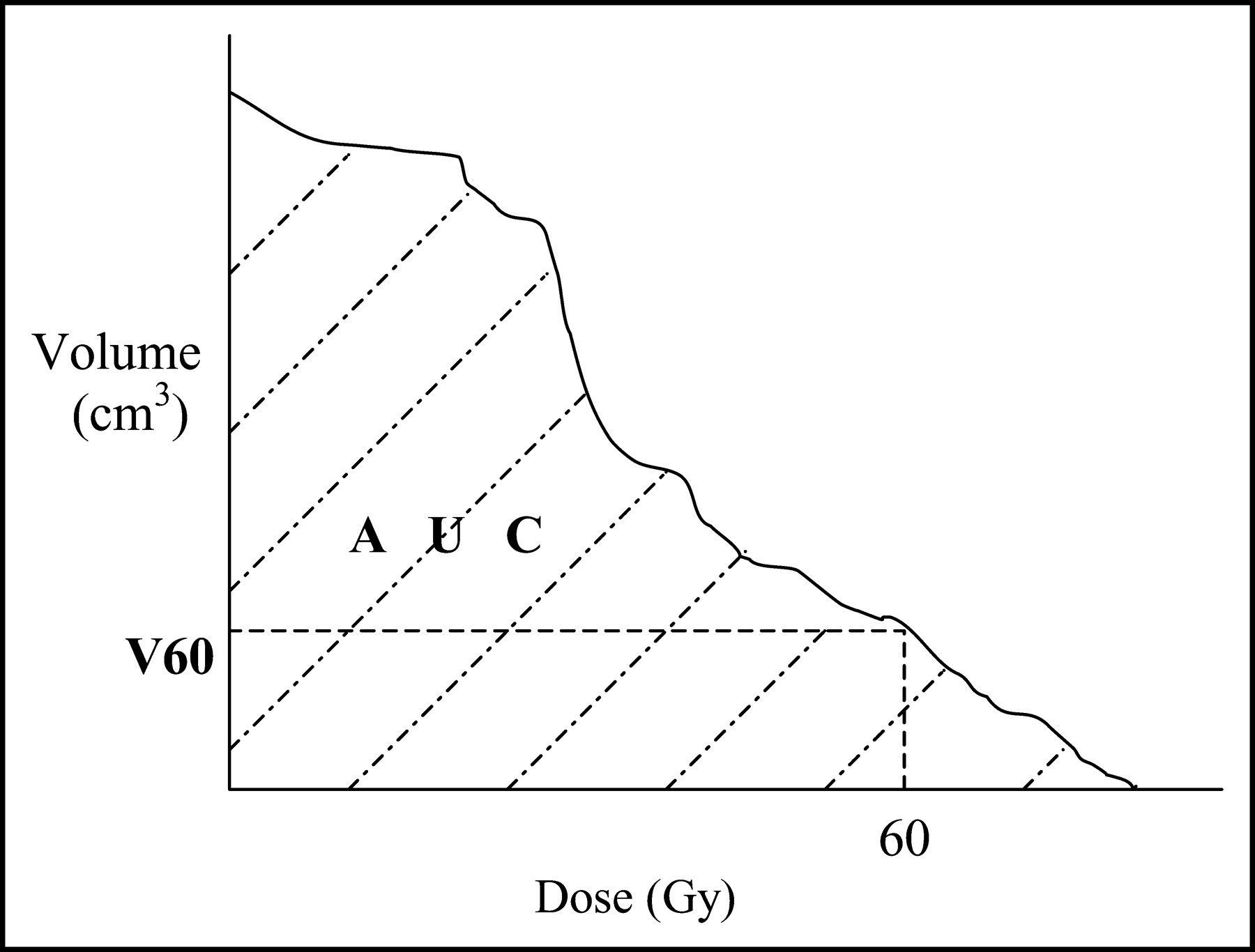
  • FIGURE 3.
    • Download figure
    • Open in new tab
    • Download powerpoint
    FIGURE 3.

    Dose volume histogram dosimetric endpoints for bladder and rectum. AUC = hatched area.

  • FIGURE 4.
    • Download figure
    • Open in new tab
    • Download powerpoint
    FIGURE 4.

    Kaplan–Meier curve displaying biochemical failure-free survival for the cohort.

Tables

  • Figures
    • View popup
    TABLE 1

    Patient Characteristics

    CharacteristicNo. of patients* (n = 25)
    Race
     Caucasian14
     African-American7
     Hispanic2
     Other2
    Prostatectomy findings
     Pathologic T stage
      pT1/T26
      pT316
      pT42
      pTx1
     Grade (Gleason Score)
      GS 65
      GS 714
      GS 82
      GS 92
      Uncharted2
     Margins
      Positive12
      Negative11
      Uncharted2
     Seminal vesicle invasion
      Positive6
      Negative18
      Uncharted1
     Extracapsular extension
      Yes17
      No7
      Uncharted1
     Pelvic lymph node involvement:
      Yes0
      No23
      Unsampled2
    Postprostatectomy course
     Postprostatectomy nadir (ng/mL)
      PSA ≤ 0.110
      0.1 < PSA ≤ 0.22
      0.2 < PSA ≤ 0.36
      0.3 < PSA ≤ 0.51
      0.5 < PSA ≤ 1.02
      1.0 < PSA1
      Uncharted3
     Highest postprostatectomy PSA before RT consultation (ng/mL)†
      PSA ≤ 0.12
      0.1 < PSA ≤ 0.23
      0.2 < PSA ≤ 0.35
      0.3 < PSA ≤ 0.53
      0.5 < PSA ≤ 1.05
      1.0 < PSA ≤ 2.05
      2.0 < PSA2
      Uncharted0
    RIS findings
     Uptake only in prostate fossa23
     Prostate fossa and pelvic node uptake2
     Extrapelvic uptake0
     No uptake0
    Treatment information‡
     Volume
      Whole pelvis initially2
      Prostate bed for entire duration23
     Prostate bed treatment technique
      4-field2
      6-field13
      IMRT10
     Final dose (Gy)§
      64.02
      64.81
      65.01
      66.018
      66.41
      68.02
    • ↵* Age: mean, 65 y; range, 50–82 y.

    • ↵† Time from prostatectomy to RT consultation ranged from 1.0 to 144 mo (mean, 29 mo).

    • ↵‡ Follow-up interval after RT: mean, 14.1 mo; median, 12.0 mo; range: 1.0–49.0 mo.

    • ↵§ Mean, 65.9 Gy.

    • View popup
    TABLE 2

    Volumetric Analysis

    Patient no.Treatment volume
    CTVpre (cm3)CTVpost (cm3)
    124.316.1
    247.8107.8
    338.634.2
    425.641.5
    539.859.6
    620.638.4
    723.842.6
    833.214.2
    924.59.9
    1035.845.5
    1110.722.0
    1238.851.8
    1317.517.2
    1428.149.3
    1524.231.3
    1619.115.5
    1715.116.8
    1818.128.0
    1914.523.6
    2013.435.1
    2125.538.8
    2228.653.3
    2315.624.9
    2421.748.6
    256.28.0
    Mean ± SD*24.4 ± 10.235.0 ± 21.2
    • ↵* P = 0.032 and was obtained using a 2-tailed t-test.

    • View popup
    TABLE 3

    Dosimetric Analyses for Rectum and Bladder

    Patient no.RectumBladder
    Volume (cm3)AUCpre (Gy × cm3)AUCpost (Gy × cm3)V60pre (cm3)V60post (cm3)Volume (cm3)AUCpre (Gy × cm3)AUCpost (Gy × cm3)V60pre (cm3)V60post (cm3)
    1226.15,834.33,182.836.89.581.62,683.73,034.316.524.9
    2207.13,674.610,219.817.6101.2123.54,081.06,815.729.176.1
    3191.18,538.05,527.069.735.5153.55,859.42,964.239.717.1
    478.22,102.03,552.85.526.786.72,933.63,927.915.928.0
    5155.67,049.18,572.352.088.0180.57,880.48,580.061.873.5
    6148.25,097.77,209.340.173.31064,385.35,709.132.158.3
    7167.94,923.97,235.942.881.6147.95,522.37,797.841.973.4
    8114.54,972.63,816.739.727.757.12,998.42,901.229.528.8
    9147.76,505.12,333.152.67.5287.86,929.24,429.042.325.3
    10121.66,234.06,254.258.558.5106.94,883.35,239.237.346.7
    1175.43,603.23,879.135.941.7155.55,981.66,724.942.155.1
    1273.25,008.76,160.245.471.8106.57,474.28,223.058.777.7
    13104.92,478.92,983.821.922.5155.92,677.94,990.113.237.4
    1471.42,483.43,168.918.521.71626,452.68,146.047.178.0
    1583.24,491.94,568.543.145.7118.25,116.95,278.739.843.6
    1630.71,552.51,586.015.716.5165.35,680.75,546.340.031.7
    1761.42,571.83,121.827.131.675.43,072.03,423.424.523.9
    1853.71,455.82,762.812.628.3146.14,674.16,800.230.055.8
    19101.53,590.43,867.228.036.3175.34,955.15,048.932.333.9
    2060.42,473.42,526.825.827.9151.65,200.45,204.140.340.0
    21107.24,389.94,783.330.231.6121.33,360.64,358.714.135.4
    22114.53,943.44,937.530.532.396.94,254.64,507.429.335.8
    2384.43,352.94,290.626.337.7116.83,178.34,775.722.336.9
    2489.23,421.14,951.833.254.8188.36,623.09,864.547.394.3
    2524.9921.41,409.68.315.9147.22,688.64,753.819.232.9
    Mean ± SDNA4,026.8 ± 1,877.94,516.1 ± 2,150.932.7 ± 15.941.0 ± 25.1NA4,781.9 ± 1,583.65,561.0 ± 1,873.2233.9 ± 13.046.6 ± 21.3
    P*NA0.3960.168NA0.1190.015
    • ↵* P values obtained using 2-tailed t-test.

    • AUCpre = AUC using PTVpre defined by the CTVpre; AUCpost = AUC using PTVpost; V60pre = V60 using PTVpre; V60post = V60 using PTVpost; NA = not applicable.

    • View popup
    TABLE 4

    Toxicity Analysis

    ToxicityRectumBladder
    Acute (n = 25)Late (n = 21)Acute (n = 25)Late (n = 21)
    Grade 010 (40%)16 (76%)10 (40%)12 (57%)
    Grade 16 (24%)2 (10%)13 (52%)5 (24%)
    Grade 29 (36%)3 (14%)2 (8%)4 (19%)
    Grade 30 (0%)0 (0%)0 (0%)0 (0%)
    Grade 40 (0%)0 (0%)0 (0%)0 (0%)
    Grade 50 (0%)0 (0%)0 (0%)0 (0%)
PreviousNext
Back to top

In this issue

Journal of Nuclear Medicine
Vol. 45, Issue 2
February 1, 2004
  • Table of Contents
  • Index by author
Print
Download PDF
Article Alerts
Sign In to Email Alerts with your Email Address
Email Article

Thank you for your interest in spreading the word on Journal of Nuclear Medicine.

NOTE: We only request your email address so that the person you are recommending the page to knows that you wanted them to see it, and that it is not junk mail. We do not capture any email address.

Enter multiple addresses on separate lines or separate them with commas.
Impact of Radioimmunoscintigraphy on Definition of Clinical Target Volume for Radiotherapy After Prostatectomy
(Your Name) has sent you a message from Journal of Nuclear Medicine
(Your Name) thought you would like to see the Journal of Nuclear Medicine web site.
Citation Tools
Impact of Radioimmunoscintigraphy on Definition of Clinical Target Volume for Radiotherapy After Prostatectomy
Ashesh B. Jani, Danny Spelbring, Russell Hamilton, Michael J. Blend, Charles Pelizzari, Charles Brendler, Lani Krauz, Srinivasan Vijayakumar, Bipin Sapra, Ralph R. Weichselbaum
Journal of Nuclear Medicine Feb 2004, 45 (2) 238-246;

Citation Manager Formats

  • BibTeX
  • Bookends
  • EasyBib
  • EndNote (tagged)
  • EndNote 8 (xml)
  • Medlars
  • Mendeley
  • Papers
  • RefWorks Tagged
  • Ref Manager
  • RIS
  • Zotero
Share
Impact of Radioimmunoscintigraphy on Definition of Clinical Target Volume for Radiotherapy After Prostatectomy
Ashesh B. Jani, Danny Spelbring, Russell Hamilton, Michael J. Blend, Charles Pelizzari, Charles Brendler, Lani Krauz, Srinivasan Vijayakumar, Bipin Sapra, Ralph R. Weichselbaum
Journal of Nuclear Medicine Feb 2004, 45 (2) 238-246;
Twitter logo Facebook logo LinkedIn logo Mendeley logo
  • Tweet Widget
  • Facebook Like
  • Google Plus One
Bookmark this article

Jump to section

  • Article
    • Abstract
    • MATERIALS AND METHODS
    • RESULTS
    • DISCUSSION
    • CONCLUSION
    • Acknowledgments
    • Footnotes
    • REFERENCES
  • Figures & Data
  • Info & Metrics
  • PDF

Related Articles

  • No related articles found.
  • PubMed
  • Google Scholar

Cited By...

  • Impact of 18F-Fluciclovine PET on Target Volume Definition for Postprostatectomy Salvage Radiotherapy: Initial Findings from a Randomized Trial
  • The Role of Indium-111 Radioimmunoscintigraphy in Post-Radical Retropubic Prostatectomy Management of Prostate Cancer Patients
  • Radioimmunoscintigraphy for Postprostatectomy Radiotherapy: Analysis of Toxicity and Biochemical Control
  • Influence of Radioimmunoscintigraphy on Postprostatectomy Radiotherapy Treatment Decision Making
  • Google Scholar

More in this TOC Section

  • Cardiac Presynaptic Sympathetic Nervous Function Evaluated by Cardiac PET in Patients with Chronotropic Incompetence Without Heart Failure
  • Validation and Evaluation of a Vendor-Provided Head Motion Correction Algorithm on the uMI Panorama PET/CT System
  • An Investigation of Lesion Detection Accuracy for Artificial Intelligence–Based Denoising of Low-Dose 64Cu-DOTATATE PET Imaging in Patients with Neuroendocrine Neoplasms
Show more Clinical Investigations

Similar Articles

SNMMI

© 2025 SNMMI

Powered by HighWire