


Article Figures & Data
Figures
- FIGURE 1.
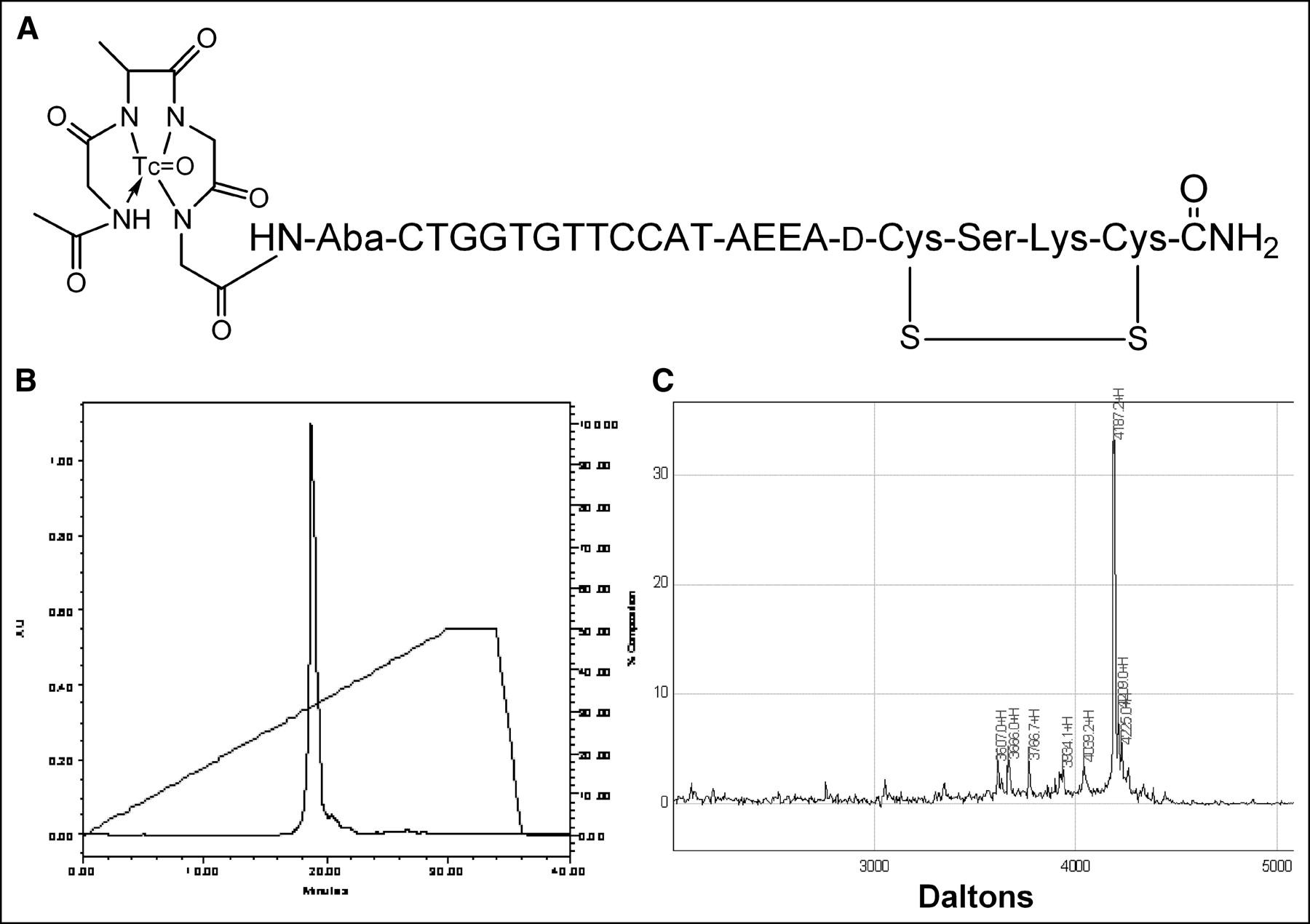
99mTc-chelator-PNA-peptide designed to bind to IGF1R, to be internalized, and to hybridize with CCND1 mRNA. Scintigraphic imaging of γ-rays emitted on decay of 99mTc will identify sites with high levels of CCND1 expression. (A) Schematic structure of 99mTc-AcGlyd(Ala)GlyGlyAba-CTGGTGTTCCAT-AEEA-d(CysSerLysCys), WT4185. (B) Preparative C18 HPLC of cyclized chimera AcGlyd(Ala)GlyGlyAba-CTGGTGTTCCAT-AEEA-d(CysSerLysCys), WT4185. (C) MALDI-TOF MS analysis of purified chimera AcGlyd(Ala)GlyGlyAba-CTGGTGTTCCAT-AEEA-d(CysSerLysCys), WT4185. Experimental mass was 4,187.2 Da; the calculated mass was 4,185.0 Da.
- FIGURE 2.
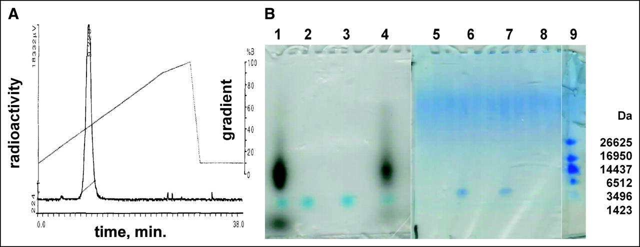
Analysis of 99mTc-AcGlyd(Ala)GlyGlyAba-CTGGTGTTCCAT-AEEA-d(CysSerLysCys), WT4185. (A) A sample from the labeling reaction mixture was analyzed by reversed-phase HPLC on a Microbond C18 column (10 × 250 mm) eluted with a gradient from 10% to 100% CH3CN in aqueous 0.1% CF3CO2H at 1 mL/min over 28 min at 25°C. %B (CH3CN) is shown on right axis, and NaI (Tl) radiometric γ-emission is shown on left axis. The single labeled peak eluted at 9.3 min. (B) Denaturing gel electrophoresis on polyacrylamide (10%–20%)–Tris–Tricine–SDS gels. Left panel is an autoradiogram; right panel is stained with Coomassie blue. Lanes 1 and 5: 99mTc labeling reaction; lanes 2 and 6: mock reaction without 99mTc; lanes 3 and 7: purified WT4185; lanes 4 and 8: 9.3-min 99mTc peak from A; lane 9: peptide mass standards.
- FIGURE 3.
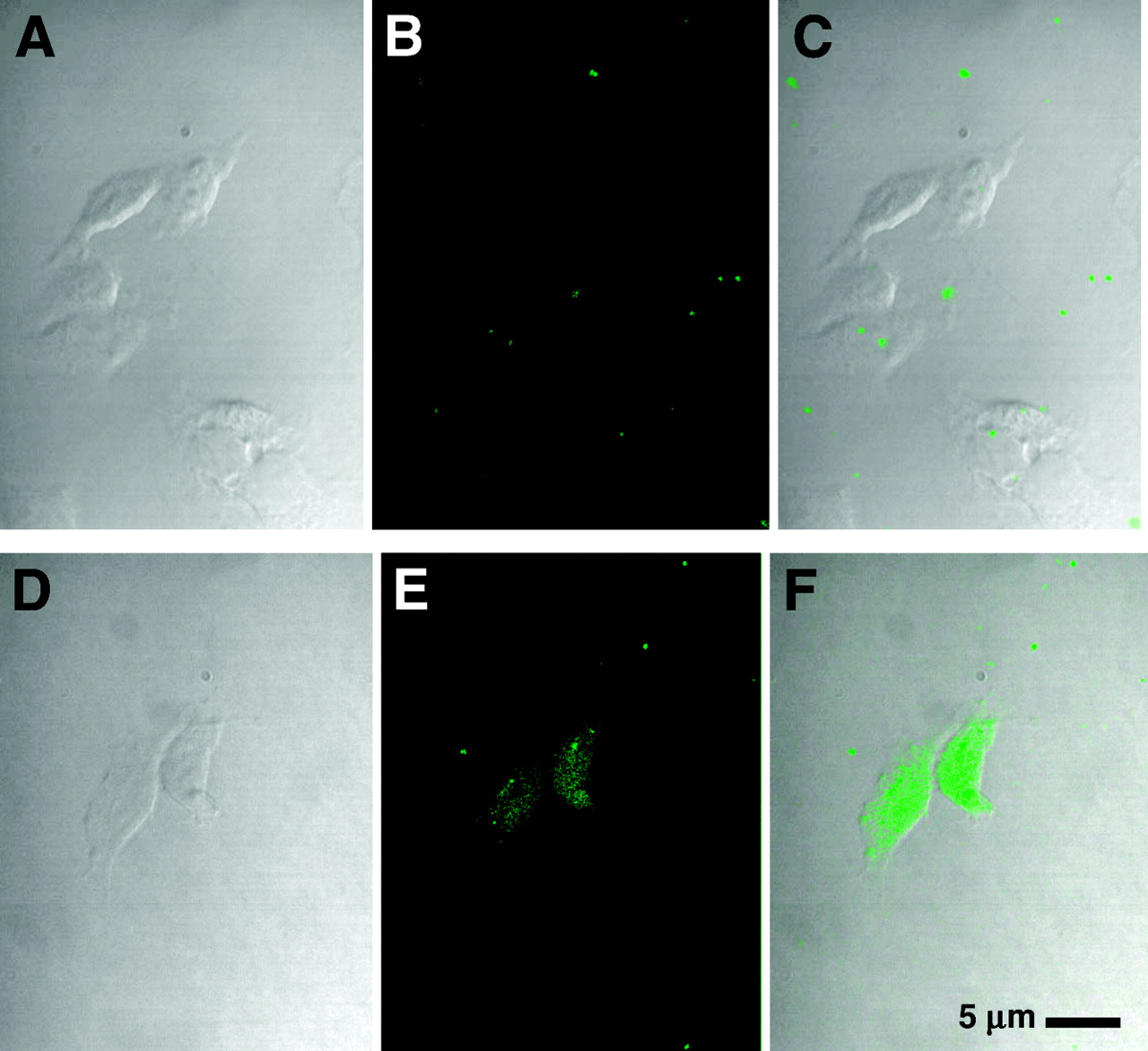
MCF7:IGF1R cell uptake of CCND1 fluoresceinyl-PNA-mismatch peptide probe, WT4361 (A–C), and CCND1 fluoresceinyl-PNA-IGF1 peptide probe, WT4433 (D–F). Cells were incubated with fluoresceinyl-PNA-peptide at 1 μmol/L for 8 h at 37°C in PRF-SFM, fixed, and examined by confocal microscopy. (Left) Phase contrast. (Middle) Fluorescence. (Right) Overlay.
- FIGURE 4.
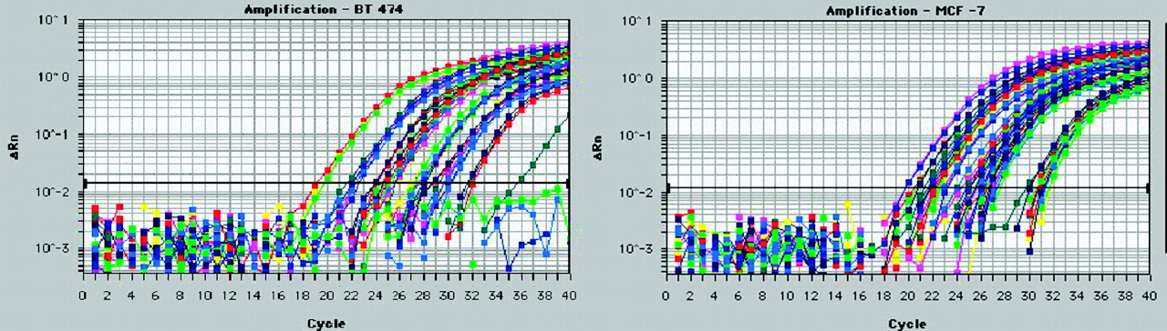
BT474 (left) and MCF7:IGF1R (right) breast cancer cell mRNAs (10, 1, and 0.1 ng) were analyzed by QRT–PCR with a Prism 7700 (Applied Biosystems) to determine the levels of expression of HER2 (blue), CCND1 (green), IGF1R (red), MYC (pink), and TATA-box binding protein (yellow). ΔRn = relative difference in fluorescence at cycle n.
- FIGURE 5.
Western blots of 100 μg of protein extracted from MCF7:IGF1R estrogen receptor-positive breast tumor cell xenografts at 24 h after direct injection of the PNA mismatch chimera, WT4172, the peptide mismatch chimera, WT4113, and the PNA antisense chimera, WT4185. CD1 = cyclin D1; B-actin = β-actin.
- FIGURE 6.
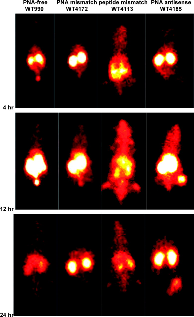
Scintigraphic images of γ-rays emitted by decaying 99mTc in nude mice carrying human MCF7:IGF1R estrogen receptor-positive breast tumor cell xenografts at 4, 12, and 24 h after injection of the PNA-free control probe, WT990, the PNA mismatch control probe, WT4172, the peptide mismatch control probe, WT4113, and the PNA antisense probe, WT4185.
- FIGURE 7.
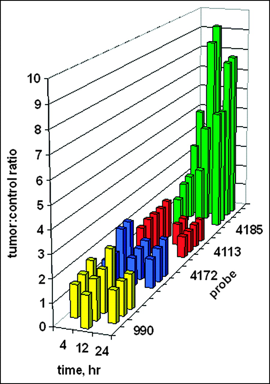
Ratios of tumor site γ-intensity to control site γ-intensity after systemic administration of 99mTc-peptide-PNA-peptide chimeras for all subjects in Figure 6. Bars indicate data for the PNA-free control probe, WT990 (yellow), the PNA mismatch control probe, WT4172 (blue), the peptide mismatch control probe, WT4113 (red), and the PNA antisense probe, WT4185 (green).


Tables
Name Sequence Label Yield (%) Mass (Da) Calculated Measured PNA-free Glyd(Ala)GlyGlyAba-(Gly)4-d(CysSerLysCys) WT990 19.0 990.0 992.0 PNA mismatch AcGlyd(Ala)GlyGlyAba-CTGGACAACCAT-AEEA-d(CysSerLysCys) WT4172 39.1 4,172.0 4,174.1 Peptide mismatch AcGlyd(Ala)GlyGlyAba-CTGGTGTTCCAT-AEEA-d(CysAlaAlaCys) WT4113 34.0 4,113.0 4,113.7 PNA antisense AcGlyd(Ala)GlyGlyAba-CTGGTGTTCCAT-AEEA-d(CysSerLysCys) WT4185 30.6 4,185.0 4,187.2 Fl-peptide mismatch SFX-AEEA-CTGGTGTTCCAT-AEEA-d(CysAlaAlaCys) WT4361 3.0 4,361.0 4,360.6 Fl-PNA antisense SFX-AEEA-CTGGTGTTCCAT-AEEA-d(CysSerLysCys) WT4433 2.8 4,433.0 4,433.8 Fl = fluoresceinyl.
- TABLE 2
Cyclin D1 Protein in Tumors Injected Intratumorally with Peptide-PNA-Peptide Chimeras
Peptide-PNA-peptide Cyclin D1 intensity Mean ± SD Median PNA mismatch, WT4172 6.39 ± 1.34 6.45 Peptide mismatch, WT4113 5.68 ± 1.51 5.57 PNA antisense, WT4185 2.93 ± 1.38 2.87 For each chimera, 4 tumors were analyzed in duplicate by Western blotting. Bands on films were quantitated by scanning.
Peptide-PNA-peptide CCND1/TBP ratio Mean ± SD Median PNA mismatch, WT4172 7.78 ± 6.31 4.62 Peptide mismatch, WT4113 6.65 ± 3.57 5.36 PNA antisense, WT4185 12.29 ± 6.94 9.13 TBP = TATA-box binding protein.
For each chimera, 3 tumors were analyzed in duplicate.
- TABLE 4
Tissue Distribution of PNA-Free Probe, WT990, After Systemic Administration (n = 5)
Tissue Tissue distribution (mean ± SD %ID/g) of WT990 at the following hours after administration: 4 12 24 Muscle 0.14 ± 0.10 0.04 ± 0.01 0.08 ± 0.02 Intestine 0.09 ± 0.01 0.15 ± 0.19 0.06 ± 0.02 Heart 0.05 ± 0.01 0.05 ± 0.00 0.07 ± 0.01 Lung 0.16 ± 0.01 0.12 ± 0.02 0.14 ± 0.02 Blood 0.11 ± 0.01 0.07 ± 0.01 0.08 ± 0.00 Spleen 0.08 ± 0.01 0.13 ± 0.05 0.41 ± 0.16 Kidney 7.82 ± 1.21 4.78 ± 0.54 2.10 ± 0.28 Liver 0.36 ± 0.03 0.41 ± 0.09 0.77 ± 0.22 Tumor 0.16 ± 0.08 0.09 ± 0.03 0.09 ± 0.01 T/M ratio 1.31 ± 0.32 2.85 ± 1.48 1.06 ± 0.10 T/B ratio 1.43 ± 0.76 1.35 ± 0.42 1.10 ± 0.17 T/M ratio = tumor distribution-to-muscle distribution ratio; T/B ratio = tumor distribution-to-blood distribution ratio.
- TABLE 5
Tissue Distribution of PNA Mismatch Probe, WT4172, After Systemic Administration (n = 5)
Tissue Tissue distribution (mean ± SD %ID/g) of WT4172 at the following hours after administration: 4 12 24 Muscle 0.15 ± 0.04 0.06 ± 0.01 0.06 ± 0.01 Intestine 0.17 ± 0.03 0.07 ± 0.01 0.05 ± 0.01 Heart 0.15 ± 0.01 0.08 ± 0.01 0.06 ± 0.01 Lung 0.39 ± 0.07 0.22 ± 0.03 0.14 ± 0.03 Blood 0.29 ± 0.02 0.12 ± 0.02 0.07 ± 0.01 Spleen 0.23 ± 0.02 0.19 ± 0.04 0.16 ± 0.02 Kidney 35.29 ± 8.41 23.07 ± 2.83 9.62 ± 2.61 Liver 0.89 ± 0.16 0.81 ± 0.10 0.47 ± 0.02 Tumor 0.23 ± 0.55 0.14 ± 0.03 0.07 ± 0.03 T/M ratio 1.63 ± 0.55 2.12 ± 0.23 1.26 ± 0.39 T/B ratio 0.80 ± 0.18 1.17 ± 0.16 1.05 ± 0.30 T/M ratio = tumor distribution-to-muscle distribution ratio; T/B ratio = tumor distribution-to-blood distribution ratio.
- TABLE 6
Tissue Distribution of Peptide Mismatch Probe, WT4113, After Systemic Administration (n = 5)
Tissue Tissue distribution (mean ± SD %ID/g) of WT4113 at the following hours after administration: 4 12 24 Muscle 0.33 ± 0.06 0.13 ± 0.02 0.13 ± 0.03 Intestine 0.39 ± 0.06 0.14 ± 0.00 0.16 ± 0.04 Heart 0.35 ± 0.02 0.29 ± 0.04 0.19 ± 0.05 Lung 0.63 ± 0.05 0.42 ± 0.08 0.31 ± 0.06 Blood 0.61 ± 0.04 0.40 ± 0.07 0.17 ± 0.03 Spleen 0.40 ± 0.04 0.53 ± 0.22 1.28 ± 0.53 Kidney 10.30 ± 1.07 4.22 ± 0.67 5.51 ± 1.15 Liver 2.00 ± 0.21 1.56 ± 0.25 2.74 ± 0.75 Tumor 0.53 ± 0.07 0.28 ± 0.04 0.20 ± 0.06 T/M ratio 1.64 ± 0.29 2.25 ± 0.69 1.74 ± 0.88 T/B ratio 0.88 ± 0.14 0.72 ± 0.22 1.23 ± 0.47 T/M ratio = tumor distribution-to-muscle distribution ratio; T/B ratio = tumor distribution-to-blood distribution ratio.
- TABLE 7
Tissue Distribution of PNA Antisense Probe, WT4185, After Systemic Administration (n = 5)
Tissue Tissue distribution (mean ± SD %ID/g) of WT4185 at the following hours after administration: 4 12 24 Muscle 0.12 ± 0.03 0.10 ± 0.05 0.05 ± 0.02 Intestine 0.12 ± 0.01 0.09 ± 0.01 0.05 ± 0.01 Heart 0.11 ± 0.01 0.07 ± 0.02 0.05 ± 0.01 Lung 0.29 ± 0.03 0.19 ± 0.03 0.09 ± 0.02 Blood 0.23 ± 0.02 0.11 ± 0.02 0.05 ± 0.01 Spleen 0.17 ± 0.02 0.17 ± 0.02 0.12 ± 0.02 Kidney 21.55 ± 2.90 19.10 ± 3.94 11.33 ± 2.74 Liver 0.52 ± 0.04 0.81 ± 0.10 0.39 ± 0.09 Tumor 0.20 ± 0.06 0.17 ± 0.06 0.11 ± 0.05 T/M ratio 1.78 ± 0.53 1.85 ± 0.57 2.01 ± 0.29 T/B ratio 0.88 ± 0.20 1.49 ± 0.34 1.92 ± 0.58 T/M ratio = tumor distribution-to-muscle distribution ratio; T/B ratio = tumor distribution-to-blood distribution ratio.
In this issue


Jump to section
Related Articles
Cited By...
- IGF1R-Targeted Delivery of a Bridged Nucleic Acid Oligonucleotide-Peptide Conjugate for MicroRNA-21 Inhibition in Triple-Negative Breast Cancer
- Molecular Imaging of MDM2 Messenger RNA with 99mTc-Labeled Antisense Oligonucleotides in Experimental Human Breast Cancer Xenografts
- Molecular Imaging of bcl-2 Expression in Small Lymphocytic Lymphoma Using 111In-Labeled PNA-Peptide Conjugates
- Noninvasive Imaging of Human Telomerase Reverse Transcriptase (hTERT) Messenger RNA with 99mTc-Radiolabeled Antisense Probes in Malignant Tumors
- PET Imaging of CCND1 mRNA in Human MCF7 Estrogen Receptor Positive Breast Cancer Xenografts with Oncogene-Specific [64Cu]Chelator-Peptide Nucleic Acid-IGF1 Analog Radiohybridization Probes
- Receptor-mediated internalization of chelator-PNA-peptide hybridization probes for radioimaging or magnetic resonance imaging of oncogene mRNAs in tumours
- Cyclin D1 Is Necessary for Tamoxifen-Induced Cell Cycle Progression in Human Breast Cancer Cells
- Initial Mechanistic Studies of Antisense Targeting in Cells
- Meeting Report: The International Conference on Tumor Progression and Therapeutic Resistance