


Article Figures & Data
Figures
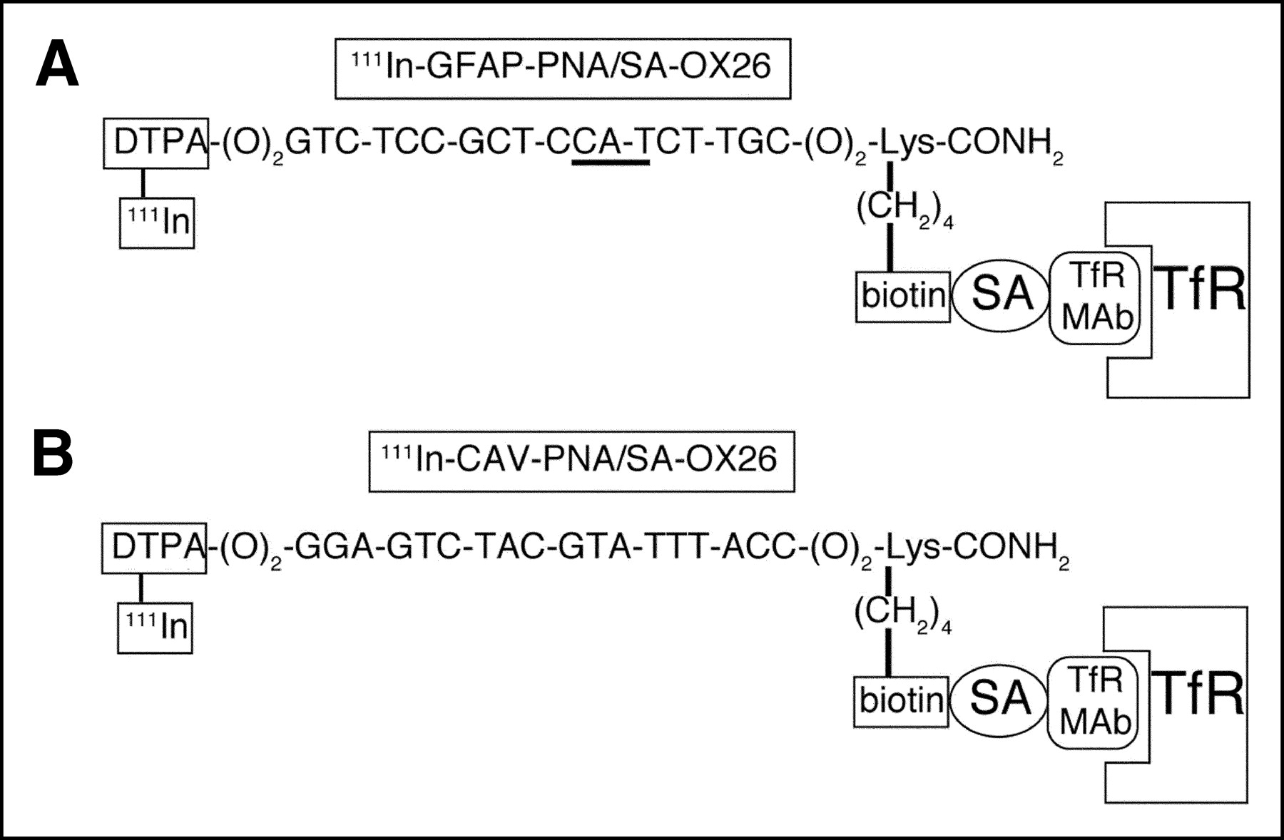
- FIGURE 1.
(A) An 18-mer PNA that is antisense to nucleotides 20–37 of rat GFAP mRNA (accession number NM_017009). The triplet that is antisense to GFAP methionine initiation codon (AUG) is underlined. The amino terminus has 2 linkers (O) separating PNA base sequence from amino-terminal DTPA group, where O = (CH2-O-CH2-CH2-O-CH2-CH2-O-CH2). The amino terminus was conjugated with DTPA dianhydride while PNA was still attached to the solid-phase resin during synthesis. At the carboxyl terminus of PNA, which is amidated, there are 2 linkers separating the base sequence from a carboxyl-terminal lysine residue, and the ε-amino group of the lysine residue is conjugated with biotin. Upon mixing, there is immediate attachment of biotinylated PNA to SA conjugated to a mAb to the rat TfR. The TfRMAb and the SA are conjugated with a thiol-ether linkage and this conjugate is designated SA-OX26. (B) An 18-mer PNA that is antisense to nucleotides 10–27 of rat CAV mRNA (accession number AF439778). Apart from the base sequence, CAV-PNA has a structure identical to that of GFAP-PNA shown in A, as both are designed for labeling with 111In.
- FIGURE 2.
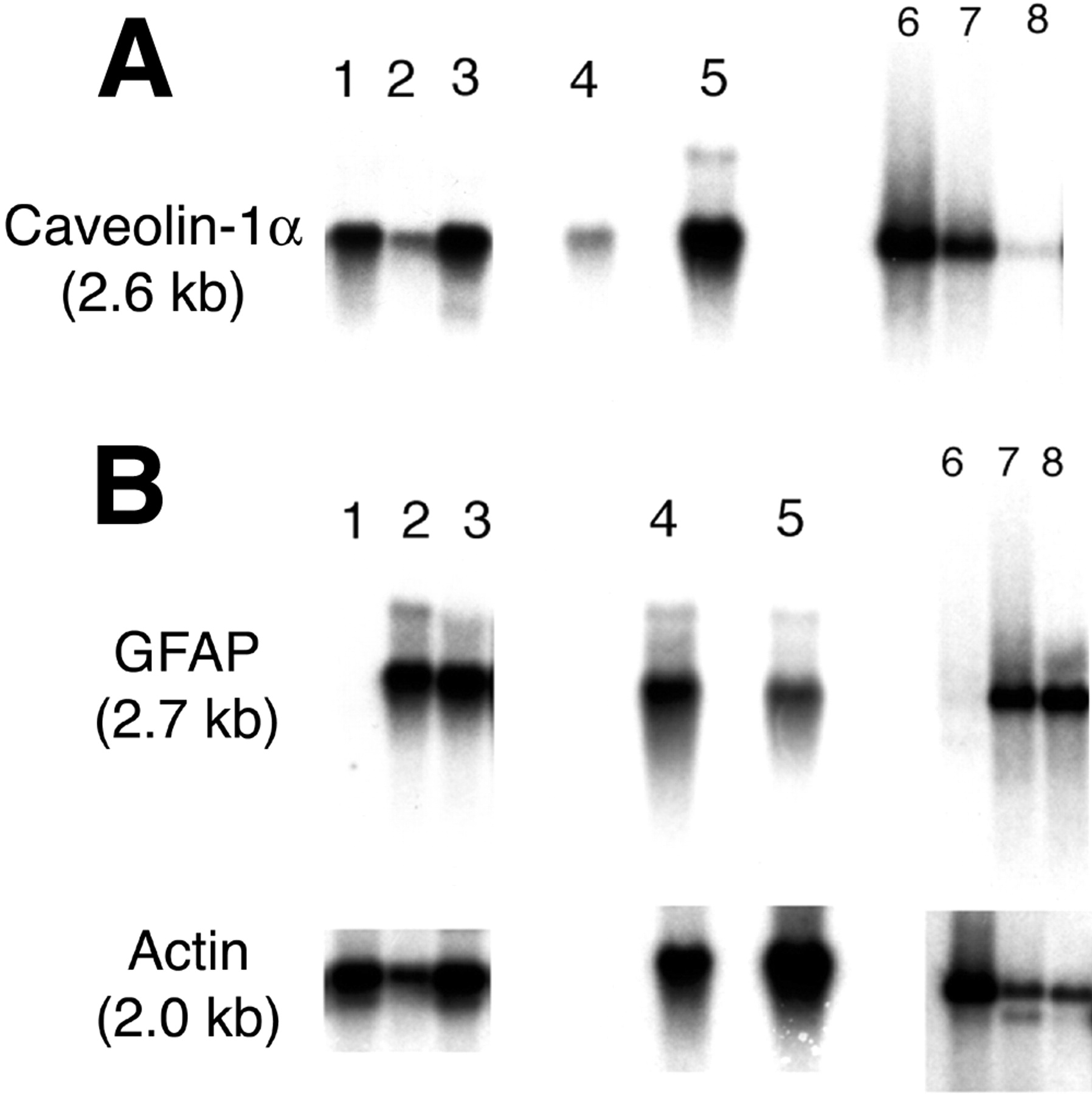
Northern blot produced with cRNA derived by IVT of transcription plasmids encoding either rat GFAP cDNA or rat LAT1 cDNA. Each lane was spotted with 10, 20, or 50 ng of GFAP cRNA or LAT1 cRNA. After electrophoresis with parallel RNA molecular size standards, and blotting, the hybridization was performed with 111In-GFAP-PNA/SA-OX26. The film was developed overnight at −20°C. 111In-GFAP-PNA/SA-OX26 selectively hybridized to 2.6-kb GFAP mRNA but did not hybridize to LAT1 mRNA.
- FIGURE 3.
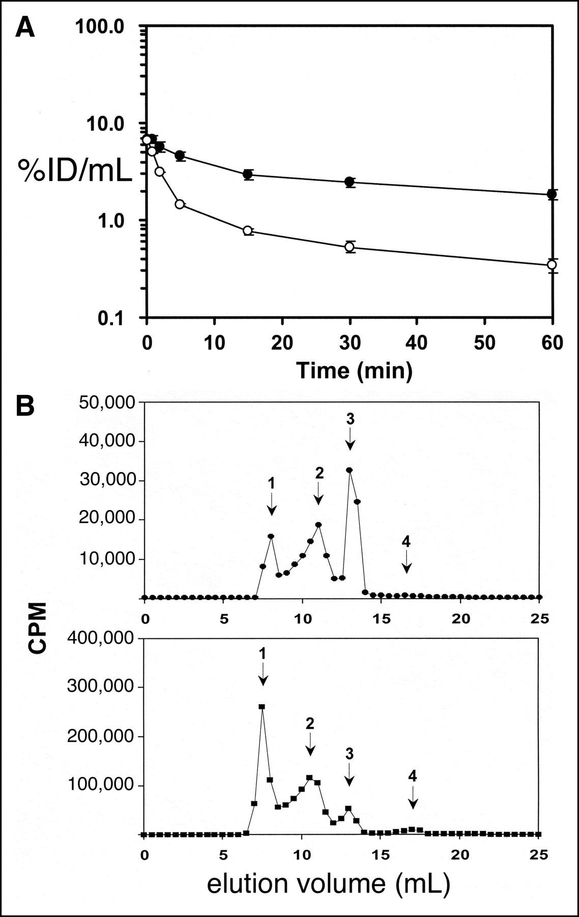
(A) Plasma radioactivity, expressed as percentage of injected dose (%ID)/mL plasma at 0.25–60 min after intravenous injection of either unconjugated 111In-GFAP-PNA (○) or 111In-GFAP-PNA/SA-OX26 (•) in adult rats. Data are mean ± SE (n = 3 rats per time point). These data were used to generate the pharmacokinetic parameters shown in Table 1. (B) Gel-filtration FPLC using a Superose 12HR10/30 column with elution in PBST. (Top) Elution of serum removed 60 min after intravenous injection of 111In-GFAP-PNA/SA-OX26. (Bottom) Elution of uninjected 111In-GFAP-PNA/SA-OX26 premixed in control uninjected rat serum. Peak 1, 111In-GFAP-PNA/SA-OX26 bound to lipoprotein fraction of serum; peak 2, 111In-GFAP-PNA/SA-OX26; peak 3, 111In-GFAP-PNA bound to unconjugated SA; peak 4, free 111In-GFAP-PNA.
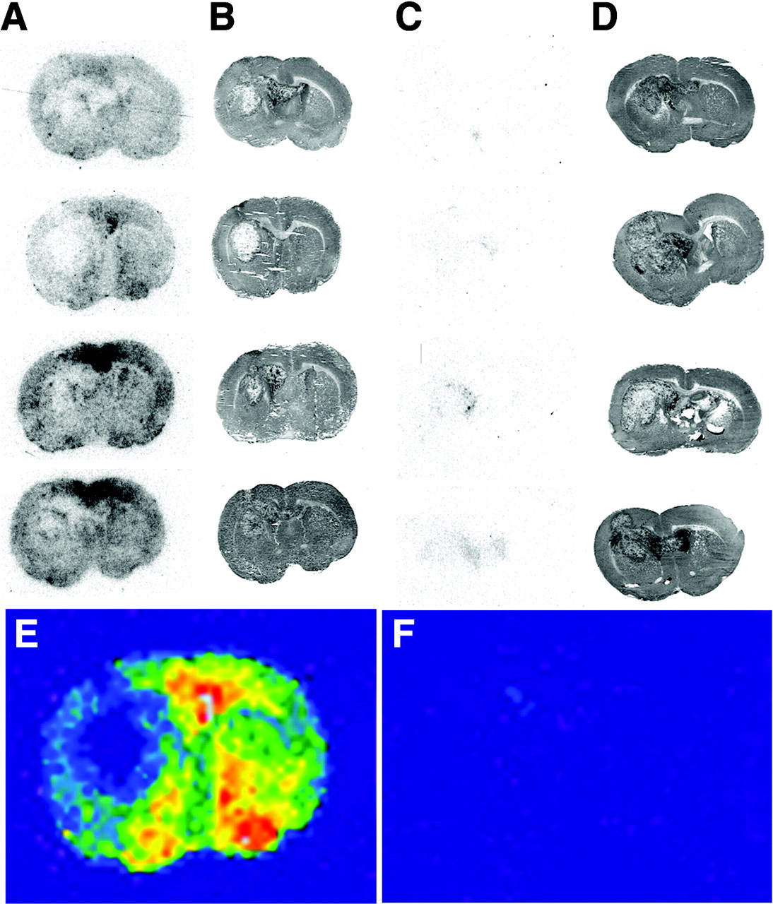
- FIGURE 4.
111In-GFAP-PNA/SA-OX26 conjugate or unconjugated 111In-GFAP-PNA (3.7 MBq [100 μCi] per rat) was injected into RG2 brain tumor–bearing rats at 15 d after implantation of 350,000 RG2 cells per brain, and rats were sacrificed at 60 min. The Biomax film was developed for 12 d at −20°C. The film autoradiograms for 4 rats injected with 111In-GFAP-PNA/SA-OX26 conjugate are shown in A and the corresponding hematoxylin autopsy stains indicating the size of tumor are shown in B. The film autoradiograms for 4 rats injected with unconjugated 111In-GFAP-PNA are shown in C, and the corresponding hematoxylin autopsy stains are shown in D. The gray-scale autoradiograms from the second images in A and C were colorized, as shown in E and F, respectively.
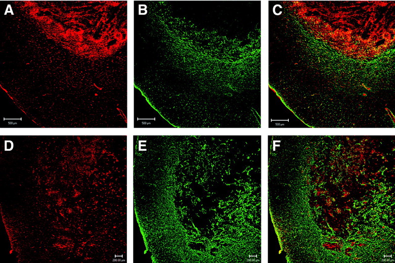
- FIGURE 5.
Confocal microscopy of sections of brain taken from rats implanted with either C6 glioma cells (A–C) or RG2 glioma cells (D–F). (A and D) Section immunostained with rabbit polyclonal antiserum to rat CAV. (B and E) Section immunostained with a mAb to GFAP. (C and F) Overlap image with parallel staining of both GFAP (green) and CAV (red). Bar in A–C is 500 μm; bar in D–F is 200 μm.
- FIGURE 6.
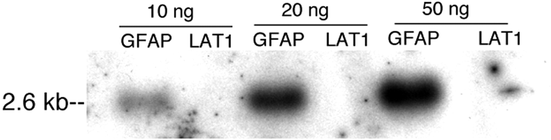
Northern blot of tissue mRNA probed with a cDNA to either rat CAV (A) or rat GFAP or actin (B). Lanes 1–3, 4 and 5, and 6–8 are 3 different blots: lanes 1–3, RG2 tumor cells in tissue culture, contralateral rat brain, and RG2 rat brain tumor in vivo, respectively; lanes 4 and 5, contralateral rat brain and C6 rat brain tumor in vivo, respectively; lanes 6–8, C6 glioma cells in tissue culture, freshly isolated rat brain capillaries, and control rat brain, respectively. Films of actin and CAV Northern blots were developed for 3 d. The GFAP Northern autoradiogram for lanes 1–5 was developed overnight and for lanes 6–8 was developed for 10 d; despite this overexposure of the film, the signal representing the GFAP transcript expressed in C6 glioma cells in tissue culture is not detectable (B, lane 6).
- FIGURE 7.
(A, C, E, and G) Colorized film autoradiograms of sections removed from Fisher CD344 rat brain implanted with RG2 tumors; rats were sacrificed at 6 h after intravenous injection of 111In unconjugated or conjugated PNA. (A) RG2 tumor-bearing rat injected with unconjugated 111In-GFAP-PNA. (C) RG2 tumor-bearing rat injected with 111In-GFAP-PNA/SA-OX26. (E) RG2 tumor-bearing rat injected with unconjugated 111In-CAV-PNA. (G) RG2 tumor-bearing rat injected with 111In-CAV-PNA/SA-OX26. (B, D, F, and H) Hematoxylin autopsy stains show size of tumor corresponding to A, C, E, and G, respectively. (I) Integrated density of autoradiogram signal over the brain tumor (tumor) or contralateral brain (brain) for each of 4 different antisense radiopharmaceutical formulations: 1 and 2, 111In-CAV-PNA/SA-OX26 conjugate (CAV-PNA CONJ.); 3 and 4, 111In-GFAP-PNA/SA-OX26 conjugate (GFAP-PNA CONJ.); 5 and 6, unconjugated 111In-CAV-PNA (CAV-PNA); 7 and 8, unconjugated 111In-GFAP-PNA (GFAP-PNA). Data are mean ± SE (n = 4 rats in each of 4 groups).


Tables
Parameter (units) 111In-GFAP-PNA 111In-GFAP-PNA/SA-OX26 A1 (%ID/mL) 6.3 ± 0.4 4.0 ± 0.3 A2 (%ID/mL) 0.94 ± 0.07 3.2 ± 0.3 k1 (min−1) 0.49 ± 0.04 0.20 ± 0.04 k2 (min−1) 0.017 ± 0.002 0.0095 ± 0.0018 t1/21 (min) 1.4 ± 0.1 3.5 ± 0.7 t1/22 (min) 40 ± 4 73 ± 14 AUC(0–60) (%ID · min/mL) 48 ± 1 167 ± 3 AUCSS (%ID · min/mL) 67 ± 3 360 ± 43 VSS (mL/kg) 243 ± 17 89 ± 6 Cl (mL/min/kg) 5.2 ± 0.3 0.89 ± 0.11 MRT (min) 47 ± 5 100 ± 19 A1 = first intercept; A2 = second intercept; k1 = first exponential rate constant; k2 = second exponential rate constant; t1/21 = half-time of first exponential decay; t1/22 = half-time of second exponential decay; AUC(0–60) = plasma area under the concentration curve (AUC) for 0–60 min; AUCSS = steady state AUC; VSS = steady state whole-body volume of distribution; Cl = plasma clearance; MRT = plasma mean residence time.
Parameters were determined from plasma profiles shown in Figure 3A.
Organ Uptake (%ID/g) 111In-GFAP-PNA 111In-GFAP-PNA/SA-OX26 Liver 0.11 ± 0.03 4.5 ± 0.2 Lung 0.38 ± 0.12 0.73 ± 0.05 Kidney 2.2 ± 0.3 0.33 ± 0.04 Heart 0.061 ± 0.013 0.21 ± 0.03 Brain 0.0061 ± 0.0008 0.083 ± 0.014 Mean ± SE (n = 3 rats per group); determined at 60 min after intravenous injection.
In this issue


Jump to section
Related Articles
Cited By...
- Selective targeting of oncogenic KRAS G12D using peptide nucleic acid oligomers attached to cell-penetrating peptides
- Systemic antibody-oligonucleotide delivery to the central nervous system ameliorates mouse models of spinal muscular atrophy
- Preparation and Evaluation of 99mTc-Epidermal Growth Factor Receptor (EGFR)-Peptide Nucleic Acid for Visualization of EGFR Messenger RNA Expression in Malignant Tumors