


Article Figures & Data
Figures
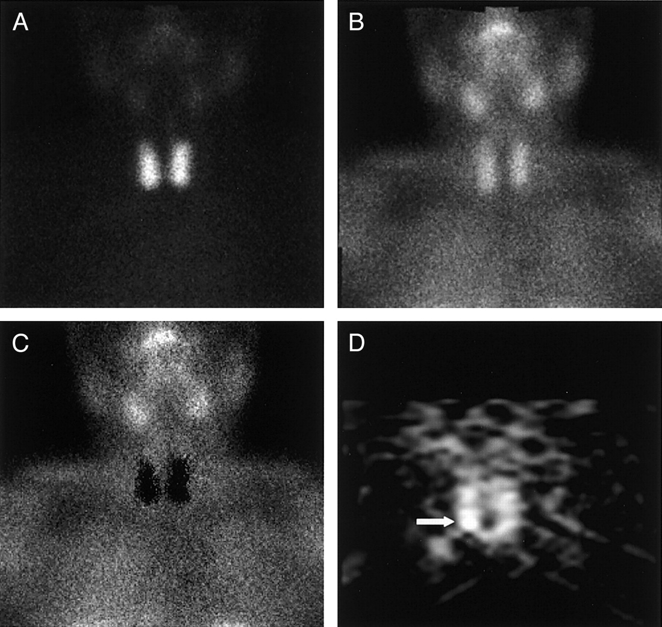
- FIGURE 1.
A 55-y-old female patient affected by pHPT with small parathyroid adenoma (size, 6.1 mm; weight, 261 mg) sited behind inferior pole of right thyroid lobe, negative on planar 99mTc-pertechnetate (A), 99mTc-tetrofosmin (B), and subtraction (C) scintigraphy and clearly revealed (arrow) on coronal P-SPECT (D).
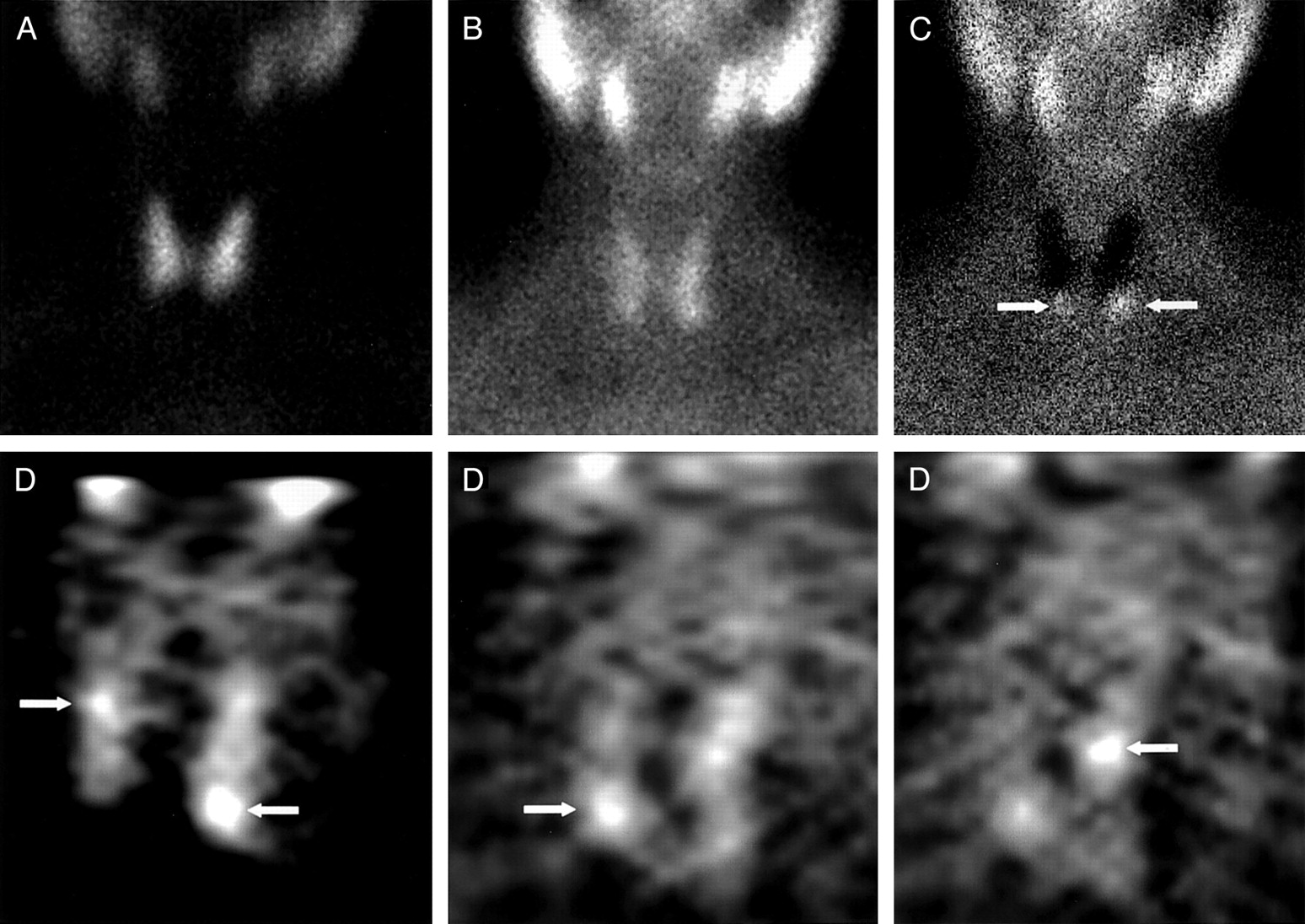
- FIGURE 2.
A 67-y-old male patient affected by sHPT with 4 hyperplastic glands sited behind upper two thirds of right thyroid lobe (size, 6 mm; weight, 274 mg), below inferior pole of right thyroid lobe (size, 15 mm; weight, 685 mg), behind upper two thirds of left thyroid lobe (size, 9 mm; weight, 429 mg), and below inferior pole of left thyroid lobe (size, 17 mm; weight, 850 mg). All glands were clearly revealed (arrows) on different multiple coronal P-SPECT slices (D), whereas planar 99mTc-pertechnetate (A), 99mTc-tetrofosmin (B), and subtraction (C) scintigraphy identified only the 2 sited below inferior pole of 2 thyroid lobes (arrows), missing the other 2, which were smaller in size and weight and were sited behind upper two thirds of 2 thyroid lobes.
- FIGURE 3.
A 65-y-old female patient affected by pHPT with small parathyroid adenoma (size, 6.8 mm; weight, 290 mg) sited behind upper two thirds of left thyroid lobe, positive (arrow) on both early (A) and delayed (B) planar 99mTc-MIBI scintigraphy and on coronal P-SPECT (C), but more clearly visualized by the latter.


Tables
- TABLE 1
Histologic Findings in 48 Patients with pHPT (49 Lesions) and in 19 Patients with sHPT (51 Lesions)
Histology No. of lesions Maximum diameter range (mm) Weight range (mg) pHPT Solitary adenoma 43 6.1–30 261–4,000 Carcinoma 1 40 10,000 Hyperplastic glands 5 6–18 300–900 sHPT Solitary adenoma 2 15–20 750–1,060 Hyperplastic glands 49 6–40 210–3,000 - TABLE 2
Planar Parathyroid Scintigraphy and Neck P-SPECT Results Related to Histopathologic Findings in Patients with pHPT
Parameter Overall (n = 49) Adenoma/carcinoma (n = 44) Hyperplasia (n = 5) Planar P-SPECT Planar P-SPECT Planar P-SPECT TP findings 43 48 40 44 3 4 FN findings 6 1 4 0 2 1 Sensitivity (%) 87.7 97.9* 90.9 100 60 80 ↵* P > 0.05 when compared with corresponding planar value (McNemar test results).
TP = true-positive; FN = false-negative.
- TABLE 3
Planar Parathyroid Scintigraphy and Neck P-SPECT Results Related to Histopathologic Findings in Patients with sHPT
Parameter Overall (n = 51) Adenoma/carcinoma (n = 2) Hyperplasia (n = 49) Planar P-SPECT Planar P-SPECT Planar P-SPECT TP findings 40 47 1 2 39 45 FN findings 11 4 1 0 10 4 Sensitivity (%) 78.4 92.1* 50 100 79.6 91.8 ↵* P < 0.05 when compared with corresponding planar value (McNemar test results).
TP = true-positive; FN = false-negative.
In this issue


Jump to section
Related Articles
Cited By...
- Parathyroid Imaging: The Importance of Dual-Radiopharmaceutical Simultaneous Acquisition with 99mTc-Sestamibi and 123I
- Parathyroid Imaging and Localization Using SPECT/CT: Initial Results
- Improved Delineation of Parathyroid Lesions in Patients with Chronic Renal Failure Using Magnified Pinhole Imaging
- Subtraction SPECT for Parathyroid Scintigraphy Based on Maximization of Mutual Information
- BEST PRACTICE NO 183: Examination of parathyroid gland specimens
- The Value of 99mTc-Sestamibi SPECT/CT over Conventional SPECT in the Evaluation of Parathyroid Adenomas or Hyperplasia