


Article Figures & Data
Figures
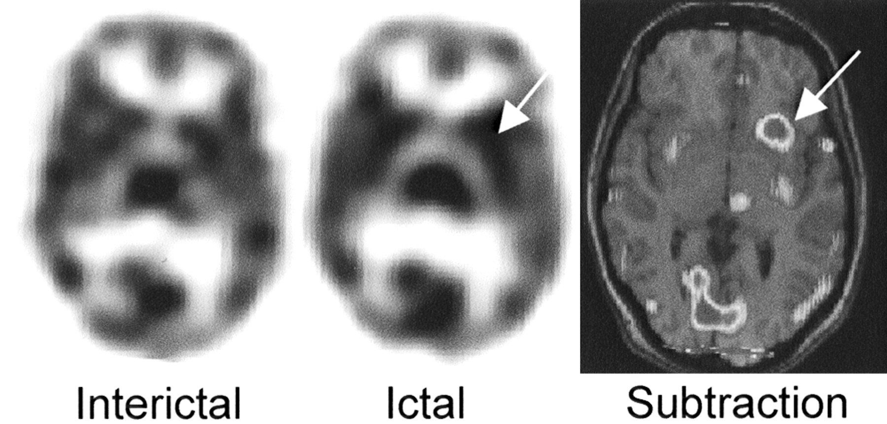
- FIGURE 1.
Example of ipsilateral basal ganglia lateralization. Asymmetric basal ganglia activation (left > right) in patient with left frontal focus (not visualized here) seen on transaxial ictal and subtraction SPECT images. Arrows indicate activation of left caudate.
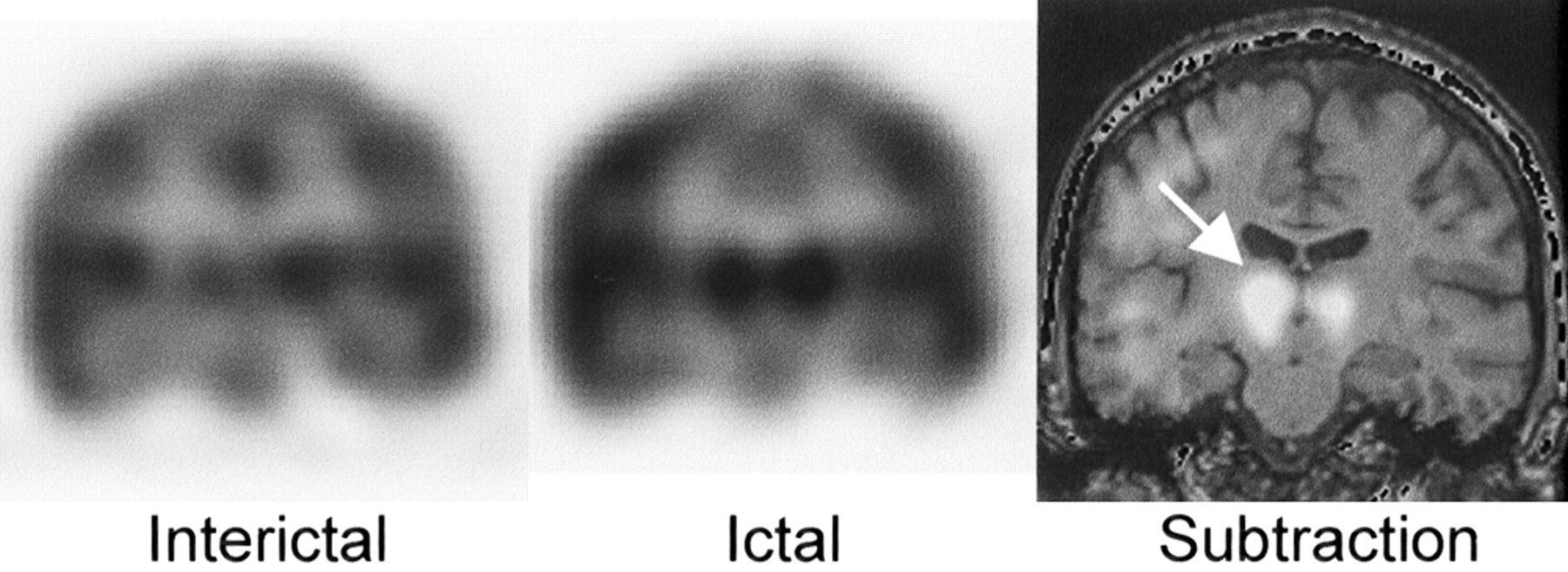
- FIGURE 2.
Example of ipsilateral thalamic lateralization. Asymmetric thalamus activation (right > left) in patient with right mesiotemporal focus seen on coronal ictal and subtraction SPECT images. Arrow indicates right thalamic activation.
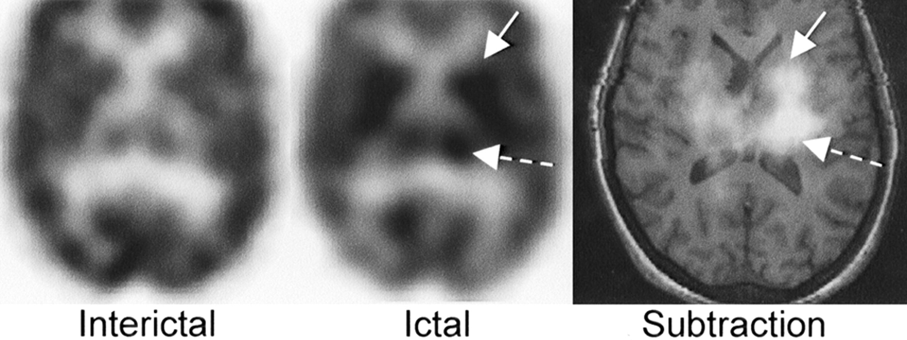
- FIGURE 3.
Example of contralateral basal ganglia activation. Transaxial SPECT images of patient with final focus in right temporal neocortex focus (not shown), who was found to have left basal ganglia uptake (solid arrow), right thalamic uptake (dashed arrow), and increased uptake in left temporal neocortex in study with injection time of 8 s.
- FIGURE 4.
Example of contralateral thalamic activation. Transaxial SPECT images show left thalamic (dashed arrow) and basal ganglia (solid arrow) activation. Left cortical activation observed in this study (presumably due to generalization of seizure) is not visualized. Patient’s final focus was located in right temporal lobe.


Tables
Subcortical structure No. of studies Studies with asymmetric subcortical activation Activation ipsilateral to final cortical focus Activation contralateral to final cortical focus Basal ganglia 72 22 (30.6) 17 (77.3) 5 (21.7) Thalamus 72 15 (20.8) 12 (80.0) 3 (20.0) -
Values in parentheses are percentages.
-
- TABLE 2
Lateralized Subcortical Activation with Respect to Final Localization of Foci on 72 Ictal SPECT Studies
Seizure focus* Basal ganglia Thalamus Asymmetric Ipsilateral Contralateral Asymmetric Ipsilateral Frontal 8/27 (29.6) 7 (87.5) 1 (12.5) 6/27 (22.2) 6 (75) SMA† 4/13 (30.8) 3 (75) 1 (25) 0/13 (0) 0 (0) Parietal 0/7 (0) 0 (0) 0 (0) 0/7 (0) 0 (0) Temporal neocortex 3/18 (16.7) 1 (33.3) 2 (66.6) 4/18 (22.2) 3 (75) Occipital 2/5 (40) 1 (50) 1 (50) 1/5 (20) 1 (100) Mesiotemporal 5/10 (50) 5 (100) 0 (0) 3/10 (30) 3 (100) -
↵* Patients with final foci considered to have seizure originating from combination of lobes (e.g., frontoparietal) were included in analysis of both lobes (n = 6 patients, 8 studies).
-
↵ † SMA = supplementary motor area. Patients with SMA focus were counted separately and not included in group of patients with frontal seizure localization.
-
Values in parentheses are percentages.
-
- TABLE 3
Comparison Between Seizure Frequency in Patients With and Without Asymmetric Subcortical Findings on Any Ictal Study at 1 Year After Surgery Follow-Up
Risk of persistent seizures at 1-y follow-up Patients with mesial TLE* and neocortical epilepsy (n = 43) (%) Subgroup of patients with neocortical epilepsy (n = 3) (%) Asymmetric thalamus 7.7 (0.4–37.9) 10.0 (0.5–45.9) Symmetric thalamus 50.0 (30.4–69.6) 61.9 (3.7–81.0) Asymmetric basal ganglia 7.1 (0.4–35.8) 11.1 (0.6–40.2) Symmetric basal ganglia 52.0 (31.8–71.7) 25.0 (6.7–57.2) -
↵* None of 8 patients with mesial temporal epilepsy who underwent surgery had seizures at 1 y or at last follow-up.
-
Values in parentheses are 95% confidence intervals.
-
- TABLE 4
Relationship Between Postsurgical Outcome of Patients Who Underwent Surgery at Our Institution as Determined by Engel’s Classification and Asymmetric Basal Ganglia and Thalamic Uptake on Ictal SPECT Studies
Postsurgical outcome* (n = 37) Basal ganglia Thalamus Asymmetric* Symmetric Asymmetric* Symmetric Engel’s class I (n = 24) 10 12 9 13 Engel’s class II (n = 10) 4 6 4 6 Engel’s class III (n = 5) 0 5 0 5 Engel’s class IV (n = 0)* 0 0 0 0 -
↵* Additional 2 patients (1 from class III and 1 from class IV, both of whom without asymmetric uptake in thalamus or basal ganglia) underwent surgical treatment elsewhere and were referred to our facility for further evaluation.
-
Engel’s classification (27).
-


