


Abstract
In 1925, Hermann Blumgart performed the first diagnostic procedure using radioactive indicators on humans; this first is well recognized. Less well recognized is the fact that Blumgart and his coworker Otto C. Yens, then a medical student, developed the first instrumentation used in a diagnostic procedure involving radioactive indicators. The instrumentation, a modified Wilson cloud chamber, turned out to be the detector most suitable for their purpose. Blumgart also showed remarkable foresight in outlining the requirements both for a satisfactory indicator (tracer) and for a satisfactory detector—requirements that still hold true today. The Blumgart–Yens modified cloud chamber was the birth of nuclear medicine instrumentation.
If one takes a closer look at one of the most famous of all radiotracer experiments on humans, the well-known study of arm-to-arm circulation time (1) done by Hermann L. Blumgart and his coworker Otto C. Yens in 1925, a new insight emerges. Hermann Ludwig Blumgart (1895–1977), chair of the Department of Medicine at Beth Israel Hospital from 1928 to 1962, was an early advocate of 131I therapy of cardiac disease in euthyroid patients. Historical accounts variously label his arm-to-arm circulation measurement as blood flow or blood velocity. Blumgart actually measured circulation time (in seconds) and not what we would today call blood flow (in mL/s or mL/min) or blood velocity (in cm/sec). Otto Christian Yens (1901–1969), as a first- or second-year medical student, assisted in the development and construction of the modified cloud chamber. Yens had a background in physiology; he later became a pediatrician. As we all know, this was the first diagnostic radiotracer, or radioindicator, procedure done on a human—the first nuclear medicine procedure. But there is more to it than that: It was also the birth of nuclear medicine instrumentation.
The year 1925 saw the discovery of cosmic rays (Millikan); the discovery of element 75, rhenium (Noddack); the Scopes trial in Tennessee; and many artistic milestones: The Great Gatsby (Fitzgerald), Arrowsmith (Lewis), and The Gold Rush (Chaplin)—and the Charleston was the dance.
Hermann Blumgart was physician-in-chief at the Beth Israel Hospital in Boston and professor of medicine at Harvard Medical School. As a Harvard medical student working under the physiology pioneer Walter Cannon, he had acquired a deep appreciation of the dynamics of physiology. At Harvard, William Duane had developed the radium–radon “cow” for cancer therapy with radon seeds. Blumgart noticed that Duane was measuring the internal radon from outside the patient, using a radiation detector, and it occurred to him that some other radiation-emitting substance might be used to gain information about what was happening inside the body. This idea was the birth of diagnostic clinical nuclear medicine.
Blumgart was especially interested in the circulation and knew that despite many ingenious attempts, no one had succeeded in measuring circulation time between any 2 points accurately and convincingly. There were many clever approaches, most of which were so invasive that they altered the very hemodynamics they were attempting to measure.
Almost exactly 300 y before Blumgart’s experiment, William Harvey had shown that blood flows in a closed circuit, and the question naturally arose as to how fast it flowed. In 1733, Stephen Hales used an anatomic/geometric approach based on measurements of the heart and aorta in the horse. In 1827, Eduard Hering tried his hand at it by injecting a solution of potassium ferrocyanide (not very toxic despite its name), which binds with iron in the blood to form the deep blue pigment Prussian blue (ferric ferrocyanide), which he tested for in a series of blood samples.
Several other ingenious approaches followed, but all were technically unsatisfactory; most involved placing some kind of device within a blood vessel, which would inherently interfere with the free flow of blood. Almost all depended on the use of some substance as an indicator.
Blumgart reviewed the requirements that a satisfactory substance must fulfill (except for the fourth, all of Blumgart’s requirements still hold for us):
The substance must be nontoxic.
The substance must not be present in the body before the study.
The substance must have no effect on the process being measured.
The substance must disappear rapidly, allowing for repeated measurements.
The substance must be readily detectable in minute amounts.
You are already way ahead of me—yes, the idea of using a radioactive tracer occurred to Blumgart. In 1925, only naturally occurring radionuclides were available. Blumgart looked for one that would satisfy all the requirements and came up with radium C, which is 20-min 83Bi214, a β-decay product of 27-min 82Pb214 (radium B), a radium decay product. Radium C (called active deposit in those days, actually a mixture of 82Pb214 and 83Bi214) emitted β- and γ-rays. It had been used therapeutically in 1,850- to 2,775-MBq doses with no ill effects, so Blumgart assumed it would be nontoxic in the 37- to 222-MBq doses he used. (Some historical accounts say Blumgart used radium; others, radon. He used neither; the confusion arises because of the terminology [radium C = a mixture of 82Pb214 and 83Bi214, both radium decay products].)
Preparing this primordial “radiopharmaceutical” involved exposing solid sterile sodium chloride to radon (“radium emanation”) for a while, during which the following sequence of nongaseous daughter radionuclides would accumulate on it: 3-min 84Po218 (radium A) (α) → 27-min 82Pb214 (radium B) (β,γ) → 20-min 83Bi214 (radium C) (β,γ). The salt was then dissolved in sterile water, assayed with an electroscope, and held for 20 min to allow the short-lived α-emitting 84Po218 to decay. The substance emits both β- and γ-radiation.
Selecting a detector proved to be a far greater challenge. Blumgart laid out the requirements that a satisfactory detection method must fulfill:
The method must be objective, requiring little or no cooperation on the part of the patient.
The method must be noninvasive except for injection of the substance.
The method must be rapid enough to indicate the time of arrival of the substance automatically.
The detector would have to indicate the arrival of the radiotracer immediately; an electroscope would have been far too slow. The Geiger counter of 1925 was a capricious, unstable device, and Blumgart was apprehensive about the high voltage (several hundred volts) so near the patient. He tried various ionization chambers, also without success. Blumgart enlisted the aid of Yens, who, like Blumgart, had a background and special interest in physiology.
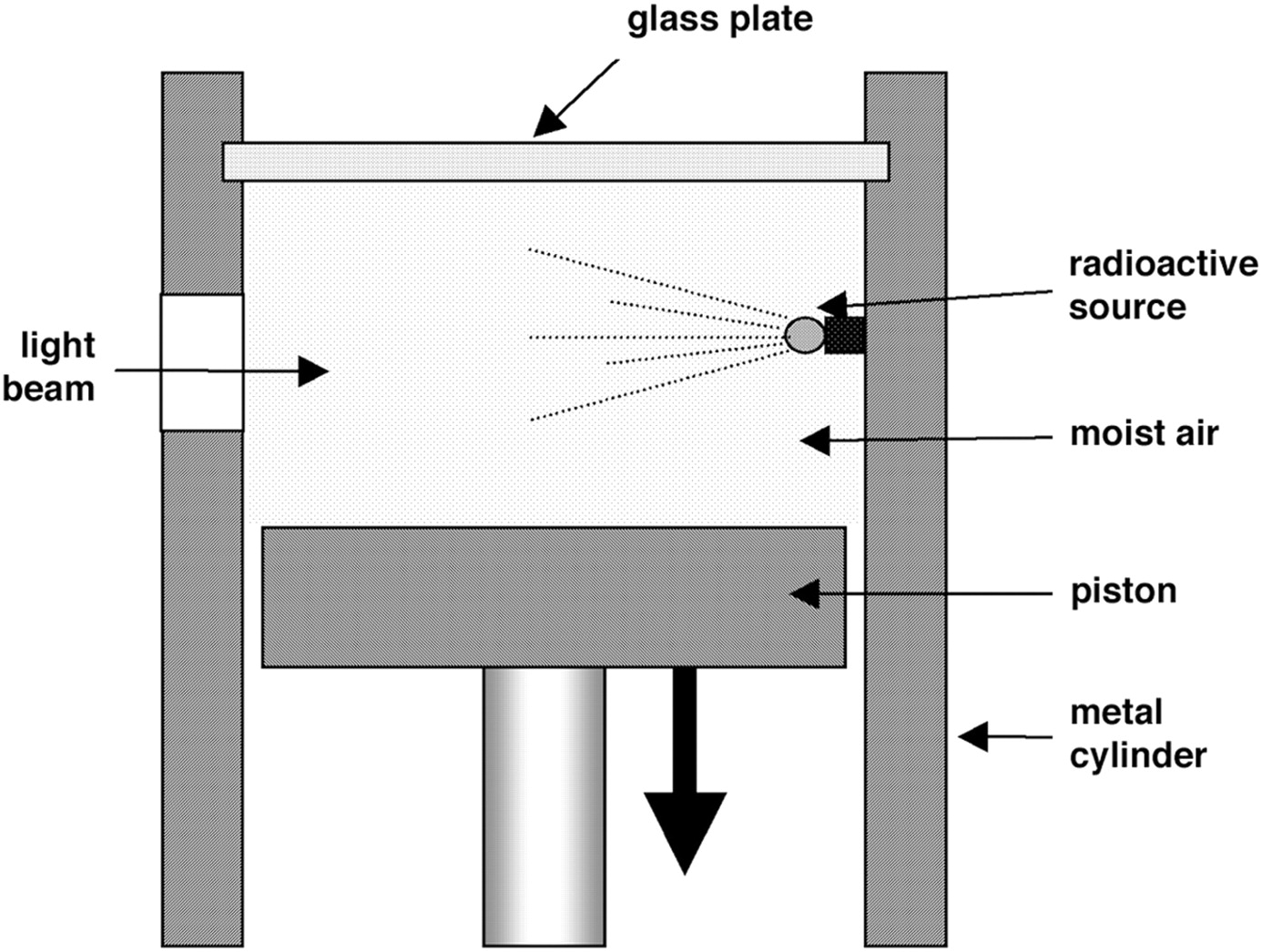
Enter C.T.R. Wilson and his fascination with thunderstorms. Charles Thomson Rees Wilson—a pupil of J.J. Thomson at the Cavendish Laboratory at Cambridge—while studying cloud formation, noted that ions in the air act as condensation nuclei for water vapor. His cloud chamber was actually built to study just that, clouds. His work ultimately led to a great number of advances in physics, including a greater understanding of weather phenomena, and the investigation of cosmic rays and several elementary particles. The Wilson cloud chamber (Fig. 1) is a transparent compartment containing saturated water vapor. When the chamber pressure is suddenly lowered (by a falling piston, which suddenly increases the chamber volume), the vapor becomes supersaturated and water droplets condense on any ions they find. They are easily seen under side illumination. The cloud chamber of that time was only briefly sensitive after the piston fell (∼1/30 s) and required at least 20 s to regain sensitivity.
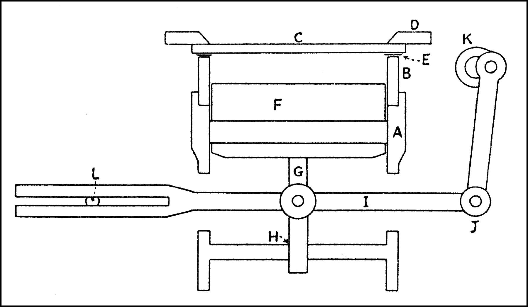
The Wilson cloud chamber offered Blumgart the advantages of automatic detection, speed, and electric safety, and it could detect both β- and γ-radiation; but since it was sensitive for only a fraction of a second every 20 s, it was useless to him in its original form. Enter now the Japanese physicist Takeo Shimizu, who was working with Wilson at Rutherford’s Cavendish Laboratory. Shimizu was dissatisfied with the long recovery time and designed a reciprocating piston that made the chamber sensitive from 1 to 5 times every second (Fig. 2) (3). Shimizu’s improvement made it possible to observe events in the cloud chamber almost continuously.
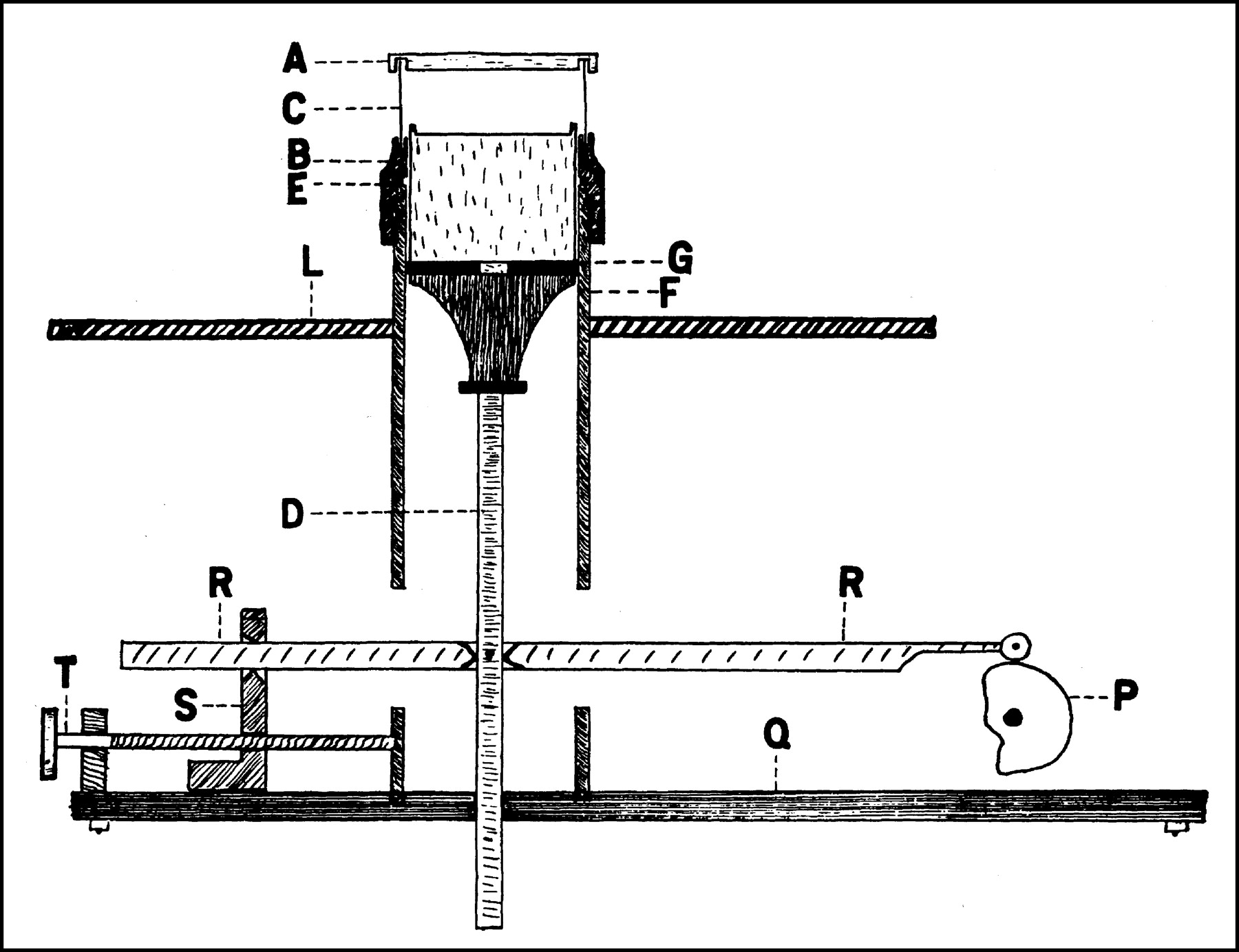
Blumgart and Yens modified Shimizu’s dynamic Wilson cloud chamber in at least 3 significant respects. In the first place, Shimizu’s reciprocating piston was driven in a sinusoidal pattern, so that the decompression within the chamber was never sudden, although it was adequate for his purposes. Blumgart and Yens replaced the rotating shaft with a highly eccentric cam (Fig. 3) that caused the piston to drop suddenly, causing a rapid decompression—thereby making the chamber a little more efficient while also providing a somewhat longer decompression (sensitive) phase.
Second, Shimizu placed a voltage of 200 V across the chamber during the compression phase, to dispel the tracks formed during the previous decompression. Blumgart and Yens—perhaps with electric safety in mind—reduced this potential to 50 V.
Finally, in the cloud chambers of Wilson and Shimizu it was necessary to place the radioactive source inside the chamber. Blumgart needed to use the cloud chamber as a detector and therefore replaced the glass cylinder (which was the chamber itself) with a celluloid one, which would allow both β- and γ-rays to enter the chamber from the outside.
I am stressing these points because this seems to be the first adaptation of an instrument for use in a diagnostic nuclear medicine application. Before this, electroscopes, Geiger counters, and cloud chambers had been used in various applications in physics, radiotherapy, and radiation safety, but as far as I can determine, this instrumentation—an integral part of the first human radiotracer procedure—was in itself a true first.
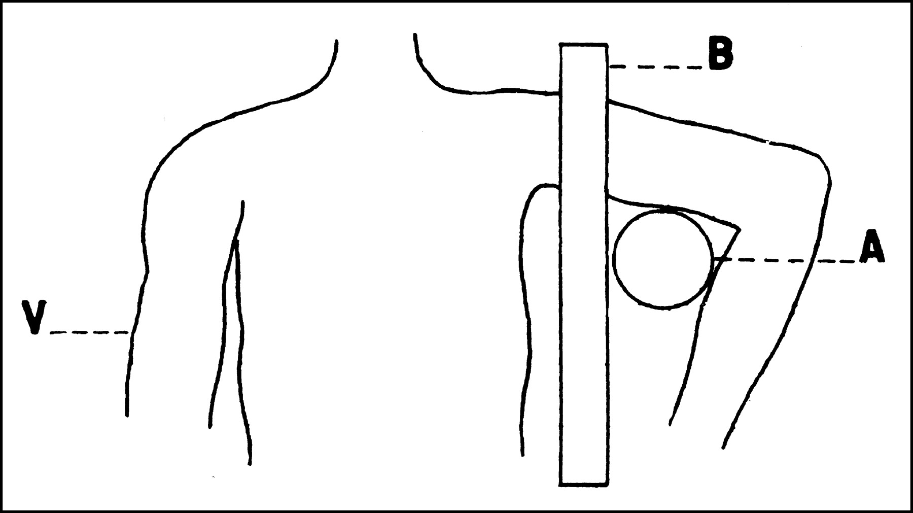
The inaugural subject was Blumgart himself (in February 1925). The radiotracer was injected into one arm (presumably by Yens), and the cloud chamber was placed beneath the other arm, shielded from the body by a lead slab (Fig. 4). Blumgart did not report his own circulation time, but the arm-to-arm circulation time was found to be 15–21 s in patients with a normal cardiovascular system and 50–65 s in patients with cardiac decompensation.
How fitting it is that the primal nuclear medicine procedure was a dynamic, functional one—requiring instrumentation specially modified for dynamic detection—a herald of the dynamic, functional field of nuclear medicine that was to come!
Herman Blumgart (Fig. 5) is rightly called the father of diagnostic nuclear medicine, in that he performed the first radiotracer test on a human (himself). His foresight in defining the requirements for a satisfactory indicator and a satisfactory detector show a highly perceptive, original mind. But he and his coworker, Otto C. Yens, should also be recognized as introducing the first device to be modified specifically for this application—the birth of nuclear medicine instrumentation.

Wilson cloud chamber. Sudden descent of piston supersaturates chamber, and water droplets condense on any ions present.

Shimizu modification. Rotating shaft K causes reciprocating motion in piston, providing repeated chamber decompression (1–5 cycles per second) allowing almost continuous observation. A = brass cylinder; B = glass cylinder (chamber itself); C = top glass plate for observation; D = brass retaining ring; E = copper ring with electric lead; F = brass piston; G = piston rod; H = base plate with hole for piston; I = piston-driving rod; J = coupling; K = motor-driven rotating shaft; L = adjustable fulcrum. (Reprinted with permission of (3).)

Blumgart–Yens modification. A = top glass plate for observation; B = threaded brass collar; C = celluloid cylinder (chamber itself); D = duralumin piston; E = rubber washer; F = brass cylinder; G = leather washer; L = shelf for armrest during experiments; P = eccentric cam; Q = steel bottom plate; R = piston-driving duralumin shaft; S = support and bearing for shaft; T = adjusting screw. (Reprinted from (1).)

Blumgart’s experimental setup. Injection is given to supine subject. A = cloud chamber detector; B = lead shield with opening for left arm; V = right arm, into which radioactive indicator is injected. (Reprinted from (1).)
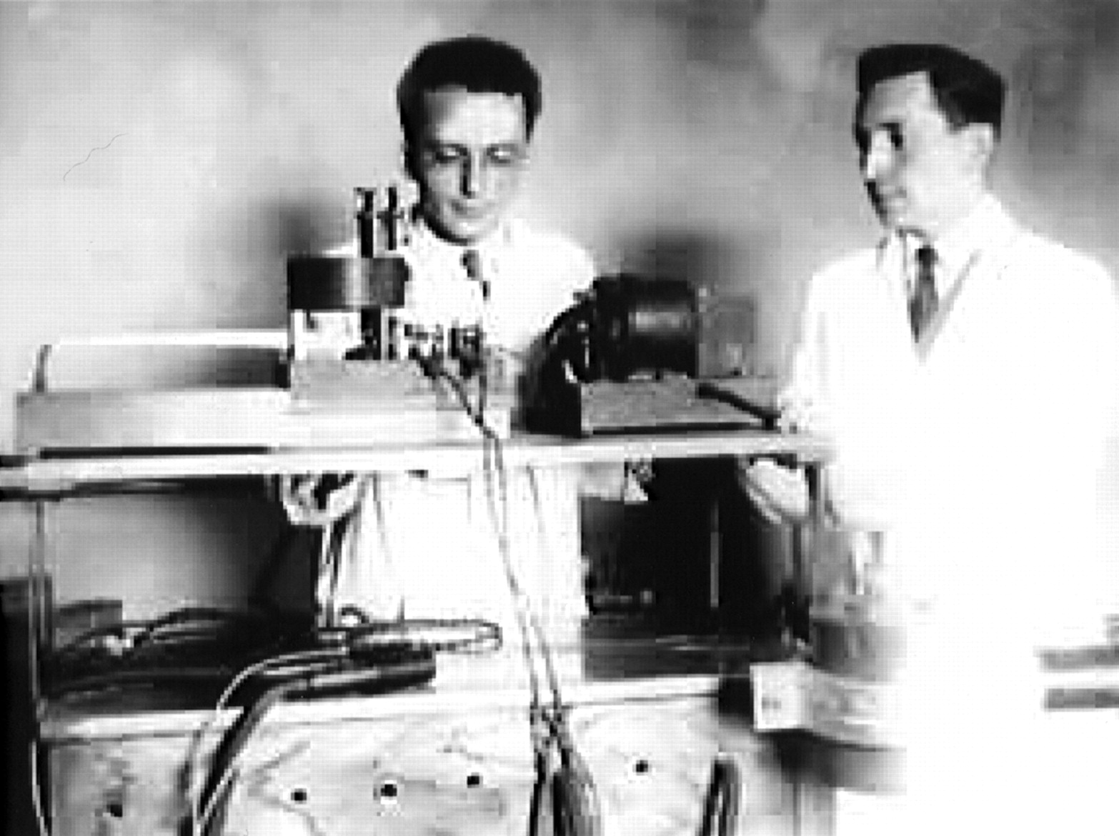
Possible prototype. Soma Weiss, MD (left), and Hermann Blumgart, MD (right), are pictured with apparatus that might be incomplete prototype of Blumgart–Yens detector. Role of Dr. Weiss is not documented. (Reprinted with permission of (4).)
Footnotes
Received Jan. 27, 2003; revision accepted April 2, 2003.
For correspondence or reprints contact: Dennis D. Patton, MD, Division of Nuclear Medicine, Department of Radiology, University Medical Center, 1501 N. Campbell Ave., Tucson, AZ 85724-5068.
E-mail: dpatton{at}radiology.arizona.edu
In this issue


Jump to section
Related Articles
Cited By...
- No citing articles found.