


Article Figures & Data
Figures
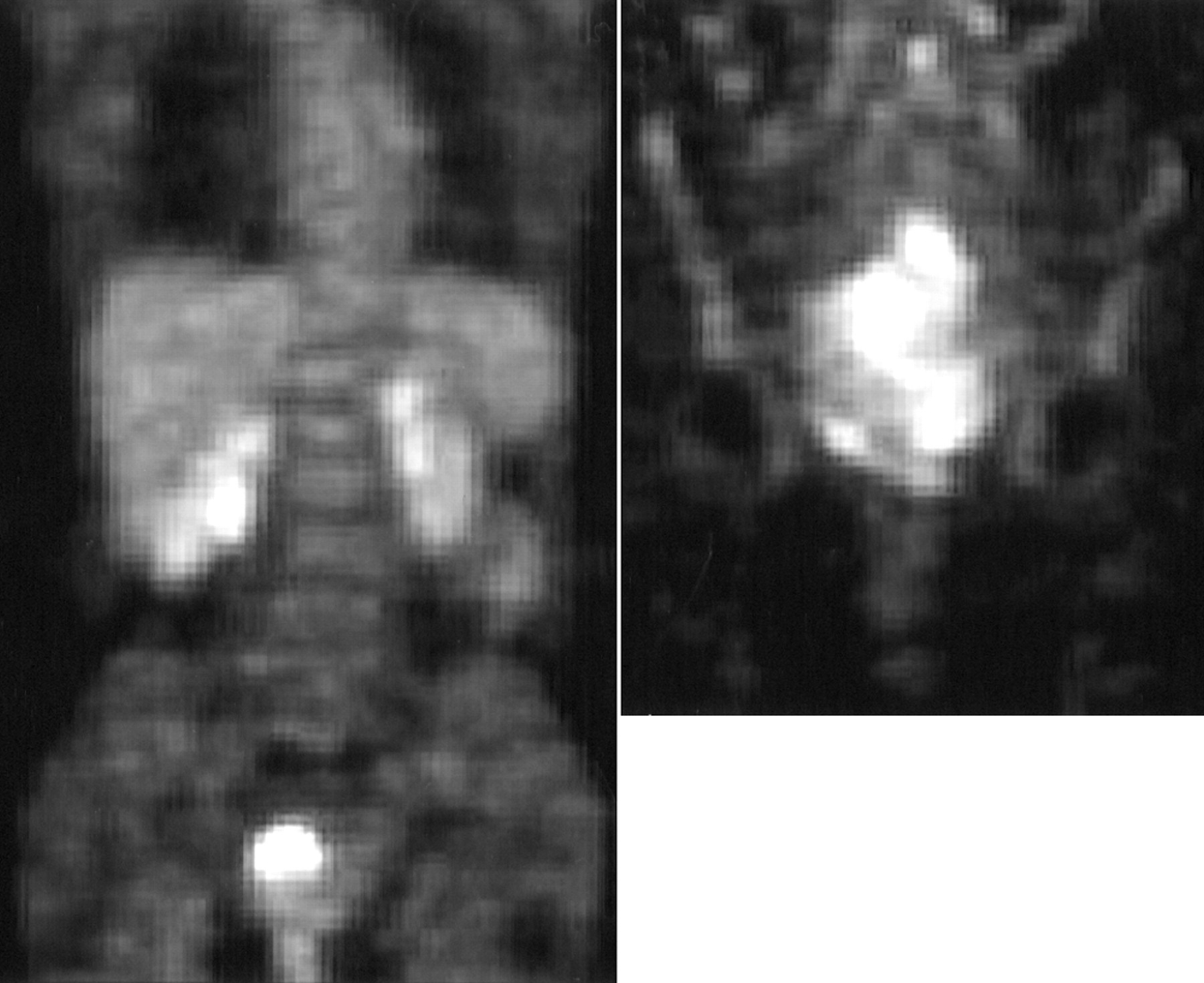
- FIGURE 1.
On left is coronal image of patient with small, spherical, and homogeneous primary tumor and no lymph node disease (score = 0), who was alive without disease at 703 d. On right is coronal image of patient with large, nonspherical, and markedly heterogeneous primary tumor (score = 2 + 1 + 2 + 2, or 7), who was dead of her disease at 149 d. Patient on right also had paraaortic lymph node disease (not shown).
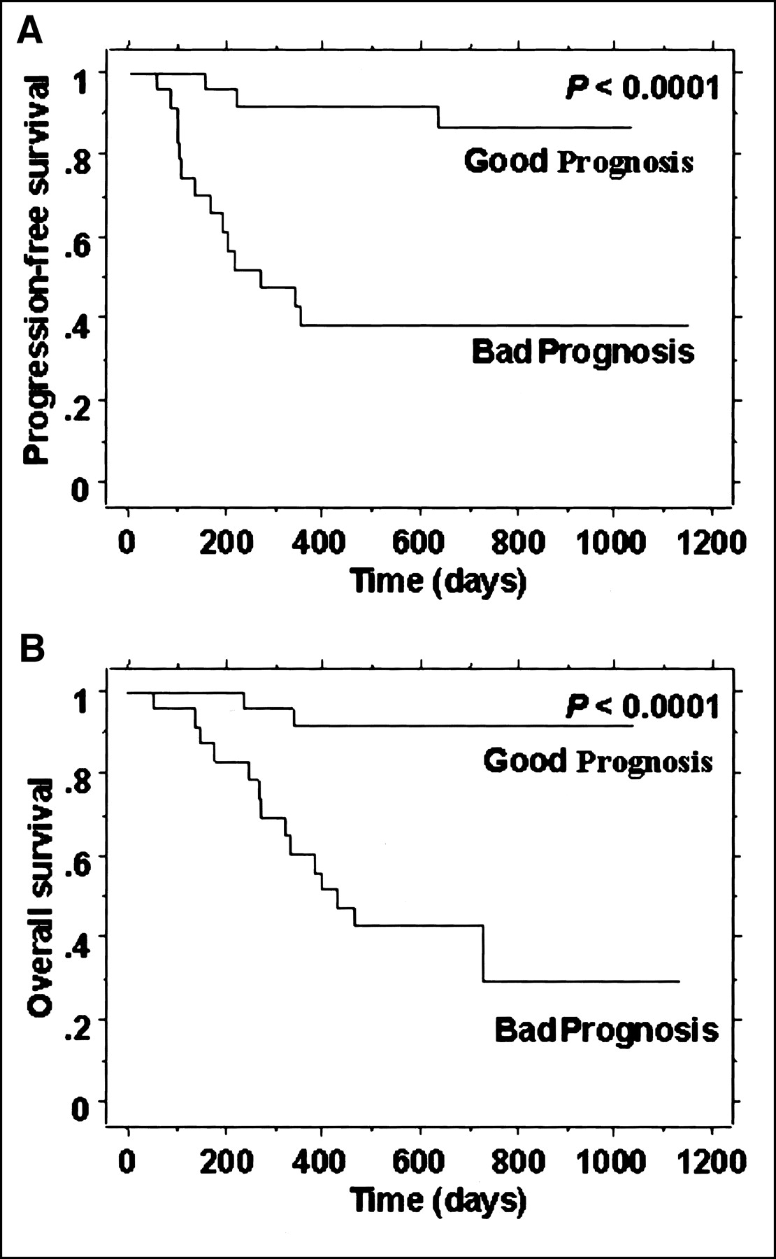
- FIGURE 2.
(A) PFS for scoring of observer 1. (B) OS for observer 1. Good Prognosis = patients with total score < 4; Bad Prognosis = patients with score ≥ 4.
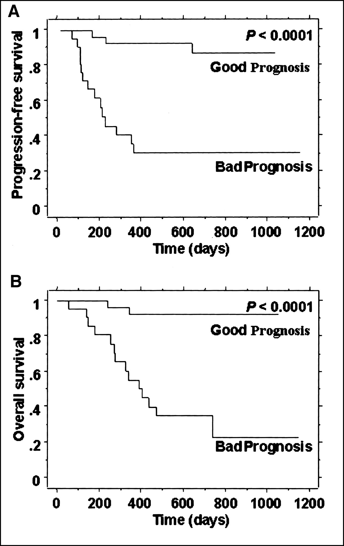
- FIGURE 3.
(A) PFS superimposed for the 3 observers. (B) OS for the 3 observers. Good Prognosis = patients with total score < 4; Bad Prognosis = patients with score ≥ 4.
- FIGURE 4.
(A) PFS when only lymph node status was considered. (B) OS when only lymph node status was considered. LN negative = no lymph node disease; LN positive = disease at any site.
- FIGURE 5.
(A) PFS for scoring of observer 1 when only visual characteristics of tumor were considered. (B) OS for visual characteristic scoring of observer 1. Good Prognosis = patients with total score < 3; Bad Prognosis = patients with score ≥ 3.


Tables
Patient no. Stage Histology Age (y) Status PFS (d) OS (d) 1 IIb Squamous 43 AWD 622 832 2 IIIb Squamous 45 DOD 96 322 3 IIb Squamous 61 DOD 151 342 4 IIb Adenosq 44 DOD 95 466 5 IIb Squamous 27 DOD 211 275 6 Ib2 Adenosq 56 DOD 337 397 7 IIIb Squamous 52 DOD 103 384 8 Ib2 Squamous 24 DOD 188 270 9 IVb Squamous 38 DOD 132 149 10 IIIb Squamous 40 DOD 80 138 11 IIb Squamous 67 DOD 196 252 12 IIIb Squamous 47 DOD 55 55 13 IIb Squamous 43 DOD 160 177 14 IIb Squamous 52 DOD 213 239 15 Ib2 Squamous 45 DOD 265 730 16 IIIb Squamous 34 DOD 99 334 17 IIIb Squamous 48 DOD 345 429 18 IIb Squamous 47 NED 1,017 1,017 19 Ib2 Squamous 28 NED 498 498 20 IIb Squamous 28 NED 615 615 21 IIIb Squamous 31 NED 616 616 22 IIIb Squamous 47 NED 1,131 1,131 23 IIb Squamous 54 NED 894 894 24 IIIb Adenosq 49 NED 704 704 25 IIb Squamous 58 NED 732 732 26 IIIb Squamous 27 NED 752 752 27 Ib1 Squamous 47 NED 961 961 28 IIb Squamous 55 NED 703 703 29 Ia1 Adeno 53 NED 968 968 30 IIb Squamous 47 NED 643 643 31 IIb Adeno 45 NED 883 1,040 32 IIb Squamous 83 NED 661 661 33 IIb Squamous 42 NED 571 571 34 Ib2 Squamous 47 NED 703 703 35 IIb Squamous 48 NED 1,015 1,015 36 IIIb Squamous 71 NED 596 596 37 Ib2 Squamous 29 NED 799 799 38 IIb Squamous 43 NED 826 826 39 IIIb Squamous 44 NED 761 761 40 IIb Squamous 56 NED 830 830 41 IIb Squamous 57 NED 730 730 42 IIb Squamous 54 NED 614 614 43 Ib2 Squamous 60 NED 902 902 44 IIb Squamous 87 NED 154 154 45 IIb Squamous 63 NED 622 622 46 Ib1 Squamous 71 NED 788 788 47 Ib2 Squamous 36 NED 382 382 AWD = alive with disease; DOD = dead of disease; Adenosq = adenosquamous; NED = no evidence of disease; Adeno = adenocarcinoma.
Cutoff PFS OS 3 4.8 9.0 4 5.3 9.5 5 3.9 5.4 6 3.6 4.0 - TABLE 4
Comparison of Numbers of Patients Scored as Having Bad Prognosis and Good Prognosis by the 3 Pairs of Observers
Observer pair Bad prognosis Good prognosis 2/1 Bad prognosis 20 1 Good prognosis 1 25 3/1 Bad prognosis 17 4 Good prognosis 0 26 3/2 Bad prognosis 17 4 Good prognosis 0 26
In this issue


Jump to section
Related Articles
Cited By...
- Comprehensive analysis of lung cancer pathology images to discover tumor shape features that predict survival outcome
- 18F-FDG PET Uptake Characterization Through Texture Analysis: Investigating the Complementary Nature of Heterogeneity and Functional Tumor Volume in a Multi-Cancer Site Patient Cohort
- Relationship Between 18F-FDG Accumulation and Lactate Dehydrogenase A Expression in Lung Adenocarcinomas
- Visual Versus Quantitative Assessment of Intratumor 18F-FDG PET Uptake Heterogeneity: Prognostic Value in Non-Small Cell Lung Cancer
- Intratumoral Metabolic Heterogeneity of Cervical Cancer
- Selection of Response Criteria for Clinical Trials of Sarcoma Treatment
- PET in Cervical Cancer -- Implications for `Staging,' Treatment Planning, Assessment of Prognosis, and Prediction of Response
- Tumor 18F-FDG Incorporation Is Enhanced by Attenuation of P53 Function in Breast Cancer Cells In Vitro
- Expanding Role of Positron Emission Tomography in Cancer of the Uterine Cervix
- Microvessel Density: Correlation with 18F-FDG Uptake and Prognostic Impact in Lung Adenocarcinomas
- In Vitro Proton Magnetic Resonance Spectroscopic Lactate and Choline Measurements, 18F-FDG Uptake, and Prognosis in Patients with Lung Adenocarcinoma
- Quantification of 18F-FDG Uptake in Non-Small Cell Lung Cancer: A Feasible Prognostic Marker?