
Main menu

  • Home
  • Content
    • Current
    • Ahead of print
    • Past Issues
    • JNM Supplement
    • SNMMI Annual Meeting Abstracts
    • Continuing Education
    • JNM Podcasts
  • Subscriptions
    • Subscribers
    • Institutional and Non-member
    • Rates
    • Journal Claims
    • Corporate & Special Sales
  • Authors
    • Submit to JNM
    • Information for Authors
    • Assignment of Copyright
    • AQARA requirements
  • Info
    • Reviewers
    • Permissions
    • Advertisers
  • About
    • About Us
    • Editorial Board
    • Contact Information
  • More
    • Alerts
    • Feedback
    • Help
    • SNMMI Journals
  • SNMMI
    • JNM
    • JNMT
    • SNMMI Journals
    • SNMMI

User menu

  • Subscribe
  • My alerts
  • Log in
  • My Cart

Search

  • Advanced search
Journal of Nuclear Medicine
  • SNMMI
    • JNM
    • JNMT
    • SNMMI Journals
    • SNMMI
  • Subscribe
  • My alerts
  • Log in
  • My Cart
Journal of Nuclear Medicine

Advanced Search

  • Home
  • Content
    • Current
    • Ahead of print
    • Past Issues
    • JNM Supplement
    • SNMMI Annual Meeting Abstracts
    • Continuing Education
    • JNM Podcasts
  • Subscriptions
    • Subscribers
    • Institutional and Non-member
    • Rates
    • Journal Claims
    • Corporate & Special Sales
  • Authors
    • Submit to JNM
    • Information for Authors
    • Assignment of Copyright
    • AQARA requirements
  • Info
    • Reviewers
    • Permissions
    • Advertisers
  • About
    • About Us
    • Editorial Board
    • Contact Information
  • More
    • Alerts
    • Feedback
    • Help
    • SNMMI Journals
  • View or Listen to JNM Podcast
  • Visit JNM on Facebook
  • Join JNM on LinkedIn
  • Follow JNM on Twitter
  • Subscribe to our RSS feeds
OtherClinical Investigations

Confirmation of the Early Prognostic Value of Bone Scanning and Pinhole Imaging of the Hip in Legg-Calvé-Perthes Disease

Frédéric Comte, Vicenzo De Rosa, Hakim Zekri, Marie Claude Eberlé, Alain Dimeglio, Michel Rossi and Denis Mariano-Goulart
Journal of Nuclear Medicine November 2003, 44 (11) 1761-1766;
Frédéric Comte
  • Find this author on Google Scholar
  • Find this author on PubMed
  • Search for this author on this site
Vicenzo De Rosa
  • Find this author on Google Scholar
  • Find this author on PubMed
  • Search for this author on this site
Hakim Zekri
  • Find this author on Google Scholar
  • Find this author on PubMed
  • Search for this author on this site
Marie Claude Eberlé
  • Find this author on Google Scholar
  • Find this author on PubMed
  • Search for this author on this site
Alain Dimeglio
  • Find this author on Google Scholar
  • Find this author on PubMed
  • Search for this author on this site
Michel Rossi
  • Find this author on Google Scholar
  • Find this author on PubMed
  • Search for this author on this site
Denis Mariano-Goulart
  • Find this author on Google Scholar
  • Find this author on PubMed
  • Search for this author on this site
  • Article
  • Figures & Data
  • Info & Metrics
  • PDF
Loading

Article Figures & Data

Figures

  • Tables
  • FIGURE 1.
    • Download figure
    • Open in new tab
    • Download powerpoint
    FIGURE 1.

    Scintigraphic stages of Conway classification of LCP. Stage I (total avascularity of proximal femoral epiphysis) is observed at initial stage of disease. Appearance of lateral column formation (a) characterizes A pathway (stages IIA and IIIA). In B pathway, as neovascularization progresses, extension of activity from metaphysis is observed (stages IIB and IIIB), without lateral column formation (b).

  • FIGURE 2.
    • Download figure
    • Open in new tab
    • Download powerpoint
    FIGURE 2.

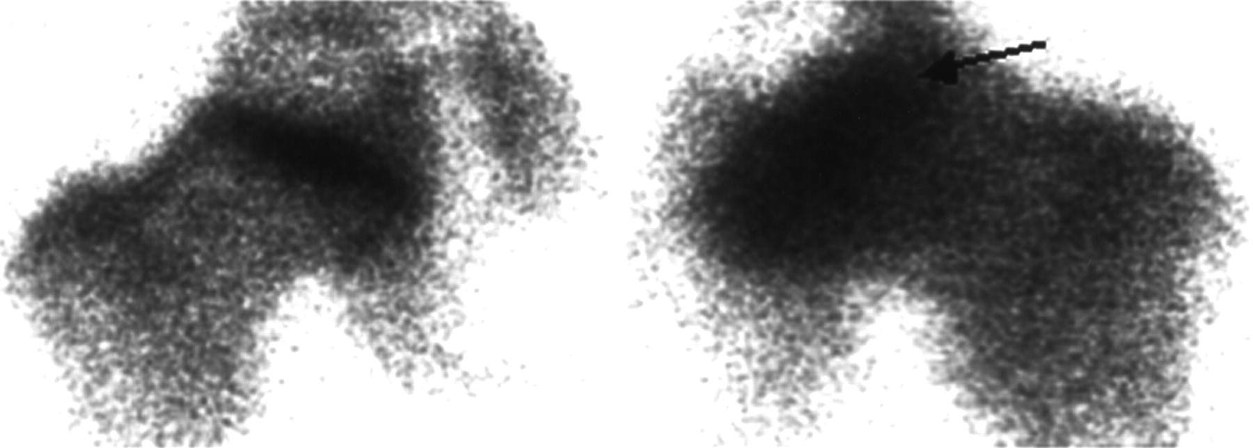
    Hyperactivity (arrow) of left metaphyseal growth plates compared with other hip, which appears normal.

Tables

  • Figures
    • View popup
    TABLE 1

    Scintigraphic Stages of the Conway Classification

    StagePathway APathway B
    INo radioactivityNo radioactivity
    IIAppearance of lateral column formationAppearance of activity from base of epiphysis
    IIIExtension of lateral column formation activityExtension of activity to capital femoral epiphysis
    IVExtension of lateral column formation activity to whole epiphysisExtension of activity to whole epiphysis
    • View popup
    TABLE 2

    MRI and Arthrography Results for Patients in Catterall Stage III or IV

    Patient no.SexAge at diagnosis (y)Time from initial symptoms to staging (mo)StagePresence of severity signs at
    MRIArthrography
    1M6.26IIIYes—*
    2F5.96IIIYes—
    3M8.78IIIYes—
    4M7.68IIIYesYes
    5M7.08IIIYesYes
    6M9.15IIIYesYes
    7M9.56IIIYesYes
    8M6.67IIIYesYes
    9M5.57IIIYesYes
    10M5.94IIINo—
    11M6.45IIINo—
    12M4.15IIINo—
    13F6.87IIINo—
    14M8.47IIINo—
    15M7.56IIINo—
    16M6.28IIINo—
    17M3.48IIINo—
    18M6.47IVYes—
    19M8.46IVYes—
    20M5.17IVYes—
    21M8.97IVYes—
    22M5.67IVYes—
    23M4.26IVYes—
    24M4.28IVYes—
    25F10.77IVYes—
    26M7.46IVYes—
    27M3.87IV—Yes
    28M7.39IVYesYes
    29M3.58IVYesYes
    30M7.76IVYesYes
    31M6.08IVYesYes
    32M4.18IVYesYes
    33M4.58IVYesYes
    34M4.18IV—Yes
    35M6.45IVYesYes
    36M4.510IVYesYes
    37M6.08IVYesYes
    38M6.07IVYesYes
    39M7.27IVYesYes
    40M7.26IVYesYes
    41F9.99IVYesYes
    42M4.05IVYesYes
    43F4.47IVYesYes
    44M8.17IVYesYes
    • ↵* Not determined.

    • View popup
    TABLE 3

    Number of Patients with Good or Poor Final Outcome Depending on Pathway

    Disease severityPathway APathway B
    Good final outcome231
    Poor final outcome431
PreviousNext
Back to top

In this issue

Journal of Nuclear Medicine
Vol. 44, Issue 11
November 1, 2003
  • Table of Contents
  • Index by author
Print
Download PDF
Article Alerts
Sign In to Email Alerts with your Email Address
Email Article

Thank you for your interest in spreading the word on Journal of Nuclear Medicine.

NOTE: We only request your email address so that the person you are recommending the page to knows that you wanted them to see it, and that it is not junk mail. We do not capture any email address.

Enter multiple addresses on separate lines or separate them with commas.
Confirmation of the Early Prognostic Value of Bone Scanning and Pinhole Imaging of the Hip in Legg-Calvé-Perthes Disease
(Your Name) has sent you a message from Journal of Nuclear Medicine
(Your Name) thought you would like to see the Journal of Nuclear Medicine web site.
Citation Tools
Confirmation of the Early Prognostic Value of Bone Scanning and Pinhole Imaging of the Hip in Legg-Calvé-Perthes Disease
Frédéric Comte, Vicenzo De Rosa, Hakim Zekri, Marie Claude Eberlé, Alain Dimeglio, Michel Rossi, Denis Mariano-Goulart
Journal of Nuclear Medicine Nov 2003, 44 (11) 1761-1766;

Citation Manager Formats

  • BibTeX
  • Bookends
  • EasyBib
  • EndNote (tagged)
  • EndNote 8 (xml)
  • Medlars
  • Mendeley
  • Papers
  • RefWorks Tagged
  • Ref Manager
  • RIS
  • Zotero
Share
Confirmation of the Early Prognostic Value of Bone Scanning and Pinhole Imaging of the Hip in Legg-Calvé-Perthes Disease
Frédéric Comte, Vicenzo De Rosa, Hakim Zekri, Marie Claude Eberlé, Alain Dimeglio, Michel Rossi, Denis Mariano-Goulart
Journal of Nuclear Medicine Nov 2003, 44 (11) 1761-1766;
Twitter logo Facebook logo LinkedIn logo Mendeley logo
  • Tweet Widget
  • Facebook Like
  • Google Plus One
Bookmark this article

Jump to section

  • Article
    • Abstract
    • MATERIALS AND METHODS
    • RESULTS
    • DISCUSSION
    • CONCLUSION
    • Acknowledgments
    • Footnotes
    • REFERENCES
  • Figures & Data
  • Info & Metrics
  • PDF

Related Articles

  • No related articles found.
  • PubMed
  • Google Scholar

Cited By...

  • Perthes' disease: PROGNOSIS IN CHILDREN UNDER SIX YEARS OF AGE
  • Google Scholar

More in this TOC Section

  • Feasibility of Ultra-Low-Activity 18F-FDG PET/CT Imaging Using a Long–Axial-Field-of-View PET/CT System
  • Cardiac Presynaptic Sympathetic Nervous Function Evaluated by Cardiac PET in Patients with Chronotropic Incompetence Without Heart Failure
  • Validation and Evaluation of a Vendor-Provided Head Motion Correction Algorithm on the uMI Panorama PET/CT System
Show more Clinical Investigations

Similar Articles

SNMMI

© 2025 SNMMI

Powered by HighWire