


Article Figures & Data
Figures
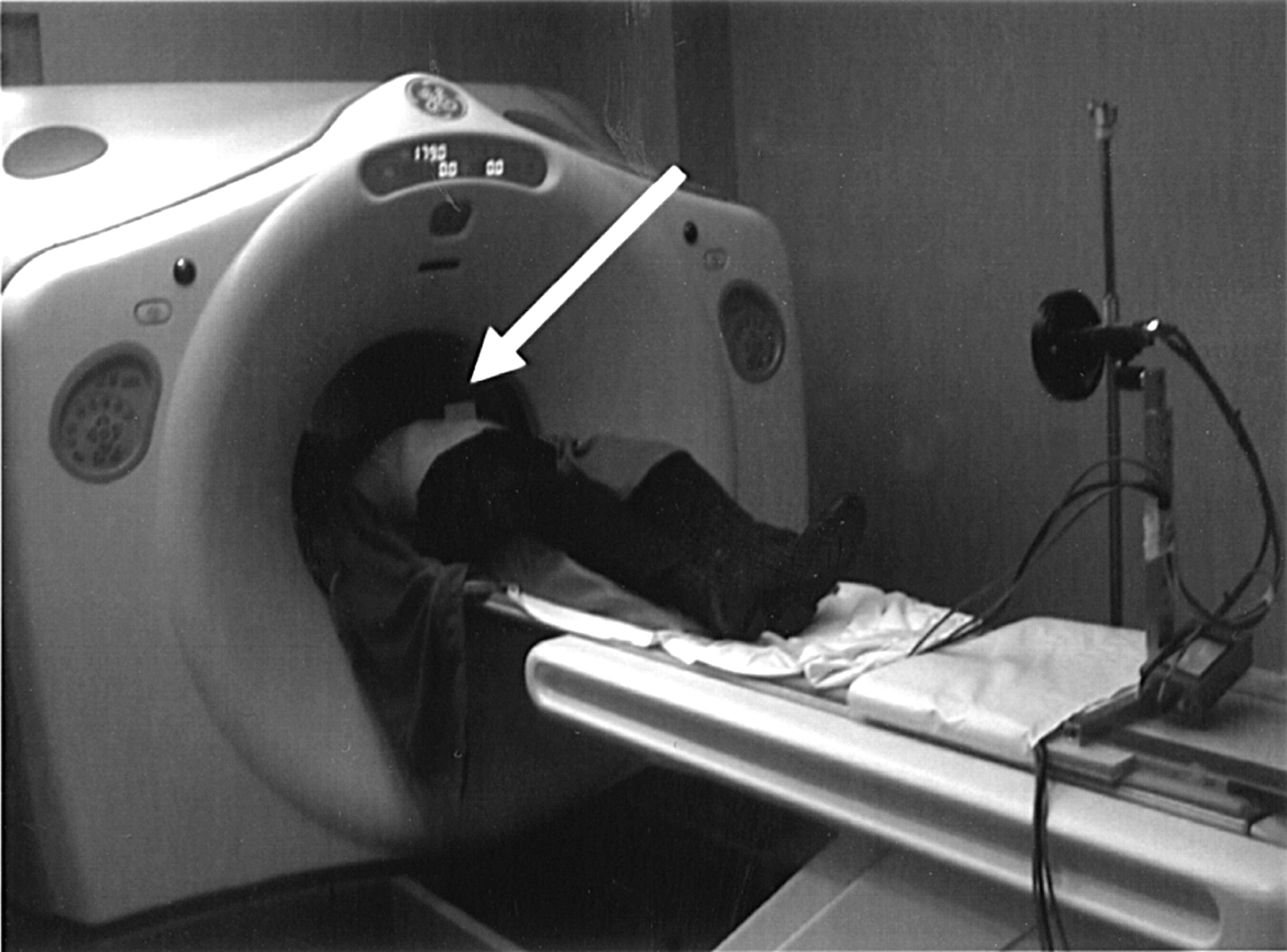
- FIGURE 1.
Patient setup in RCDPET acquisition mode. Point source is at end of low-density rod, extending into lesion FOV, and rigidly attached to Styrofoam (The Dow Chemical Co.) block positioned on abdomen of patient.
- FIGURE 2.
Patient setup in RGPET acquisition mode. Plastic block (arrow) with 2 infrared passive reflectors is positioned on abdomen of patient. Infrared camera, positioned on PET table, is used to trace motion of reflectors and, thus, patient breathing motion.
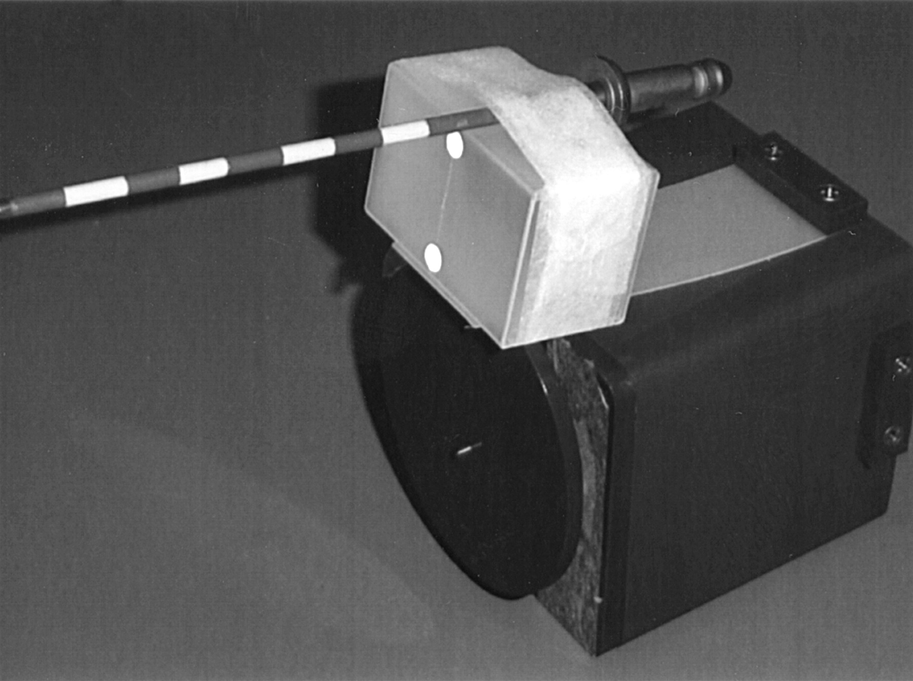
- FIGURE 3.
68Ge rod source is mounted to Varian breathing phantom, pivoted at phantom side, and other end is free to oscillate vertically. Rod source has activity of 9.25 MBq and outer diameter of 4.7 mm.
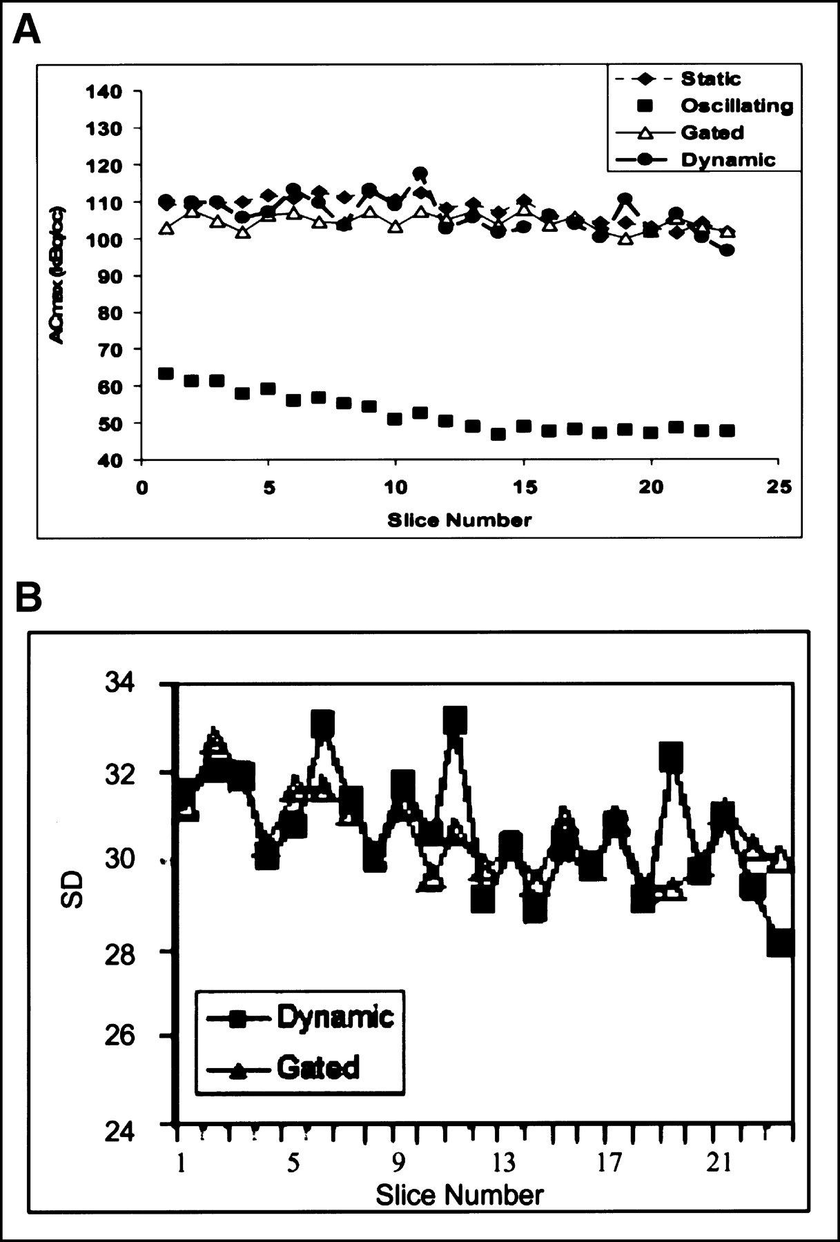
- FIGURE 4.
(A) Act_Concmax versus slice number (amplitude). RCDPET technique shows capability comparable to that of RGPET technique in recovering correct Act_Concmax, which is distorted because of respiratory motion. (B) SD of activity concentration within multiple transaxial slices of lesion (thus motion amplitudes), measured in both RCDPET and RGPET techniques, shows comparable noise level. ACmax = Act_Concmax.
- FIGURE 5.
Position of point source as it moves in and out of user-selected reference position (e.g., as defined by the 2 lines according to position of point source in first frame) in consecutive 1-s time frames. Position of point source defines fourth dimension (time) of lesion coordinates.
- FIGURE 6.
Transaxial view of lesion (arrow) in RGPET (A) and RCDPET (B).


In this issue


Jump to section
Related Articles
Cited By...
- Data-Driven Motion Correction in Clinical PET: A Joint Accomplishment of Creative Academia and Industry
- Measurement of Regional Specific Lung Volume Change Using Respiratory-Gated PET of Inhaled 13N-Nitrogen
- Deep-Inspiration Breath-Hold PET/CT of Lung Cancer: Maximum Standardized Uptake Value Analysis of 108 Patients
- The role of PET/CT scanning in radiotherapy planning
- Influence of Reconstruction Iterations on 18F-FDG PET/CT Standardized Uptake Values