


Article Figures & Data
Figures
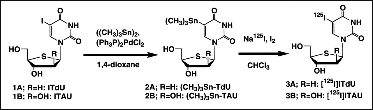
- FIGURE 1.
Synthetic pathway for preparation of radiolabeled ITdU and ITAU by destannylation.
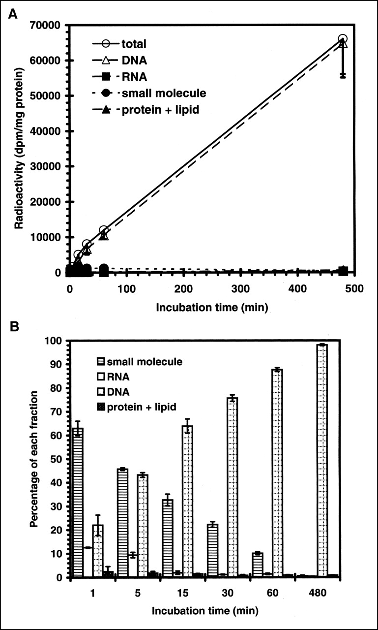
- FIGURE 2.
Time-dependent incorporation of radioactivity into Lewis lung carcinoma cells. (A) Radioactivity of acid-soluble small molecule, DNA, RNA, and protein fractions is shown. (B) Time-dependent percentage of radioactivity distribution in each fraction of Lewis lung carcinoma. Data are expressed as mean ± SD for 3 experiments.
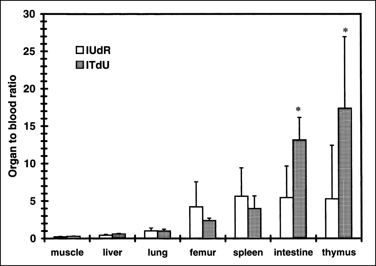
- FIGURE 3.
Uptake of iodonucleosides, expressed as organ-to-blood ratio of radioactivity. Animals were killed at 18 h after injection. Data are expressed as mean ± SD for 3 experiments. Iodonucleosides showed higher uptake in proliferation organs (femur, spleen, intestine, and thymus) than in nonproliferating organs (muscle, liver, and lung). Statistical significance between 125I-IUdR uptake and 125I-ITdU uptake was observed in intestine and thymus (P < 0.05; Student t test).


Tables
Nucleoside Tracer uptake* (relative uptake ratio) LL/2 L-M L-M (TK−) IUdR 539.7 ± 81.7 77.8 ± 7.4† 27.9 ± 2.9 (6.9) (2.8) ITdU 226.7 ± 13.1 10.9 ± 1.5† 3.9 ± 0.6 (20.8) (2.8) ITAU 1.1 ± 0.0 1.7 ± 0.3‡ 1.2 ± 0.2 (0.6) (1.4) ↵* Tracer uptake = pmol/mg protein/h.
↵† P < 0.0005 compared with L-M (TK−).
↵‡ P < 0.05 compared with L-M (TK−) (Student t test).
Data in parentheses in LL/2 row and L-M row indicate relative uptake ratios of LL/2 compared with those of L-M cells and relative uptake ratios of L-M compared with those of L-M (TK−), respectively.
Organ 1 h 1.5 h 2 h 8 h 18 h Blood 7.6 ± 0.8 5.4 ± 1.2 5.3 ± 0.5 1.1 ± 0.1 0.8 ± 1.1 Spleen 4.7 ± 0.6 4.6 ± 1.2 4.7 ± 2.2 3.1 ± 1.5 1.7 ± 0.9 Intestine 5.0 ± 0.2 3.3 ± 0.7 4.5 ± 0.5 2.4 ± 0.2 1.4 ± 0.4 Thymus 6.6 ± 1.1 4.4 ± 1.6 4.3 ± 1.0 1.8 ± 0.3 1.8 ± 1.5 Femur 4.5 ± 0.5 3.6 ± 0.8 3.7 ± 0.5 1.6 ± 0.1 1.1 ± 0.4 Thyroid 113.0 ± 30.8 80.7 ± 51.9 112.1 ± 22.3 32.9 ± 13.5 9.7 ± 8.0 Lung 5.1 ± 0.4 3.6 ± 0.8 3.6 ± 0.3 0.9 ± 0.4 0.6 ± 0.7 Heart 2.8 ± 0.5 2.1 ± 0.5 1.9 ± 0.1 0.4 ± 0.0 0.3 ± 0.3 Muscle 1.4 ± 0.2 1.5 ± 0.3 1.4 ± 0.5 0.3 ± 0.1 0.1 ± 0.2 Liver 2.7 ± 0.4 2.1 ± 0.5 1.9 ± 0.2 0.4 ± 0.1 0.3 ± 0.3 Brain 0.3 ± 0.0 0.2 ± 0.1 0.2 ± 0.0 0.0 ± 0.0 0.0 ± 0.0 Kidney 5.9 ± 0.4 4.8 ± 1.5 3.4 ± 0.3 0.7 ± 0.1 0.6 ± 0.8 Data are expressed as percentage injected dose per gram (mean ± SD).
Organ 1 h 1.5 h 2 h 8 h 18 h Blood 2.5 ± 0.3 2.6 ± 0.6 1.6 ± 0.7 0.7 ± 0.2 0.1 ± 0.0 Spleen 1.1 ± 0.1 1.7 ± 0.3 1.1 ± 0.3 0.5 ± 0.1 0.3 ± 0.0 Intestine 1.9 ± 0.1 1.9 ± 0.5 1.7 ± 0.4 1.7 ± 0.5 0.9 ± 0.1 Thymus 1.8 ± 0.1 1.8 ± 0.4 1.4 ± 0.7 2.2 ± 1.3 1.1 ± 0.4 Femur 1.0 ± 0.1 1.0 ± 0.2 0.9 ± 0.2 0.7 ± 0.1 0.2 ± 0.0 Thyroid 23.0 ± 6.4 23.1 ± 12.5 16.3 ± 9.7 16.0 ± 2.7 1.1 ± 0.3 Lung 1.4 ± 0.1 1.5 ± 0.3 1.1 ± 0.5 0.5 ± 0.1 0.1 ± 0.0 Heart 1.0 ± 0.1 1.3 ± 0.3 0.8 ± 0.4 0.2 ± 0.1 0.0 ± 0.0 Muscle 0.5 ± 0.2 0.7 ± 0.2 0.5 ± 0.2 0.1 ± 0.1 0.0 ± 0.0 Liver 1.1 ± 0.2 1.5 ± 0.4 0.9 ± 0.5 0.2 ± 0.1 0.0 ± 0.0 Brain 0.1 ± 0.0 0.2 ± 0.1 0.1 ± 0.0 0.0 ± 0.0 0.0 ± 0.0 Kidney 9.5 ± 3.5 5.8 ± 4.1 1.9 ± 1.7 0.5 ± 0.1 0.1 ± 0.0 Data are expressed as percentage injected dose per gram (mean ± SD).
Organ 1 h 1.5 h 2 h 8 h 18 h Blood 3.7 ± 0.4 4.5 ± 0.3 3.7 ± 0.8 0.4 ± 0.1 0.1 ± 0.1 Spleen 1.6 ± 0.1 2.3 ± 0.4 2.1 ± 0.5 0.2 ± 0.1 0.1 ± 0.0 Intestine 1.8 ± 0.2 2.4 ± 0.4 2.1 ± 0.6 0.4 ± 0.1 0.1 ± 0.1 Thymus 2.1 ± 0.5 3.0 ± 1.0 2.3 ± 0.6 0.2 ± 0.2 0.1 ± 0.0 Femur 1.4 ± 0.1 1.6 ± 0.1 1.4 ± 0.3 0.2 ± 0.1 0.1 ± 0.0 Thyroid 46.1 ± 5.2 57.2 ± 10.5 60.3 ± 27.6 18.6 ± 13.0 4.1 ± 2.2 Lung 2.3 ± 0.2 2.7 ± 0.3 2.4 ± 0.6 0.3 ± 0.1 0.1 ± 0.1 Heart 1.5 ± 0.3 2.1 ± 0.1 1.6 ± 0.3 0.2 ± 0.0 0.0 ± 0.0 Muscle 0.9 ± 0.1 1.3 ± 0.1 1.0 ± 0.2 0.1 ± 0.0 0.0 ± 0.0 Liver 1.4 ± 0.2 2.0 ± 0.2 1.4 ± 0.4 0.1 ± 0.0 0.0 ± 0.0 Brain 0.4 ± 0.0 0.5 ± 0.1 0.3 ± 0.1 0.0 ± 0.0 0.0 ± 0.0 Kidney 7.2 ± 2.3 5.0 ± 0.3 3.6 ± 0.6 0.3 ± 0.1 0.1 ± 0.1 Data are expressed as percentage injected dose per gram (mean ± SD).
Nucleoside 1 h 1.5 h 2 h 8 h 18 h IUdR 18.6 ± 6.7 21.6 ± 6.6 23.4 ± 4.5 48.4 ± 5.7 54.7 ± 9.1 ITdU 57.5 ± 4.7 53.6 ± 6.1 56.5 ± 7.7 73.1 ± 6.1 71.4 ± 14.6 ITAU 50.2 ± 5.4 40.0 ± 7.6 43.2 ± 6.8 70.6 ± 3.3 64.8 ± 5.5 Data are expressed as percentage injected dose (mean ± SD).
In this issue


Jump to section
Related Articles
Cited By...
- Breaking the Invulnerability of Cancer Stem Cells: Two-Step Strategy to Kill the Stem-like Cell Subpopulation of Multiple Myeloma
- Evaluation of 4'-[Methyl-11C]Thiothymidine in a Rodent Tumor and Inflammation Model
- Synthesis and Biologic Study of IV-14, a New Ribonucleoside Radiotracer for Tumor Visualization
- Preferential Tumor Targeting and Selective Tumor Cell Cytotoxicity of 5-[131/125I]Iodo-4'-Thio-2'-Deoxyuridine
- 123I-ITdU-Mediated Nanoirradiation of DNA Efficiently Induces Cell Kill in HL60 Leukemia Cells and in Doxorubicin-, {beta}-, or {gamma}-Radiation-Resistant Cell Lines
- Evaluation of 4'-[Methyl-14C]Thiothymidine for In Vivo DNA Synthesis Imaging
- Pharmacokinetics and Metabolism of 5-125I-Iodo-4'-Thio-2'-Deoxyuridine in Rodents