


Article Figures & Data
Figures
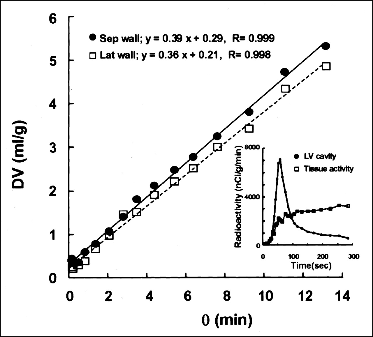
- FIGURE 1.
Graphic plotting method (Patlak plot) was applied for calculation of MBF. Representative Patlak plot shows excellent linear regression for total frame time of 5 min. Insert shows time-activity curves for LV cavity and myocardial tissue obtained from same dynamic 13NH3 PET data. θ = normalized time; DV = volume of distribution; Sep = septal; Lat = lateral.
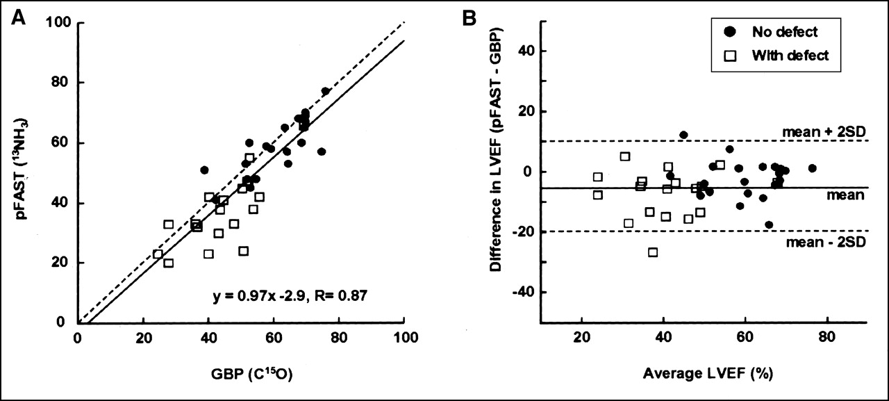
- FIGURE 2.
Representative images of MBF of healthy volunteer (NV) and of patient with OMI (with defect) in anterior wall. Images of MBF were calculated pixel by pixel on basis of graphic plotting method. Slopes of linear fit using time frames of 30–150 s in plots (Fig. 1) were converted into MBF values. Images were resliced into LV short-axis and long-axis planes.
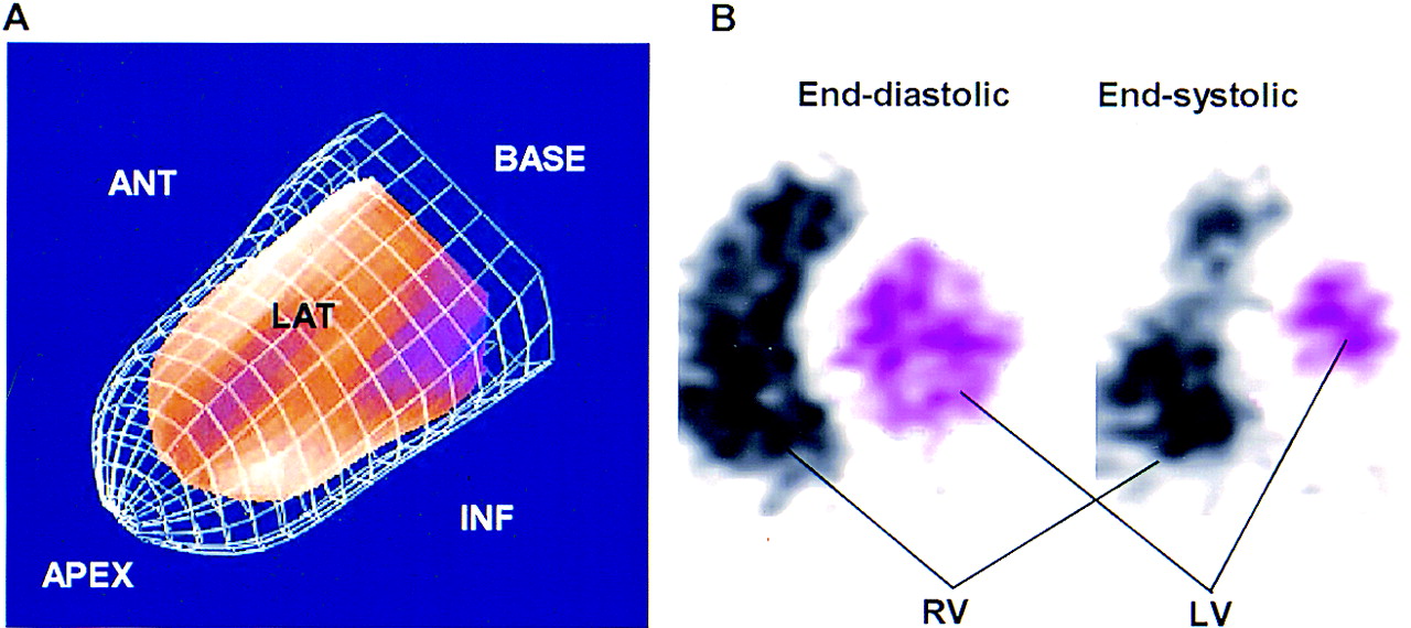
- FIGURE 3.
Images of gated 13NH3 PET in 3D mode of pFAST (A) and of GBP PET in 2D mode (B). Ratio of volumes in end-systolic phase (red part) and end-diastolic phase (meshed frame) provides LVEF. In GBP imaging, right and left ventricles (RV and LV) are clearly separated by septal myocardial wall. Bitmap images (pink transparent areas) were generated on each short-axis slice in end-diastolic and end-systolic phases of GBP PET. ANT = anterior; LAT = lateral; INF = inferior.
- FIGURE 4.
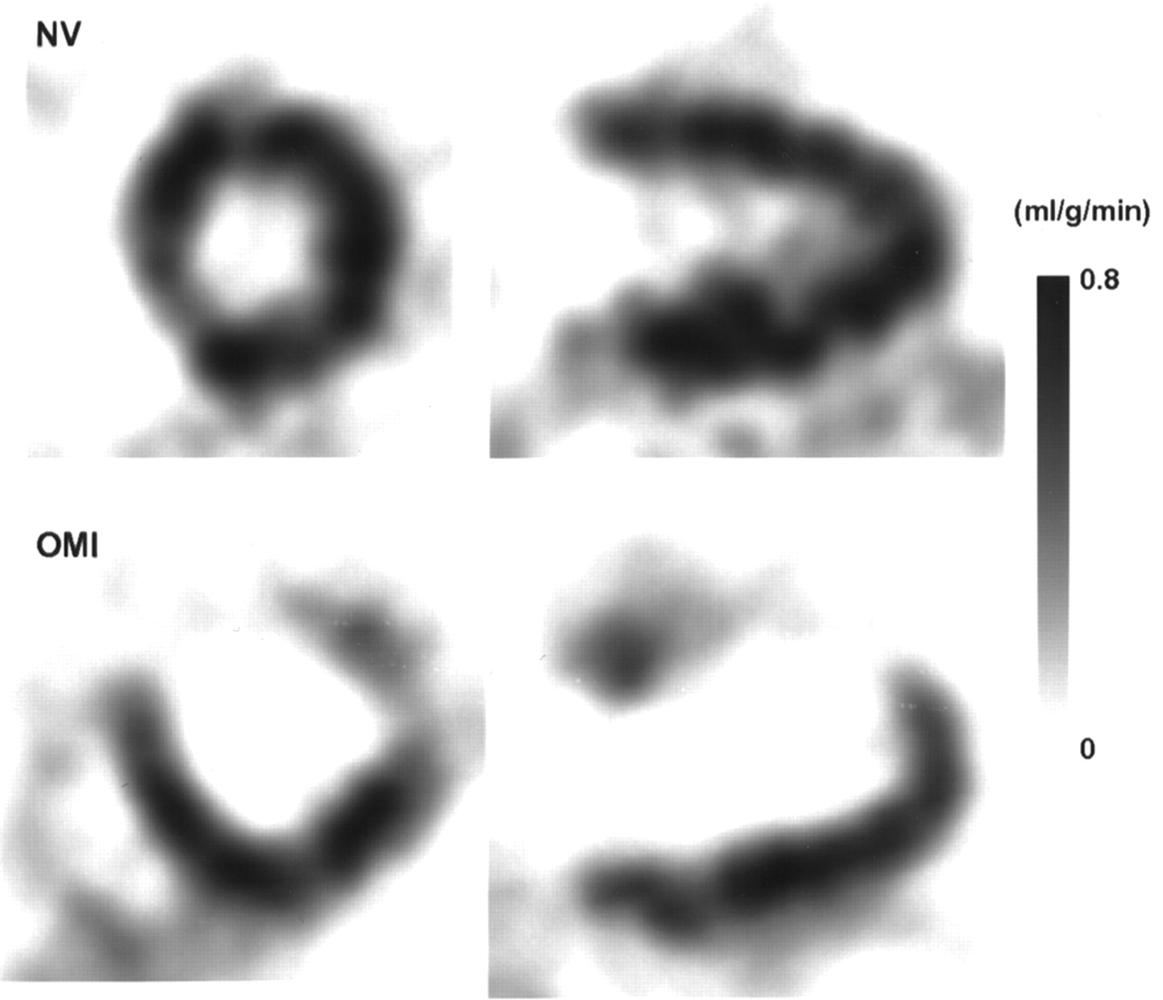
(A) Correlation of LVEF obtained from LVG and GBP PET (C15O) in 20 patients who underwent both studies. LVG and GBP PET show excellent linear correlation regardless of whether defect was (□) or was not (•) on perfusion image. Dashed line is line of identity. (B) Bland-Altman plot shows no significant degree of systematic measurement bias between 2 methods. Lines indicate mean and mean ± 2 SD.
- FIGURE 5.
(A) Correlation of LVEF obtained from GBP PET (C15O) and pFAST (13NH3 PET) in all subjects (n = 40). LVEF values from GBP and pFAST correlate well, although there was slight underestimation of LVEF with pFAST, and stronger tendency for underestimation was observed in patients with defect (□) compared with patients without defect (•). Dashed line is line of identity. (B) Bland-Altman plot also shows underestimation of LVEF by pFAST compared with GBP by −4.58% ± 7.49%. Lines indicate mean and mean ± 2 SD.


Tables
Subjects n GBP pFAST LVEF (%) ESV (mL) EDV (mL) LVEF (%) ESV (mL) EDV (mL) Healthy volunteers 6 65 ± 5 33 ± 5 93 ± 10 63 ± 6 55 ± 9* 149 ± 17* Patients with CVD 34 51 ± 14 61 ± 31 113 ± 33 46 ± 15† 96 ± 58† 168 ± 62† No defect 15 60 ± 12 43 ± 21 96 ± 25 56 ± 13 57 ± 21‡ 130 ± 32† With defect 19 45 ± 11 75 ± 32 127 ± 32 39 ± 11† 127 ± 59† 198 ± 63†
In this issue


Jump to section
Related Articles
Cited By...
- Absolute Quantification of Myocardial Blood Flow with 13N-Ammonia and 3-Dimensional PET
- Gated Cardiac 13N-NH3 PET for Assessment of Left Ventricular Volumes, Mass, and Ejection Fraction: Comparison with Electrocardiography-Gated 18F-FDG PET
- Validation of QGS and 4D-MSPECT for Quantification of Left Ventricular Volumes and Ejection Fraction from Gated 18F-FDG PET: Comparison with Cardiac MRI