


Article Figures & Data
Figures
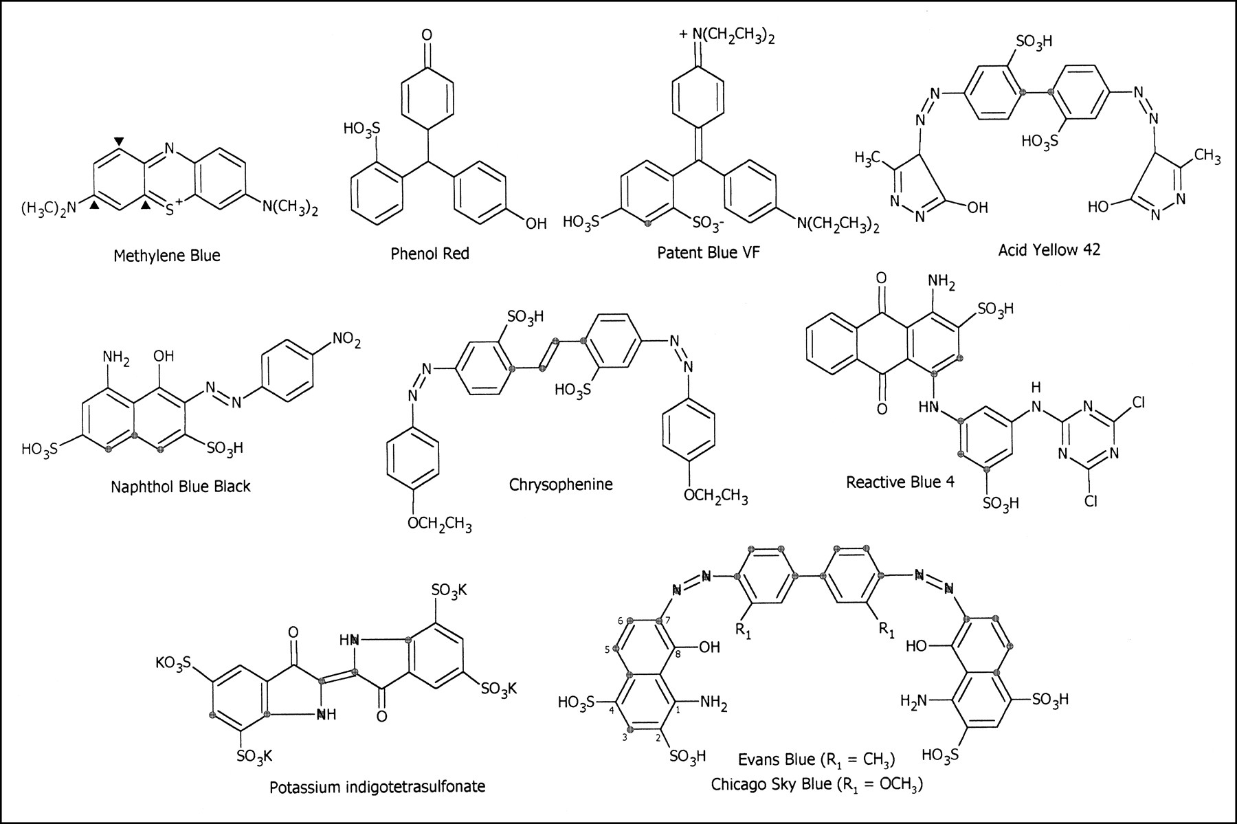
- FIGURE 1.
Molecular structures of 10 dyes used. Methylene blue with ▴ nucleophile reactive sites; Phenol red, monosulfonic acid dye; disulfonic acid dyes with
 atom spacers: Patent blue; Acid yellow 42; Naphthol blue black; Chrysophenine; Reactive blue 4; tetrasulfonic acid dyes: Potassium indigotetrasulfonate; Evans blue and Chicago sky blue, which both contain a 1-amino-8-hydroxynaphthalene ring system.
 - FIGURE 2.
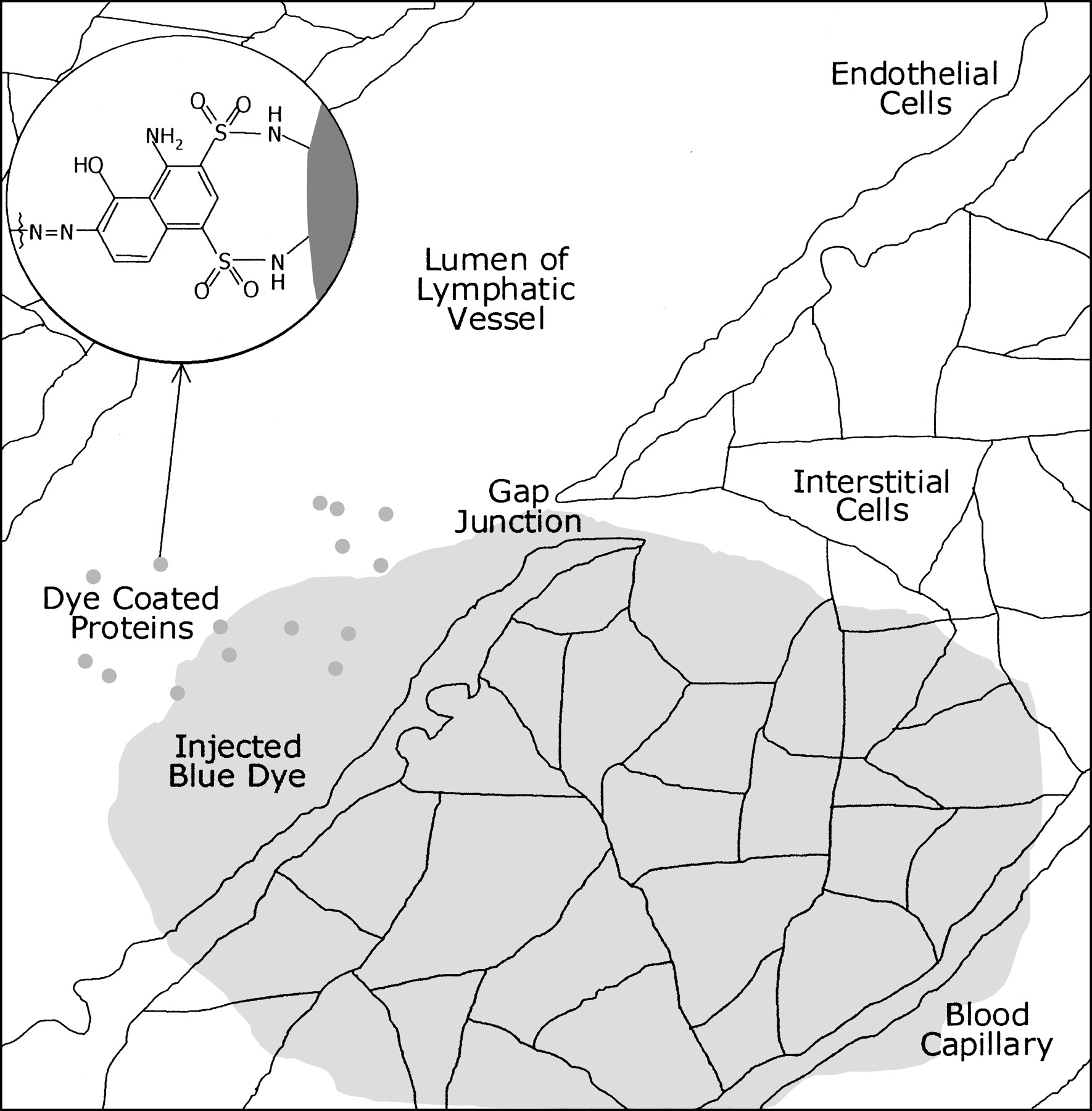
After subdermal injection of soluble blue dye such as Evans blue, there is diffusion from interstitial cells to lymphatic vessels as well as blood capillaries. Dye binds to surface of endogenous proteins by sulfonation reaction, remaining trapped inside lymphatic lumen and transported along with lymphatic flow. Dyes such as Methylene blue, which do not have sulfonic acid groups in their structure, do not bind to endogenous lymph proteins and continue migration into blood capillaries.
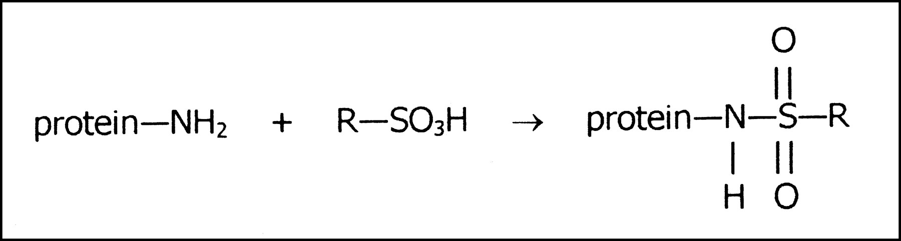
- FIGURE 3.
Dye—protein complex formed by chemical reaction of amino group on protein surface with sulfonic acid group of dye (R).

Tables
Dye No. of -SO3H groups No. of atoms between -SO3H groups % bound to plasma proteins* Methylene blue 0 0 0.0 ± 0.0 Phenol red 1 0 10.0 ± 1.2 Patent blue VF 2 1 4.7 ± 1.2 Sulforhodamine 101 2 1 14.2 ± 1.0 Orange G 2 1 15.7 ± 2.1 Acid yellow 42 2 2 84.2 ± 3.1 Naphthol blue black 2 3 99.7 ± 1.9 Nitrazine yellow 2 3 100.6 ± 1.3 Chrysophenine 2 4 96.2 ± 2.4 Direct yellow 27 2 5 97.9 ± 5.8 Reactive blue 4 2 6 102.8 ± 7.9 Indigo carmine 2 8 28.0 ± 2.3 Potassium indigotetrasulfonate 4 1 and 6 12.7 ± 0.8 Evans blue 4 1 and 20 68.1 ± 3.5 99mTc-Evans blue 4 1 and 20 69.6 ± 2.5 Chicago sky blue 4 1 and 20 69.3 ± 1.5 99mTc-Chicago sky blue 4 1 and 20 70.8 ± 0.6 Trypan blue 4 3 and 14 62.2 ± 2.0 99mTc-Trypan blue 4 3 and 14 60.5 ± 0.7 Direct yellow 4 4 and 19 58.6 ± 2.4 ↵* Mean ± SD.
99mTc-Dye Before SEC ITLC analysis After SEC analysis 99mTcO4− (%) 99mTc-Dye (%) 99mTc-Dye-protein (%) Fraction 2: 99mTc-Dye-protein (%) Fraction 3: 99mTc activity (%) Fraction 4: 99mTc-Dye (%) Column: 99mTcO2 (%) Evans blue 0.5 ± 0.2 26.0 ± 2.6 73.5 ± 2.3 69.6 ± 2.5* 1.7 ± 0.2 25.5 ± 2.9 3.2 ± 0.7 Chicago sky blue 0.1 ± 0.1 27.4 ± 1.2 72.5 ± 1.1 70.8 ± 0.6* 0.9 ± 0.3 26.6 ± 0.9 1.7 ± 0.4 Trypan blue 0.2 ± 0.1 36.8 ± 2.0 63.0 ± 1.9 60.5 ± 0.7* 1.1 ± 0.8 36.8 ± 1.5 1.6 ± 0.2 ↵* ITLC analysis gave 0.4% ± 0.0% 99mTcO4−, 3.9% ± 1.0% 99mTc-Evans blue, and 95.7% ± 1.0% 99mTc-Evans blue bound to protein; 0.3% ± 0.1% 99mTcO4−, 4.5% ± 0.4% 99mTc-Chicago sky blue, and 95.2% ± 0.4% 99mTc-Chicago sky blue bound to protein; and 0.7% ± 0.5% 99mTcO4−, 3.7% ± 0.8% 99mTc-Trypan blue, and 95.6% ± 0.9% 99mTc-Trypan blue bound to protein.
Data are expressed as mean ± SD.
In this issue


Jump to section
Related Articles
Cited By...
- Design of a Fibroblast Activation Protein-Targeted Radiopharmaceutical Therapy with High Tumor-to-Healthy-Tissue Ratios
- A Lymph Node Targeted Amphiphile Vaccine Induces Potent Cellular and Humoral Immunity to SARS-CoV-2
- Validating a semi-quantitative method to assess the degree of methylene blue staining in sentinel lymph nodes
- Enhancement of Peptide Vaccine Immunogenicity by Increasing Lymphatic Drainage and Boosting Serum Stability
- A Case of Severe Anaphylactic Reaction Secondary to Isosulfan Blue Dye Injection
- In vivo albumin labeling and lymphatic imaging
- Tumor cell entry into the lymph node is controlled by CCL1 chemokine expressed by lymph node lymphatic sinuses
- Methylene Blue Dye, an Accurate Dye for Sentinel Lymph Node Identification in Early Breast Cancer
- Formation of Sulfonamide Bonds Through Reaction of Dyes with Serum Proteins