
Main menu

  • Home
  • Content
    • Current
    • Ahead of print
    • Past Issues
    • JNM Supplement
    • SNMMI Annual Meeting Abstracts
    • Continuing Education
    • JNM Podcasts
  • Subscriptions
    • Subscribers
    • Institutional and Non-member
    • Rates
    • Journal Claims
    • Corporate & Special Sales
  • Authors
    • Submit to JNM
    • Information for Authors
    • Assignment of Copyright
    • AQARA requirements
  • Info
    • Reviewers
    • Permissions
    • Advertisers
  • About
    • About Us
    • Editorial Board
    • Contact Information
  • More
    • Alerts
    • Feedback
    • Help
    • SNMMI Journals
  • SNMMI
    • JNM
    • JNMT
    • SNMMI Journals
    • SNMMI

User menu

  • Subscribe
  • My alerts
  • Log in
  • Log out
  • My Cart

Search

  • Advanced search
Journal of Nuclear Medicine
  • SNMMI
    • JNM
    • JNMT
    • SNMMI Journals
    • SNMMI
  • Subscribe
  • My alerts
  • Log in
  • Log out
  • My Cart
Journal of Nuclear Medicine

Advanced Search

  • Home
  • Content
    • Current
    • Ahead of print
    • Past Issues
    • JNM Supplement
    • SNMMI Annual Meeting Abstracts
    • Continuing Education
    • JNM Podcasts
  • Subscriptions
    • Subscribers
    • Institutional and Non-member
    • Rates
    • Journal Claims
    • Corporate & Special Sales
  • Authors
    • Submit to JNM
    • Information for Authors
    • Assignment of Copyright
    • AQARA requirements
  • Info
    • Reviewers
    • Permissions
    • Advertisers
  • About
    • About Us
    • Editorial Board
    • Contact Information
  • More
    • Alerts
    • Feedback
    • Help
    • SNMMI Journals
  • View or Listen to JNM Podcast
  • Visit JNM on Facebook
  • Join JNM on LinkedIn
  • Follow JNM on Twitter
  • Subscribe to our RSS feeds
OtherBASIC SCIENCE INVESTIGATIONS

Preferential Labeling of Glial and Meningial Brain Tumors with [2-14C]Acetate

Gerald A. Dienel, David Popp, Paul D. Drew, Kelly Ball, Ali Krisht and Nancy F. Cruz
Journal of Nuclear Medicine August 2001, 42 (8) 1243-1250;
Gerald A. Dienel
  • Find this author on Google Scholar
  • Find this author on PubMed
  • Search for this author on this site
David Popp
  • Find this author on Google Scholar
  • Find this author on PubMed
  • Search for this author on this site
Paul D. Drew
  • Find this author on Google Scholar
  • Find this author on PubMed
  • Search for this author on this site
Kelly Ball
  • Find this author on Google Scholar
  • Find this author on PubMed
  • Search for this author on this site
Ali Krisht
  • Find this author on Google Scholar
  • Find this author on PubMed
  • Search for this author on this site
Nancy F. Cruz
  • Find this author on Google Scholar
  • Find this author on PubMed
  • Search for this author on this site
  • Article
  • Figures & Data
  • Info & Metrics
  • PDF
Loading

Article Figures & Data

Figures

  • Tables
  • FIGURE 1.
    • Download figure
    • Open in new tab
    • Download powerpoint
    FIGURE 1.

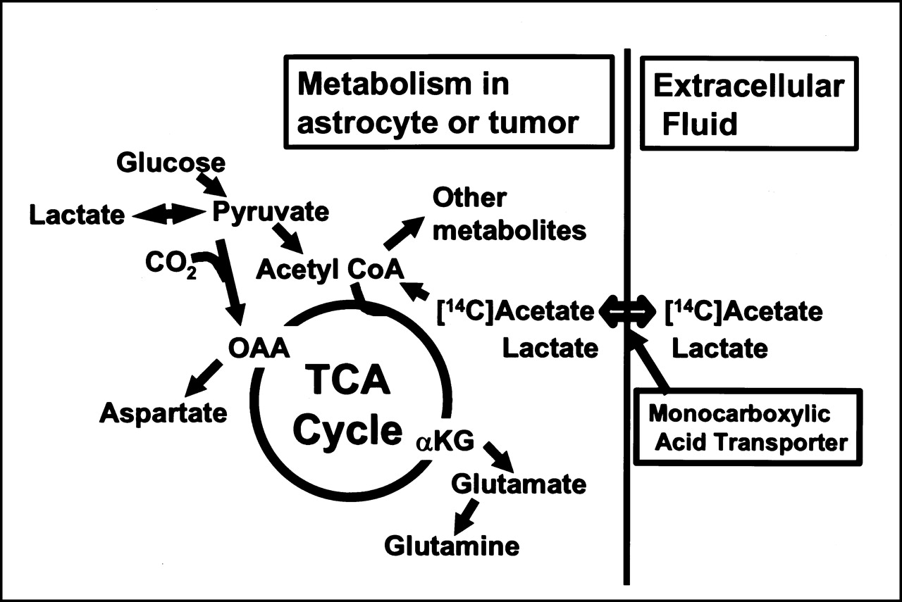
    Model for metabolic trapping of [14C]acetate involves preferential uptake into astrocyte or tumor from extracellular fluid through unidentified isoform of monocarboxylic acid transporter. Stimulation of acetate uptake into cell might be enhanced by high rate of glycolysis and lactate efflux, that is, transacceleration of acetate uptake by lactate export (6). Once in cell, acetate can be converted to acetyl coenzyme A (CoA) and metabolized further by various pathways, including entry into tricarboxylic acid (TCA) cycle, leading to incorporation of 14C into various acidic compounds, including α-ketoglutarate (αKG) and oxalacetate (OAA). Rapid transamination reactions would cause labeling of TCA cycle–derived amino acids, glutamate, glutamine, and aspartate. With longer experimental times, label derived from acetate would also be incorporated into lipid and protein.

  • FIGURE 2.
    • Download figure
    • Open in new tab
    • Download powerpoint
    FIGURE 2.

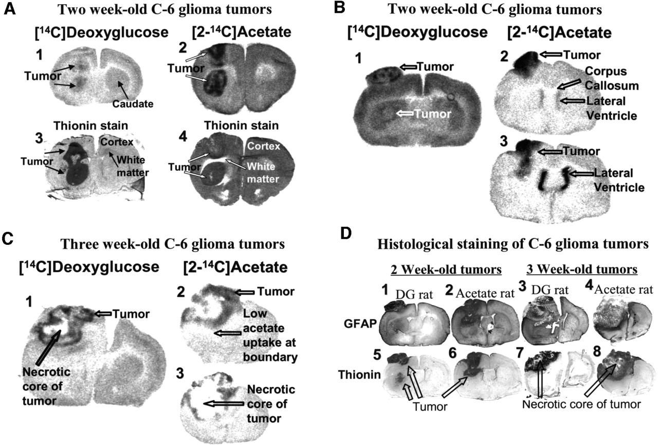
    Metabolic labeling of C6 glioma tumors with [14C]acetate is illustrated in tumors grown in rat brain in vivo for 2 (A and B) or 3 (C) wk and assessed by autoradiography (A–C) and histologic staining (A and D). Autoradiographs show heterogeneous metabolic labeling by [14C]deoxyglucose (DG) and [2-14C]acetate in 2-wk-old intracerebral (A1 and A2) and extracerebral (B1–B3) tumors and in 3-wk-old necrotic tumors (C1–C3); different serial sections of same tumor (B2 and B3; C2 and C3) illustrate varied labeling of larger tumors, particularly in necrotic tumor. Thionin staining of sections near those used for autoradiography detects regions of densely packed cells in tumor (A3 and A4; D5–D8) compared with normal tissue. GFAP is marker for reactive astrocytes and also stains portions of C6 glioma tumors and zones surrounding tumors (D1–D4). Stained sections in D correspond to autoradiograph sections taken from same animals (B and C). Note low labeling of white matter (i.e., corpus callosum and subcortical white matter) by deoxyglucose and acetate (A–C), high labeling of lateral ventricle by acetate (B2 and B3), and relatively low labeling by acetate in boundary zone ventral to necrotic tumor (C2), which appears to coincide with GFAP-rich zone in 3-wk-old tumor (D4). Values for glucose use and net acetate uptake are presented in Table 1.

  • FIGURE 3.
    • Download figure
    • Open in new tab
    • Download powerpoint
    FIGURE 3.

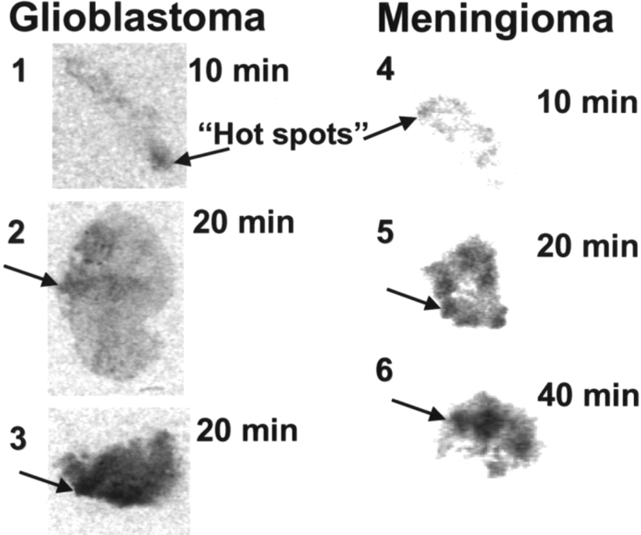
    Representative 20-μm-thick sections of slices of human glioblastoma (1–3) or meningioma (4–6) tumors that were incubated in vitro with [14C]acetate for 10–40 min show heterogeneous labeling. Arrows indicate local areas of more intense uptake of tracer.

Tables

  • Figures
    • View popup
    TABLE 1

    Glucose Use and Acetate Uptake in Tumors and Rat Brain Tissue In Vivo

    Brain regionGlucose use (μmol/100 g/min)Net acetate uptake (mL/100 g/ min)
    Rat C6 glioma111.0 ± 34.0*9.9 ± 2.1†
     Contralateral brain tissue81.0 ± 5.03.9 ± 1.0
     Tumor/contralateral tissue1.4 ± 0.52.3 ± 0.3‡
    Human U-373 glioblastoma/astrocytoma112.0 ± 27.0*4.2 ± 0.1†
     Contralateral brain tissue86.0 ± 11.02.8 ± 0.5
     Tumor/contralateral tissue1.3 ± 0.21.5 ± 0.2
    Rat gray matter§
     Frontal cortex
      Right82.4 ± 7.44.8 ± 0.7
      Left77.3 ± 6.56.9 ± 2.0
     Sensorimotor cortex
      Right84.4 ± 7.84.6 ± 1.2
      Left75.4 ± 3.74.3 ± 1.4
     Caudate nucleus
      Right83.6 ± 8.53.3 ± 0.4
      Left76.6 ± 9.63.7 ± 0.8
     Sensory cortex
      Right87.6 ± 7.54.2 ± 0.8
      Left87.5 ± 7.94.1 ± 0.9
    Rat white matter§
     Forceps minor corpus callosum
      Right35.6 ± 2.82.2 ± 0.6
      Left35.5 ± 2.32.3 ± 0.2
     Genu of corpus callosum
      Right35.2 ± 2.42.5 ± 0.5
      Left32.0 ± 1.22.3 ± 0.4
    • ↵* P < 0.09 vs. contralateral tissue.

    • ↵† P < 0.005 vs. contralateral tissue.

    • ↵‡ P < 0.05 vs. deoxyglucose tumor-to-tissue ratio.

    • ↵§ Data are from rats implanted with C6 tumors.

    • Data are mean ± SD (C6 tumors: n = 6 for glucose use, n = 7 for acetate uptake; U-373 tumors: n = 4 per group for glucose use and acetate uptake).

    • View popup
    TABLE 2

    Metabolism of [14C]Acetate in C6 Glioma Tumors and Rat Brain

    FractionPercentage of total 14C in acid extract (mean ± SD, n = 3)
    C6 tumorContralateral cerebral cortex
    Unmetabolized [14C]acetate5.0 ± 2.95.5 ± 1.9
    Acidic + any neutral metabolites37.0 ± 2.229.1 ± 0.8
    Amino acids + any basic metabolites59.6 ± 3.168.7 ± 2.9
    Total metabolized96.6 ± 2.697.7 ± 2.7
    • Proportions of labeled compounds recovered in acidic and amino acid fractions were not significantly different in tumor and contralateral tissue (P > 0.08, t test).

    • View popup
    TABLE 3

    Net Uptake of [14C]Acetate into Human Tumor Samples Assayed In Vitro

    Tumor type/sampleAcetate uptake (μmol/100 g/min)
    Whole tumorHot spots
    Glioblastoma
     1 high grade1.902.60
     2 recurrent0.560.93
     3 low grade0.380.56
     4 recurrent1.201.60
     5 recurrent0.550.89
     6 unknown grade0.420.80
     Mean ± SD (n = 6)0.84 ± 0.601.20 ± 0.76
    Meningioma
     10.771.30
     20.520.83
     30.240.42
     41.602.40
     Mean ± SD (n = 4)0.78 ± 0.581.20 ± 0.80
    Pituitary adenoma
     10.220.36
     20.270.41
    Oligodendroglioma
     10.671.00
     20.270.39
    Gliotic tissue0.130.23
    • Data are average uptake into entire slice for all slices at all incubation times for each tumor and average for hot spots in same tumors.

    • View popup
    TABLE 4

    In Vitro Metabolism of [14C]Acetate by Human Tumors

    Tumor typeTumor sampleIncubation time (min)Percentage of total 14C in acid extract
    Unmetabolized [14C]acetateAcidic + any neutral metabolitesAmino acids + any basic metabolites
    Glioblastoma11010123
    20631229
    210761016
    20202552
    310371751
    20201759
    Meningioma110582224
    40412627
    210423029
    20223547
    310112960
    20123058
    • Protein content of acid-extracted slices was 0.06–1.2 mg per sample. Recovery of 14C in fractions of acid extract was 101% ± 4%.

PreviousNext
Back to top

In this issue

Journal of Nuclear Medicine: 42 (8)
Journal of Nuclear Medicine
Vol. 42, Issue 8
August 1, 2001
  • Table of Contents
  • Index by author
Print
Download PDF
Article Alerts
Sign In to Email Alerts with your Email Address
Email Article

Thank you for your interest in spreading the word on Journal of Nuclear Medicine.

NOTE: We only request your email address so that the person you are recommending the page to knows that you wanted them to see it, and that it is not junk mail. We do not capture any email address.

Enter multiple addresses on separate lines or separate them with commas.
Preferential Labeling of Glial and Meningial Brain Tumors with [2-14C]Acetate
(Your Name) has sent you a message from Journal of Nuclear Medicine
(Your Name) thought you would like to see the Journal of Nuclear Medicine web site.
Citation Tools
Preferential Labeling of Glial and Meningial Brain Tumors with [2-14C]Acetate
Gerald A. Dienel, David Popp, Paul D. Drew, Kelly Ball, Ali Krisht, Nancy F. Cruz
Journal of Nuclear Medicine Aug 2001, 42 (8) 1243-1250;

Citation Manager Formats

  • BibTeX
  • Bookends
  • EasyBib
  • EndNote (tagged)
  • EndNote 8 (xml)
  • Medlars
  • Mendeley
  • Papers
  • RefWorks Tagged
  • Ref Manager
  • RIS
  • Zotero
Share
Preferential Labeling of Glial and Meningial Brain Tumors with [2-14C]Acetate
Gerald A. Dienel, David Popp, Paul D. Drew, Kelly Ball, Ali Krisht, Nancy F. Cruz
Journal of Nuclear Medicine Aug 2001, 42 (8) 1243-1250;
Twitter logo Facebook logo LinkedIn logo Mendeley logo
  • Tweet Widget
  • Facebook Like
  • Google Plus One
Bookmark this article

Jump to section

  • Article
    • Abstract
    • MATERIALS AND METHODS
    • RESULTS
    • DISCUSSION
    • CONCLUSION
    • Acknowledgments
    • Footnotes
    • REFERENCES
  • Figures & Data
  • Info & Metrics
  • PDF

Related Articles

  • No related articles found.
  • PubMed
  • Google Scholar

Cited By...

  • Alcohol Decreases Baseline Brain Glucose Metabolism More in Heavy Drinkers Than Controls But Has No Effect on Stimulation-Induced Metabolic Increases
  • 1-11C-Acetate Versus 18F-FDG PET in Detection of Meningioma and Monitoring the Effect of {gamma}-Knife Radiosurgery
  • PET of Glial Metabolism Using 2-18F-Fluoroacetate
  • Google Scholar

More in this TOC Section

  • Tumor-Specific Binding of Radiolabeled PEGylated GIRLRG Peptide: A Novel Agent for Targeting Cancers
  • PET/MRI of Hypoxic Atherosclerosis Using 64Cu-ATSM in a Rabbit Model
  • Tumor Uptake of Anti-CD20 Fabs Depends on Tumor Perfusion
Show more Basic Science Investigations

Similar Articles

SNMMI

© 2025 SNMMI

Powered by HighWire