


Article Figures & Data
Figures
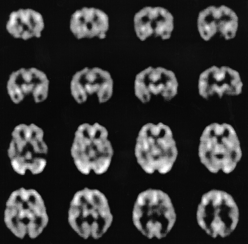
- FIGURE 1.
From top to bottom, two coronal and two transaxial slices with 99mTc-HMPAO using fanbeam collimator in healthy volunteer. Note symmetric tracer distribution in cerebral cortex. Areas with preferential perfusion include cingulate gyrus, primary visual cortex, basal ganglia, thalami, and cerebellar hemispheres.
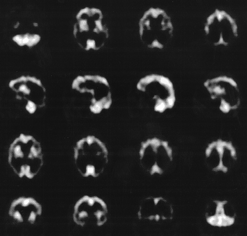
- FIGURE 2.
A 58-y-old right-handed man had 2-y history of progressive memory loss, which became worse over last 7 mo. His father and three cousins had dementia. Transaxial, sagittal, and coronal slices show marked bilateral, symmetric temporoparieto-occipital hypoperfusion, extending to frontal lobes. Basal ganglia, primary visual cortex, and cerebellum are spared.
- FIGURE 3.
A 62-y-old right-handed, hypertensive man had stroke 2 y ago and now has severe memory impairment, dysarthria, and urinary incontinence. Radionuclide cisternography showed normal findings. Transaxial, sagittal, and coronal slices show multiple scattered focal areas of hypoperfusion involving entire cerebral cortex, a pattern frequently found in vascular dementia. Head CT scan showed white matter infarcts.
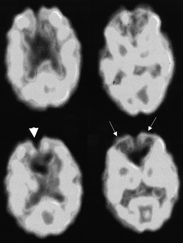
- FIGURE 4.
Transaxial slices of 73-y-old man with FD and 2-y history of progressive short-term memory loss show marked hypoperfusion of anterior cingulate gyrus (arrowhead) and mesial frontal lobes (arrows). MRI showed only mild frontal lobe atrophy, which could not explain brain SPECT findings.
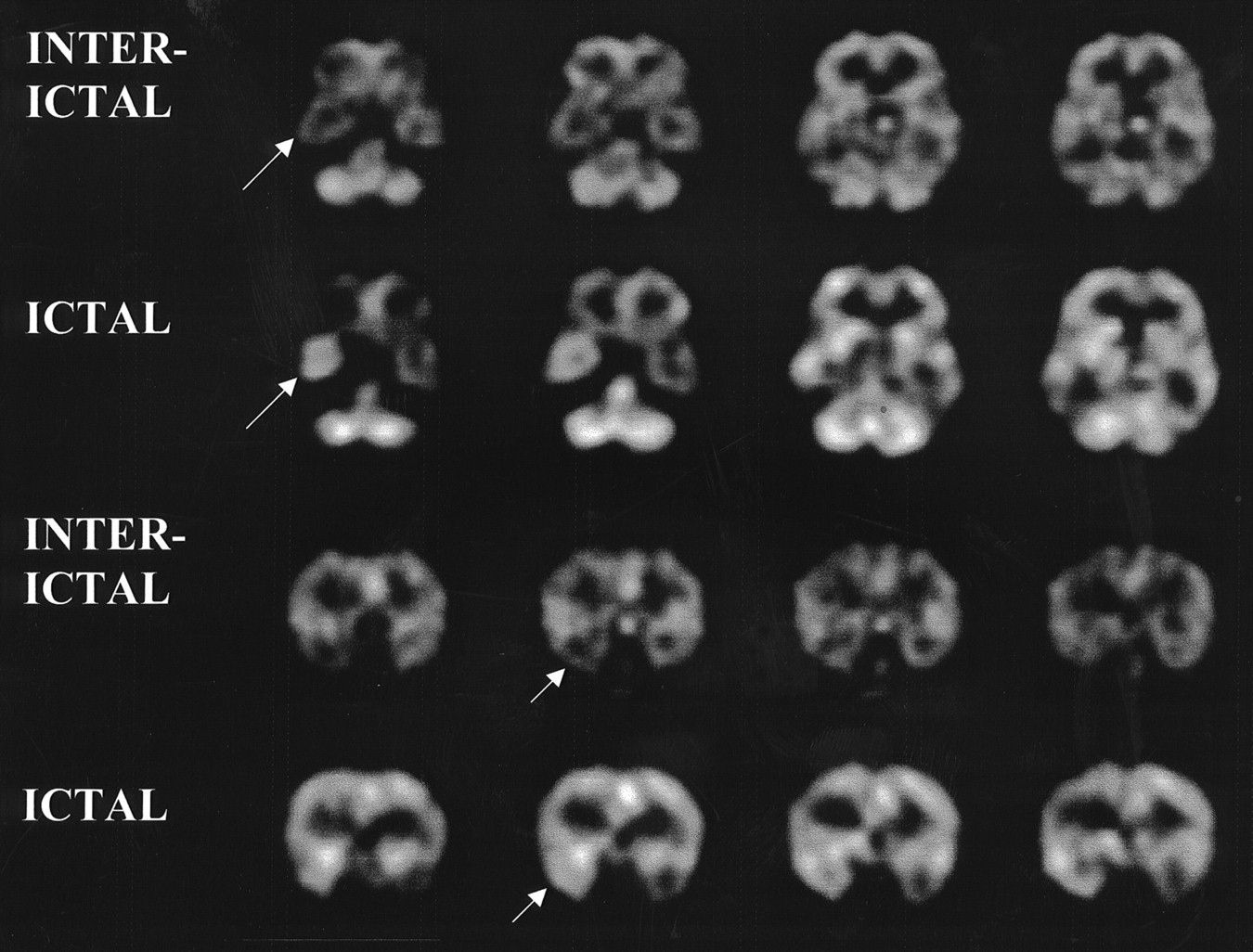
- FIGURE 5.
A 21-y-old left-handed man had history of tonic–clonic seizures since age 8. Head CT findings were normal. MRI showed T2-weighted hyperintense signal and slightly decreased size of right hippocampus. EEG showed acute waves in right frontal and temporal lobes. Interictal and ictal transaxial and coronal slices show hypoperfusion and hyperperfusion, respectively, of right temporal lobe (arrows).
- FIGURE 6.
A 13-y-old boy complained of severe anxiety and compulsions (washing hands) over last 4 y. His insight was intact. Transaxial and sagittal slices show hyperperfusion of orbitofrontal area, bilaterally (arrows).


In this issue


Jump to section
Related Articles
Cited By...
- Effectiveness of radiology modalities in diagnosing and characterizing brain disorders
- Influence of Minimum Count in Brain Perfusion SPECT: Phantom and Clinical Studies
- Effect of Prefiltering Cutoff Frequency and Scatter and Attenuation Corrections During Normal Database Creation for Statistical Imaging Analysis of the Brain
- Forensic Applications of Cerebral Single Photon Emission Computed Tomography in Mild Traumatic Brain Injury
- Semiquantitative Human Cerebral Perfusion Assessment With Ultrasound in Brain Space-Occupying Lesions: Preliminary Data
- Frontal lobe hypoperfusion and depressive symptoms in Alzheimer disease
- SPECT imaging in dementia
- Second harmonic imaging: a new ultrasound technique to assess human brain tumour perfusion