
Main menu

  • Home
  • Content
    • Current
    • Ahead of print
    • Past Issues
    • JNM Supplement
    • SNMMI Annual Meeting Abstracts
    • Continuing Education
    • JNM Podcasts
  • Subscriptions
    • Subscribers
    • Institutional and Non-member
    • Rates
    • Journal Claims
    • Corporate & Special Sales
  • Authors
    • Submit to JNM
    • Information for Authors
    • Assignment of Copyright
    • AQARA requirements
  • Info
    • Reviewers
    • Permissions
    • Advertisers
  • About
    • About Us
    • Editorial Board
    • Contact Information
  • More
    • Alerts
    • Feedback
    • Help
    • SNMMI Journals
  • SNMMI
    • JNM
    • JNMT
    • SNMMI Journals
    • SNMMI

User menu

  • Subscribe
  • My alerts
  • Log in
  • My Cart

Search

  • Advanced search
Journal of Nuclear Medicine
  • SNMMI
    • JNM
    • JNMT
    • SNMMI Journals
    • SNMMI
  • Subscribe
  • My alerts
  • Log in
  • My Cart
Journal of Nuclear Medicine

Advanced Search

  • Home
  • Content
    • Current
    • Ahead of print
    • Past Issues
    • JNM Supplement
    • SNMMI Annual Meeting Abstracts
    • Continuing Education
    • JNM Podcasts
  • Subscriptions
    • Subscribers
    • Institutional and Non-member
    • Rates
    • Journal Claims
    • Corporate & Special Sales
  • Authors
    • Submit to JNM
    • Information for Authors
    • Assignment of Copyright
    • AQARA requirements
  • Info
    • Reviewers
    • Permissions
    • Advertisers
  • About
    • About Us
    • Editorial Board
    • Contact Information
  • More
    • Alerts
    • Feedback
    • Help
    • SNMMI Journals
  • View or Listen to JNM Podcast
  • Visit JNM on Facebook
  • Join JNM on LinkedIn
  • Follow JNM on Twitter
  • Subscribe to our RSS feeds
OtherCLINICAL INVESTIGATIONS

Neuroblastoma Imaging Using a Combined CT Scanner–Scintillation Camera and 131I-MIBG

H. Roger Tang, Angela J. Da Silva, Katherine K. Matthay, David C. Price, John P. Huberty, Randall A. Hawkins and Bruce H. Hasegawa
Journal of Nuclear Medicine February 2001, 42 (2) 237-247;
H. Roger Tang
  • Find this author on Google Scholar
  • Find this author on PubMed
  • Search for this author on this site
Angela J. Da Silva
  • Find this author on Google Scholar
  • Find this author on PubMed
  • Search for this author on this site
Katherine K. Matthay
  • Find this author on Google Scholar
  • Find this author on PubMed
  • Search for this author on this site
David C. Price
  • Find this author on Google Scholar
  • Find this author on PubMed
  • Search for this author on this site
John P. Huberty
  • Find this author on Google Scholar
  • Find this author on PubMed
  • Search for this author on this site
Randall A. Hawkins
  • Find this author on Google Scholar
  • Find this author on PubMed
  • Search for this author on this site
Bruce H. Hasegawa
  • Find this author on Google Scholar
  • Find this author on PubMed
  • Search for this author on this site
  • Article
  • Figures & Data
  • Info & Metrics
  • PDF
Loading

Article Figures & Data

Figures

  • Tables
  • FIGURE 1.
    • Download figure
    • Open in new tab
    • Download powerpoint
    FIGURE 1.

    Combined CT–scintillation camera imaging system.
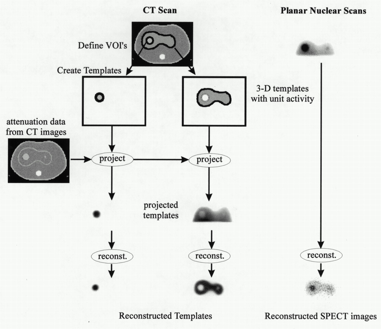
  • FIGURE 2.
    • Download figure
    • Open in new tab
    • Download powerpoint
    FIGURE 2.

    Overview of template projection reconstruction process.
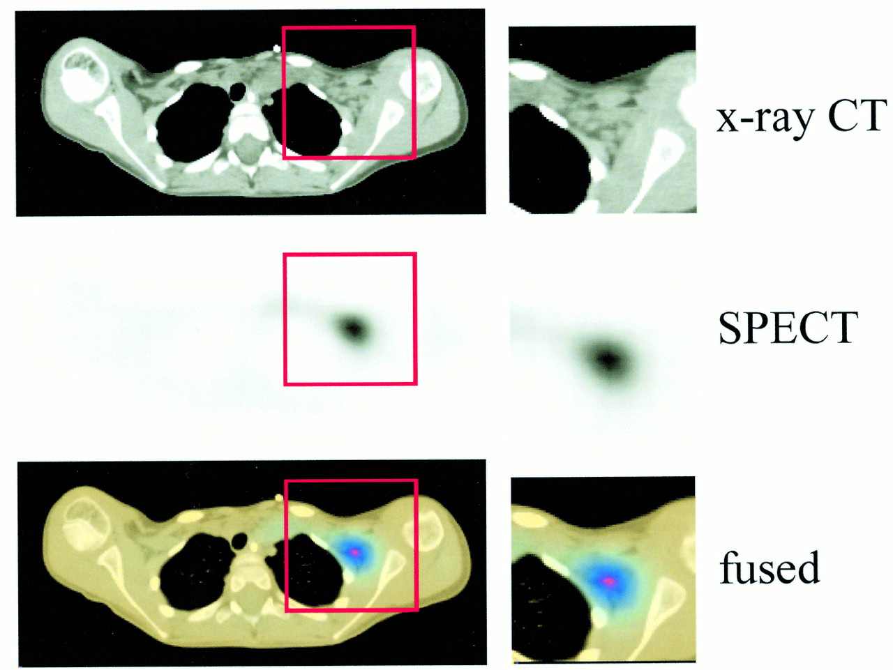
  • FIGURE 3.
    • Download figure
    • Open in new tab
    • Download powerpoint
    FIGURE 3.

    Example of fused image available from combined CT–scintillation camera imaging system. CT image is shown along with spatially correlated SPECT image indicating 131I-MIBG uptake in lymph node in patient’s left axilla.
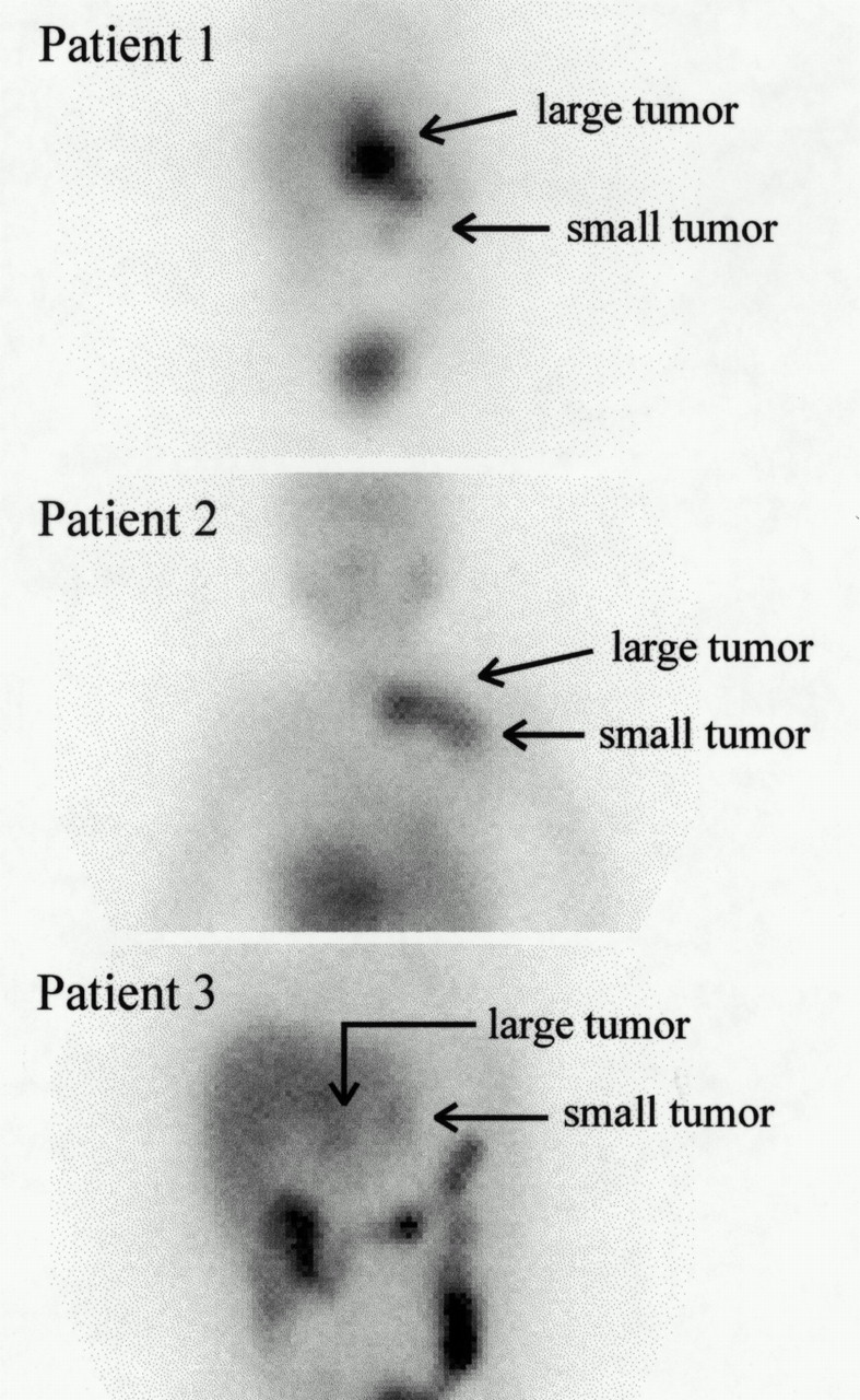
  • FIGURE 4.
    • Download figure
    • Open in new tab
    • Download powerpoint
    FIGURE 4.

    Anterior planar 131I-MIBG scans of three neuroblastoma patients. Patient 1 shows two separate lesions in abdomen along with normal liver uptake (left side of image) and excreted bladder activity (lower portion of image). Patient 2 shows one lesion in left clavicle and one near left axilla along with liver uptake (lower portion of image) and diffuse low-level background tissue uptake. Patient 3 shows two (faint) lesions in abdomen along with normal liver uptake (left side of image) and intense activity in large bowel.
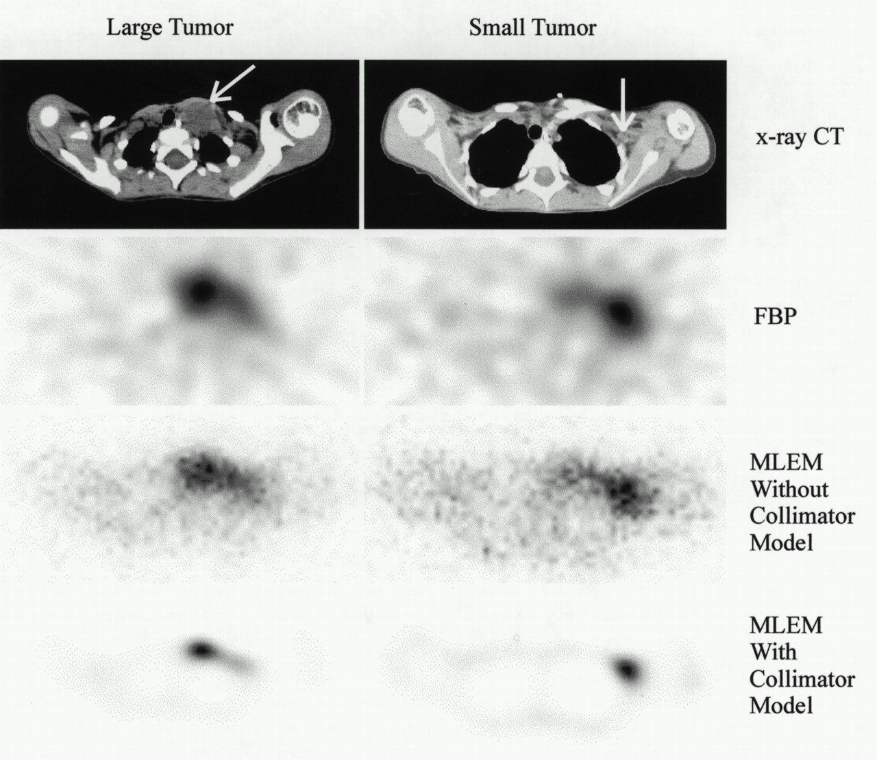
  • FIGURE 5.
    • Download figure
    • Open in new tab
    • Download powerpoint
    FIGURE 5.

    Coregistered CT and reconstructed SPECT images of patient 1.

  • FIGURE 6.
    • Download figure
    • Open in new tab
    • Download powerpoint
    FIGURE 6.

    Coregistered CT and reconstructed SPECT images of patient 2.

  • FIGURE 7.
    • Download figure
    • Open in new tab
    • Download powerpoint
    FIGURE 7.

    Coregistered CT and reconstructed SPECT images of patient 3. In images on right, central focus is small lesion that does not appear to anatomically correspond to left adrenal; left focus is part of large lesion; and right focus is part of bowel uptake.

Tables

  • Figures
    • View popup
    TABLE 1.

    Tumor Volume Measurement Results for Three Patients Using Different Measurement Methods

    Patient no.Method*
    CT (mL)CVMVOILSTCWLS
    No collW/collMLFBPMLFBP
    Patient 1
     Large48.51.770.4400.9141.221.221.591.541.29
     Small2.10.670.1350.2541.270.983.342.890.400
    Patient 2
     Large12.70.3180.07700.2550.3350.3270.4700.4290.329
     Small0.672.570.09920.3703.6253.506.8455.113.922
    Patient 3
     Large1170.03480.04330.06140.06810.07550.1470.1390.0722
     Small1.30.7050.05920.1481.531.753.132.941.132
    • ↵* Measurements in MBq/mL unless otherwise noted.

    • CV = standard conjugate views; MVOI = mean volume of interest, with and without compensation for collimator (coll); LS = least-squares fitting with reconstructed SPECT data and templates; TC = template correction with reconstructed SPECT data and templates; WLS = planar weighted least-squares fitting using planar emission data and projected templates; ML = maximum-likelihood expectation maximization reconstruction; FBP = filtered backprojection reconstruction.

    • View popup
    TABLE 2.

    Comparison of CT-Derived Tumor Volumes for Separate CT Scans and Analyses of Patients

    Patient no.Our CT-defined volume (mL)Independent CT-defined volume (mL)
    148.563.4
    212.713.3
    3116.885.7
PreviousNext
Back to top

In this issue

Journal of Nuclear Medicine
Vol. 42, Issue 2
February 1, 2001
  • Table of Contents
  • Index by author
Print
Download PDF
Article Alerts
Sign In to Email Alerts with your Email Address
Email Article

Thank you for your interest in spreading the word on Journal of Nuclear Medicine.

NOTE: We only request your email address so that the person you are recommending the page to knows that you wanted them to see it, and that it is not junk mail. We do not capture any email address.

Enter multiple addresses on separate lines or separate them with commas.
Neuroblastoma Imaging Using a Combined CT Scanner–Scintillation Camera and 131I-MIBG
(Your Name) has sent you a message from Journal of Nuclear Medicine
(Your Name) thought you would like to see the Journal of Nuclear Medicine web site.
Citation Tools
Neuroblastoma Imaging Using a Combined CT Scanner–Scintillation Camera and 131I-MIBG
H. Roger Tang, Angela J. Da Silva, Katherine K. Matthay, David C. Price, John P. Huberty, Randall A. Hawkins, Bruce H. Hasegawa
Journal of Nuclear Medicine Feb 2001, 42 (2) 237-247;

Citation Manager Formats

  • BibTeX
  • Bookends
  • EasyBib
  • EndNote (tagged)
  • EndNote 8 (xml)
  • Medlars
  • Mendeley
  • Papers
  • RefWorks Tagged
  • Ref Manager
  • RIS
  • Zotero
Share
Neuroblastoma Imaging Using a Combined CT Scanner–Scintillation Camera and 131I-MIBG
H. Roger Tang, Angela J. Da Silva, Katherine K. Matthay, David C. Price, John P. Huberty, Randall A. Hawkins, Bruce H. Hasegawa
Journal of Nuclear Medicine Feb 2001, 42 (2) 237-247;
Twitter logo Facebook logo LinkedIn logo Mendeley logo
  • Tweet Widget
  • Facebook Like
  • Google Plus One
Bookmark this article

Jump to section

  • Article
    • Abstract
    • MATERIALS AND METHODS
    • RESULTS
    • DISCUSSION
    • CONCLUSION
    • Acknowledgments
    • Footnotes
    • REFERENCES
  • Figures & Data
  • Info & Metrics
  • PDF

Related Articles

  • No related articles found.
  • PubMed
  • Google Scholar

Cited By...

  • MIRD Pamphlet No. 24: Guidelines for Quantitative 131I SPECT in Dosimetry Applications
  • Localization of Metastases from Malignant Pheochromocytoma in Patients Undergoing 131I-MIBG Therapy with Manually Fused 123I-MIBG SPECT and CT Images
  • Partial-Volume Correction in PET: Validation of an Iterative Postreconstruction Method with Phantom and Patient Data
  • Determination of the Attenuation Map in Emission Tomography
  • Correlation of Tumor and Whole-Body Dosimetry with Tumor Response and Toxicity in Refractory Neuroblastoma Treated with 131I-MIBG
  • Google Scholar

More in this TOC Section

  • Cardiac Presynaptic Sympathetic Nervous Function Evaluated by Cardiac PET in Patients with Chronotropic Incompetence Without Heart Failure
  • Validation and Evaluation of a Vendor-Provided Head Motion Correction Algorithm on the uMI Panorama PET/CT System
  • An Investigation of Lesion Detection Accuracy for Artificial Intelligence–Based Denoising of Low-Dose 64Cu-DOTATATE PET Imaging in Patients with Neuroendocrine Neoplasms
Show more Clinical Investigations

Similar Articles

SNMMI

© 2025 SNMMI

Powered by HighWire