


Article Figures & Data
Figures
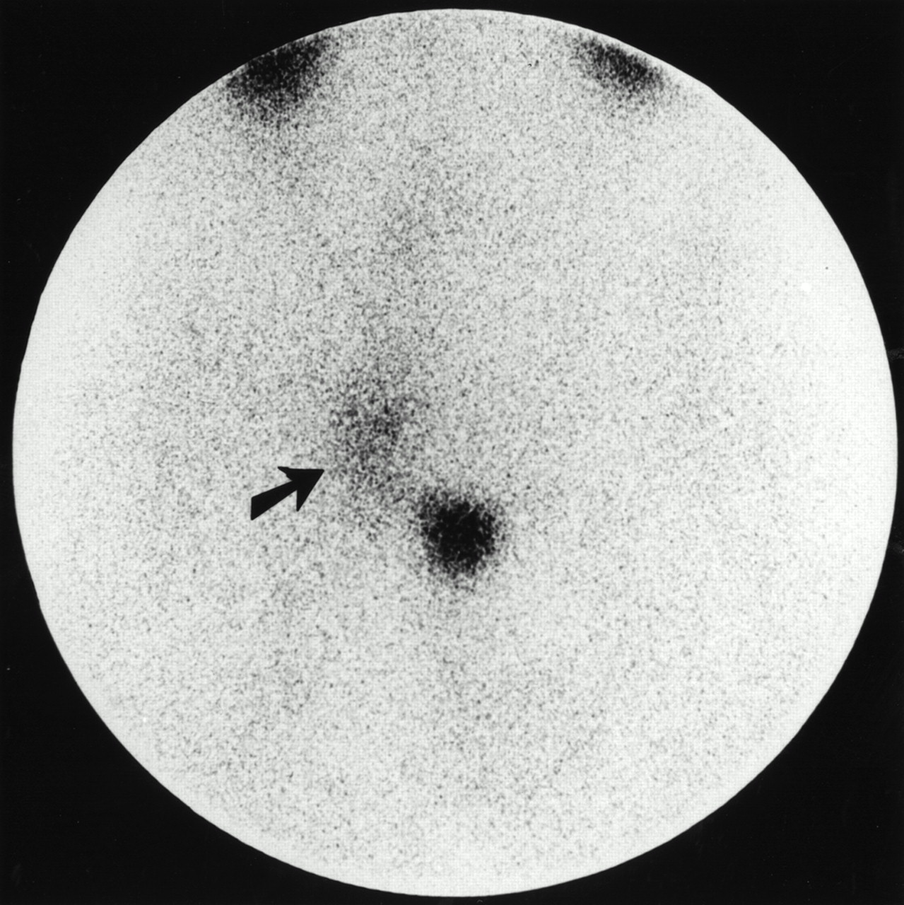
- FIGURE 1.
Visualization of submandibular lymph node in 42-y-old woman with stage IA NHL of low-grade malignancy by SS-R scintigraphy. Right lateral image of head and neck shows grade 2 pathologic uptake (arrow) within submandibular lesion. Normal uptake of radioactivity is seen in thyroid. On physical examination, lymph node of 3 cm in diameter was palpated.
- FIGURE 2.
SS-R scintigraphy in 36-y-old man with low-grade NHL shows multiple enlarged lymph nodes of 2–3 cm in diameter in right inguinal and iliac regions. Pathologic lymph nodes are clearly seen (grade 2 uptake, arrow). Normal accumulation of radioactivity is seen in bladder and kidneys.
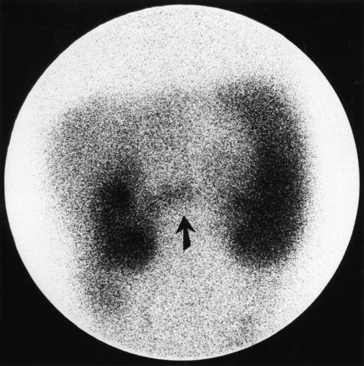
- FIGURE 3.
SS-R scintigraphy in 52-y-old woman with low-grade NHL shows pathologic uptake in abdominal region between kidneys (arrow). Normal uptake of radioactivity is seen in enlarged spleen. Normal uptake is also seen in liver and kidneys. CT scan of abdomen showed enlarged mesenteric lymph nodes.


Tables
Characteristic No. of patients (%)* Sex Male 30 (60) Female 20 (40) Ann Arbor clinical stage† I 25 (50) II 5 (10) III 7 (14) IV 13 (26) Histologic subtype (Working Formulation) A: Small lymphocytic 7 (14) B: Follicular, predominantly small cleaved cell 2 (4) C: Follicular, mixed small cleaved and large cell 30 (60) MALT 9 (18) Unclassifiable low grade 2 (4) Localization of malignant lesion % of patients Neck LN 56 Para-aortic LN 34 Mesenteric LN 22 Inguinal LN 20 Mediastinal LN 18 Axillary LN 18 Iliac LN 12 Spleen 10 Stomach 10 Orbit/eye 10 Lung hilar LN 4 Waldeyer’s ring 4 Intestine 4 Skin 4 Central nervous system 2 Urinary bladder 2 Pleura 2 Parotid gland 2 Mouth 2 * Diagnosed on basis of conventional staging procedures and SS-R scintigraphy.
LN = lymph node.
- TABLE 3.
Comparison of Results of SS-R Scintigraphy with Those of Conventional Staging in 50 Patients with Low-Grade NHL
Result No. of patients (%) SS-R scan detects new lesions 10 (20) SS-R scan misses lesions 19 (38) SS-R scan both detects new lesions and misses lesions in same patient 3 (6) Agreement between SS-R scan and conventional staging procedures 18 (36) - TABLE 4.
Clinical Stage Based on Conventional Staging Procedures Complemented with SS-R Scintigraphy Compared with Clinical Stage Based on Conventional Staging Only
Stage based on conventional staging complemented with SS-R scintigraphy Stage based on conventional staging only* I II III IV Total I 15 1 0 0 16 II 5 4 0 0 9 III 4† 0 7 0 11 IV 1† 0 0 13 14 Total 25 5 7 13 50 - TABLE 5.
Sensitivity of Various Diagnostic Approaches for Detection of Low-Grade NHL Lesions in Different Lymph Node Regions and Extranodal Sites
Localization of malignant lesion No. of malignant lesions Physical examination (%) Chest radiograph (%) CT scan (%) LAG (%) Sonography (%) SS-R scan (%) Cervical LN* 38 96 70 (10) 66 Supraclavicular LN 17 90 63 (8) 59 Axillary LN 16 94 86 (14) 63 Mediastinal LN 9 25 (8) 100 (8) 78 Orbit/eye 6 67 40 (5) 33 Lung-hilar LN 3 0 100 67 Paraaortic LN 17 82 100 (2) 43 (7) 35 Mesenteric LN 11 100 67 (6) 36 Inguinal LN 13 69 85 (13) 54 Iliac LN 10 90 30 Stomach/intestine 8 13 38 Spleen 5 100 100 100 (2) 100 ↵* Indicates occipital, submandibular, and cervical lymph nodes.
LAG = bipedal lymphangiogram; LN = lymph node.
Numbers in parentheses indicate frequency at which the test was performed and are given only if different from number of malignant lesions.


