


Article Figures & Data
Figures
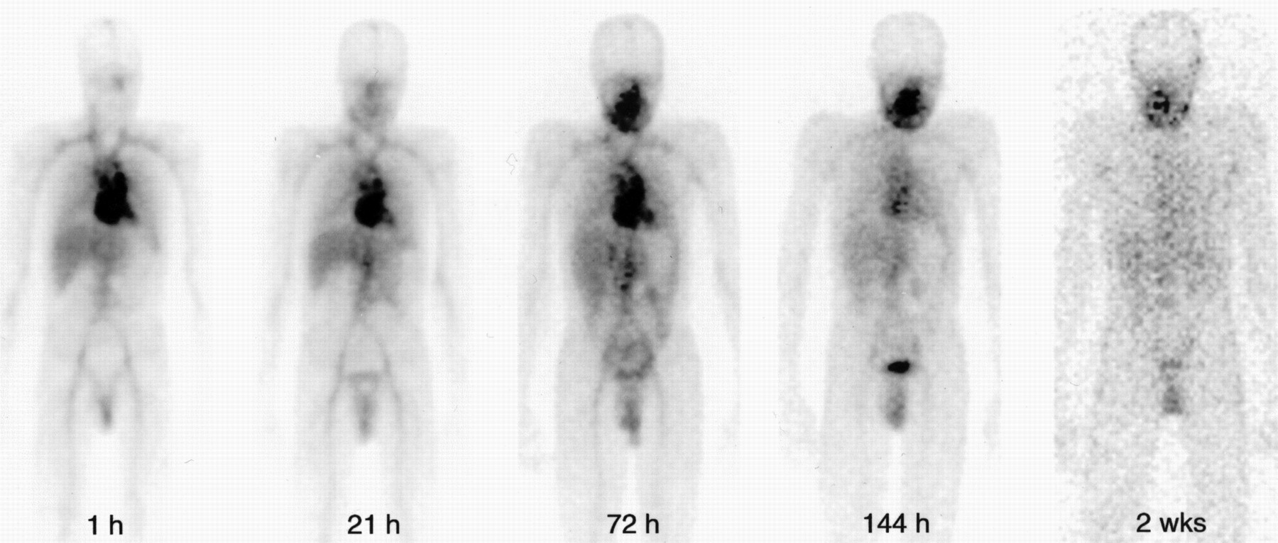
- FIGURE 1.
Whole-body scans of patient 4 acquired within 1 h after administration of 186Re-cMAb U36 and after 21, 72, and 144 h and 2 wk. Immediately after injection, most prominent activity is in blood pool. This activity remains high up to 72 h after injection. Relative uptake of radioimmunoconjugate in tumor in right oropharynx increases over time. Tumor becomes better delineated as background activity decreases.
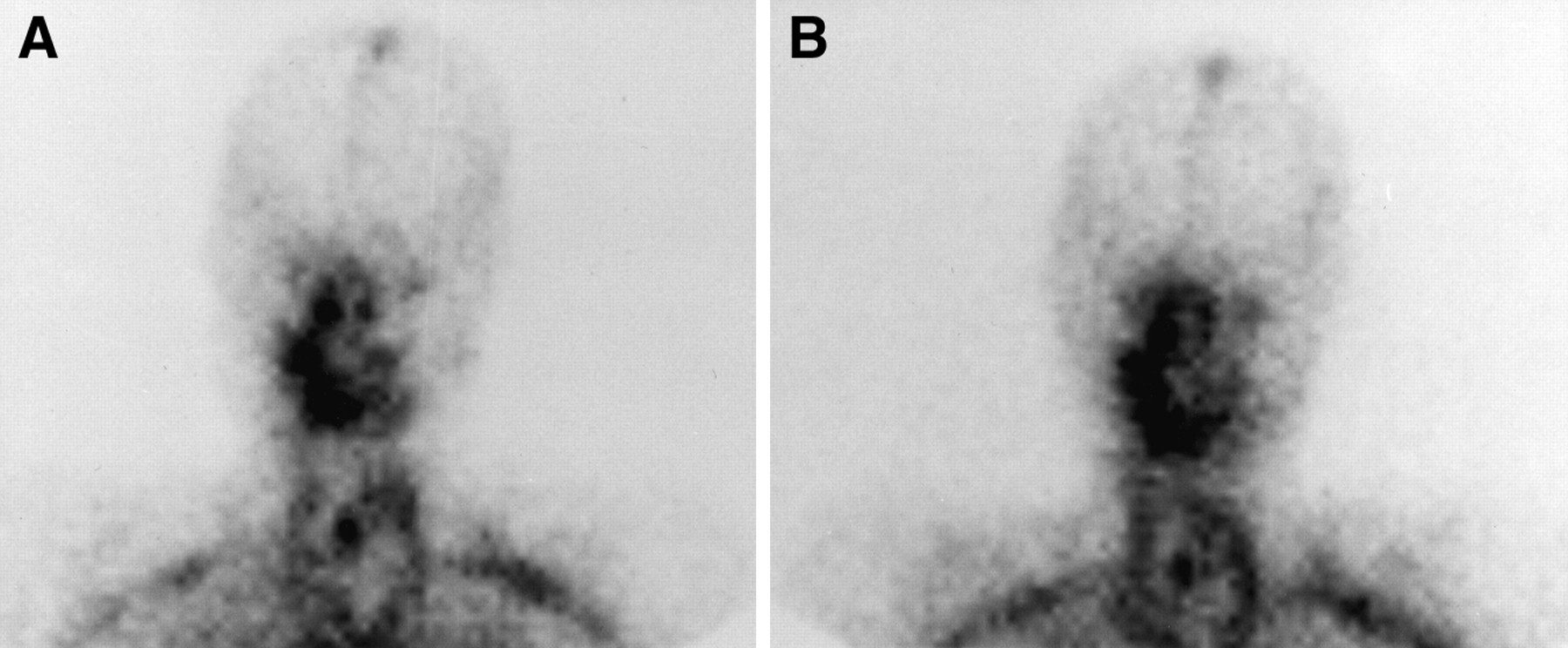
- FIGURE 2.
Comparison of planar imaging of head and neck region of patient 1 21 h after administration of 99mTc-cMAb U36 (A) and 186Re-cMAb U36 (B). Accumulation of radiolabeled cMAb U36 is visible at tumor recurrence in right oropharynx.
- FIGURE 3.
CT scan (A) and planar anterior gamma camera scan (B) of patient 7 obtained 144 h after administration of 186Re-cMAb U36 show targeting of lung metastasis in upper lobe of left lung. Accumulation of 186Re-cMAb U36 is also visible on both sides of oropharynx, where recurrent tumor is present.
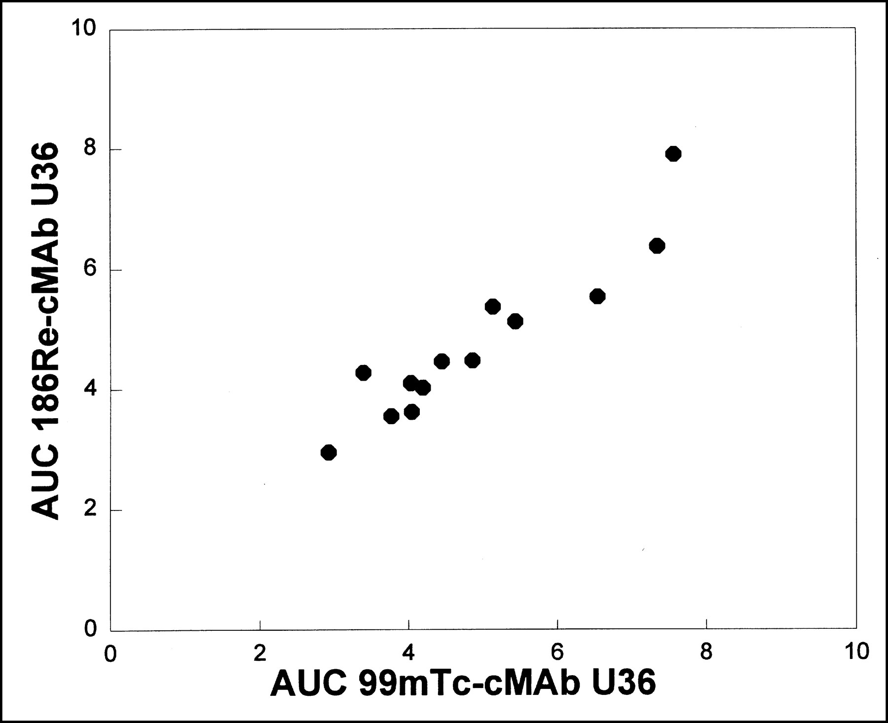
- FIGURE 4.
Relationship between AUC (expressed as %ID × h) determined for 0–25 h after injection for individual patients, both for 99mTc-cMAb U36 (horizontal axis) and for 186Re-cMAb U36 (vertical axis). Although relatively large variations are seen between patients, pharmacokinetic behavior of the 2 conjugates seems similar for individual patients (r = 0.94; P < 0.01).
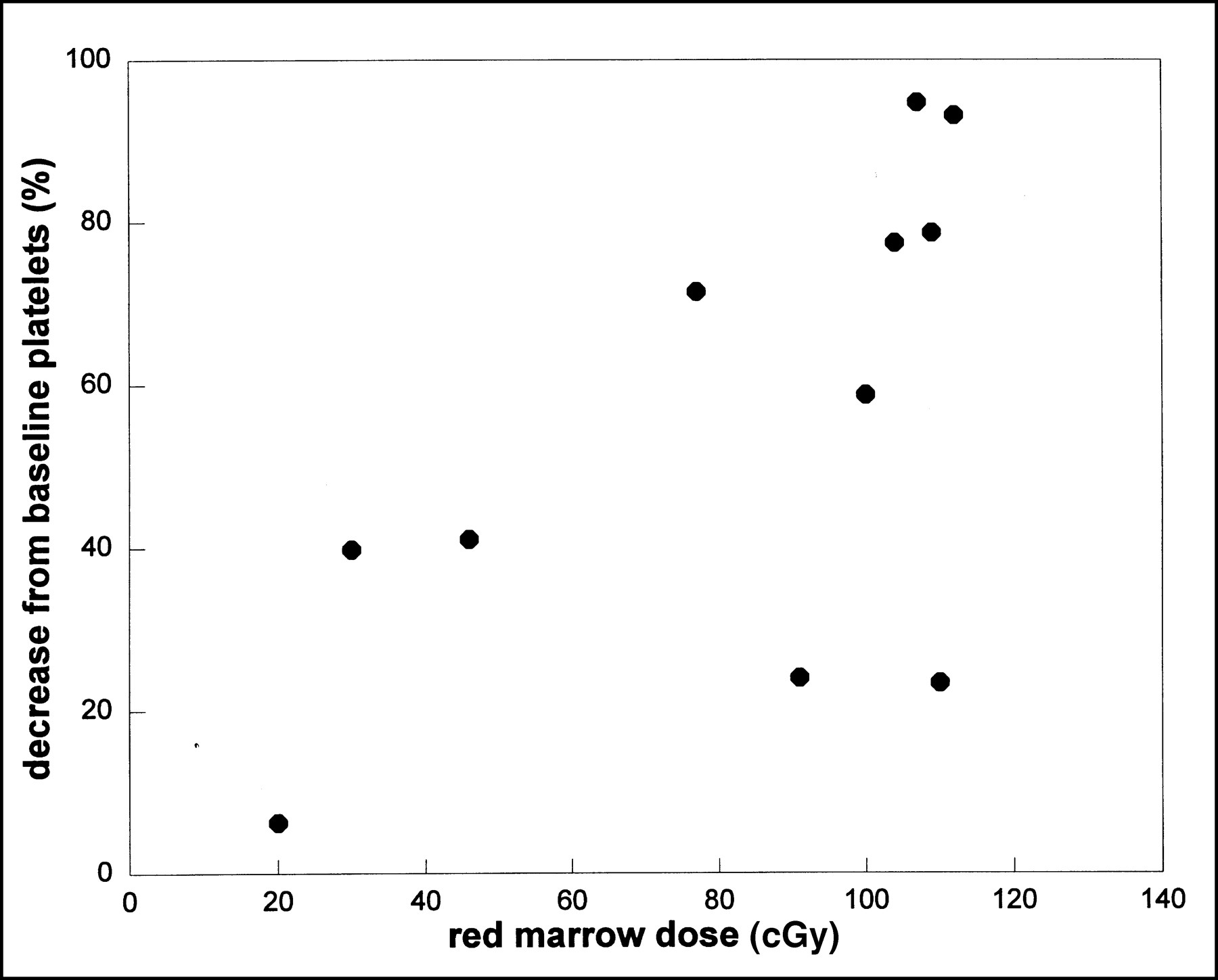
- FIGURE 5.
Relationship between red marrow dose derived from whole-blood time–activity curve for 186Re-cMAb U36 and percentage decrease from baseline platelet count (r = 0.6; P < 0.05).
- FIGURE 6.
(A) CT scan of patient 13 shows large tumor originating from esophagus compressing stent that was placed for palliation 12 mo before RIT. (B) CT scan of same patient 3 wk after administration of 2.15 GBq 186Re-labeled cMAb U36. Sixty percent decrease in tumor size was observed as well as relaxation of stent.


Tables
Patient no. Sex Age (y) Site of disease Prior treatment Radiotherapy Chemotherapy 1 M 53 Oropharynx Yes MTX 2 F 54 Posterior pharyngeal wall Yes None 3 M 54 Oropharynx Yes None 4 M 47 Oropharynx No None 5 M 56 Piriform sinus, left and right parapharyngeal area Yes Cis + 5-FU 6 M 66 Larynx and cystic lesion, neck, right side Yes Cis + Gem/MTX 7 F 56 Oropharynx, both sides Yes None 8 M 58 Hypopharynx Yes None 9 M 67 Neck recurrence, left side Yes None 10 M 68 Oropharynx, right axilla metastasis Yes None 11 M 52 Larynx Yes None 12 M 52 Floor of mouth, submental recurrence Yes Cis + Gem 13 F 59 Esophagus Yes None MTX = methotrexate; Cis = cisplatin; 5-FU = fluorouracil; Gem = gemcitabine.
Patient no. Dose (GBq/m2) Total administered dose (GBq) Absorbed dose, whole body (cGy/GBq) Absorbed dose, red marrow (cGy) Platelet nadir Toxicity grade WBC nadir Toxicity grade Granulocyte nadir Toxicity grade 1 0.4 0.48 35.1 46 230 0 3.4 1 2.3 0 2 0.4 0.59 24.3 25* NA NA NA NA NA NA 3 0.4 0.70 32.4 30 200 0 6.1 0 5.2 0 4 0.4 0.70 35.1 20 285 0 5.7 0 4.1 0 5 1.0 1.60 21.6 73* NA NA NA NA NA NA 6 1.0 2.11 21.6 109 37 3 3.6 1 2.1 0 7 1.0 1.63 NA 91 206 0 5.3 0 4.5 0 8 1.0 1.70 37.8 77 97 1 4.7 0 3.7 0 9 1.0 1.70 29.7 110 190 0 7.6 0 6.2 0 10 1.0 1.78 29.7 100 126 0 4.9 0 3.4 0 11 1.5 2.96 27.0 104 118 0 4.3 0 3.2 0 12 1.5 2.18 8.1 107 24 4 0.6 4 0.1 4 13 1.5 2.15 29.7 112 22 4 2.3 2 1.8 1 ↵* Patients 2 and 5 did not complete follow-up for evaluation of hematologic toxicity and are therefore not included for analysis of correlation between red marrow dose and development of hematologic toxicity.
WBC = white blood cell count; NA = not available.
Patient no. cMAb U36 dose (mg) Before RIS Before RIT 1 wk after RIT 6 wk after RIT HAMA titer HACA (mg/L) HAMA titer HACA (mg/L) HAMA titer HACA (mg/L) HAMA titer HACA (mg/L) 1 2 + 12 <50 <0.2 <50 1.50* <50 2.05* <50 0.33 2 2 + 12 <50 <0.2 <50 1.30* 113 5.73* ND ND 3 2 + 52 <50 <0.2 <50 <0.2 52 <0.2 55 <0.2 4 2 + 52 ND ND <50 <0.2 <50 <0.2 110 <0.2 5 2 + 52 <50 <0.2 <50 <0.2 ND ND ND ND 6 2 + 52 <50 0.7* <50 1.21* 87 5.29* 133 2.46* 7 2 + 52 <50 <0.2 <50 <0.2 <50 <0.2 510* 1.76* 8 2 + 52 <50 <0.2 <50 <0.2 <50 <0.2 <50 <0.2 9 2 + 52 <50 <0.2 67 <0.2 <50 <0.2 119 <0.2 10 2 + 52 <50 <0.2 <50 <0.2 <50 <0.2 <50 <0.2 11 2 + 52 <50 <0.2 <50 <0.2 <50 <0.2 147 <0.2 12 2 + 52 <50 <0.2 <50 <0.2 136 1.88* 94 1.32* 13 2 + 52 <50 <0.2 <50 <0.2 <50 <0.2 <50 <0.2 ↵* Positive responses.
ND = not done.
HAMA titers and HACA levels were measured before administration of 2 mg 99mTc-cMAb U36 for RIS and within 1 wk when patients received 12 or 52 mg 186Re-cMAb U36 for RIT. After start of RIT, HAMA and HACA responses were measured at 1 and 6 wk.
Patient no. Total administered dose (GBq) Tumor volume (cm3) Total tumor absorbed dose* (Gy) Tumor absorbed dose (cGy/GBq) Response Follow-up (mo) 1 0.48 34 2.0 424.3 Stable lung metastases 4 2 0.59 80 1.8 308.1 Progression 1 3 0.70 37 4.1 578.4 Progression 4 4 0.70 81 6.7 957.1 Progression 3 5 1.60 40 3.0 186.5 NA NA 6 2.11 ND ND ND Progression 5 7 1.63 32 ND ND Stable lung metastases 3 8 1.70 28 3.5 205.4 Stable disease for 6 mo 8 9 1.70 14 18.1 1064.9 Progression 4 10 1.78 74 4.5 248.6 Progression 3 11 2.96 80 16.7 262.2 Progression 6 12 2.18 135 8.3 383.8 Reduction of tumor mass 1 13 2.15 98 ND ND Reduction of tumor mass 2 ↵* Ten patients were evaluable for tumor dosimetry. Site of origin of tumors is indicated in Table 1. Only volumes of visualized tumor lesions are included; dosimetry of lung metastases was not possible.
NA = not available; ND = not done.
In this issue


Jump to section
Related Articles
Cited By...
- Antidrug Antibody Formation in Oncology: Clinical Relevance and Challenges
- Intraoperative Near-Infrared Fluorescence Tumor Imaging with Vascular Endothelial Growth Factor and Human Epidermal Growth Factor Receptor 2 Targeting Antibodies
- Radiation Dosimetry of 89Zr-Labeled Chimeric Monoclonal Antibody U36 as Used for Immuno-PET in Head and Neck Cancer Patients
- Immuno-PET: A Navigator in Monoclonal Antibody Development and Applications
- A Phase I Dose Escalation Study with Anti-CD44v6 Bivatuzumab Mertansine in Patients with Incurable Squamous Cell Carcinoma of the Head and Neck or Esophagus.
- Radioimmunotherapy of Head and Neck Cancer Xenografts Using 131I-Labeled Antibody L19-SIP for Selective Targeting of Tumor Vasculature
- Potential of immuno-positron emission tomography for tumor imaging and immunotherapy planning.
- Performance of immuno-positron emission tomography with zirconium-89-labeled chimeric monoclonal antibody u36 in the detection of lymph node metastases in head and neck cancer patients.
- Radioimmunotherapy with [131I]cG250 in Patients with Metastasized Renal Cell Cancer: Dosimetric Analysis and Immunologic Response
- Dosimetric Analysis of Radioimmunotherapy with 186Re-Labeled Bivatuzumab in Patients with Head and Neck Cancer
- Phase I Therapy Study with 186Re-labeled Humanized Monoclonal Antibody BIWA 4 (Bivatuzumab) in Patients with Head and Neck Squamous Cell Carcinoma
- 89Zr Immuno-PET: Comprehensive Procedures for the Production of 89Zr-Labeled Monoclonal Antibodies
- Reinfusion of Unprocessed, Granulocyte Colony-stimulating Factor-stimulated Whole Blood Allows Dose Escalation of 186Relabeled Chimeric Monoclonal Antibody U36 Radioimmunotherapy in a Phase I Dose Escalation Study
- Evaluation of Limited Blood Sampling in a Preceding 99mTc-Labeled Diagnostic Study to Predict the Pharmacokinetics and Myelotoxicity of 186Re-cMAb U36 Radioimmunotherapy
- How Far Have We Come with Solid (Nonhematologic) Tumor Radioimmunotherapy?