


Abstract
211At-labeled tumor-specific antibodies have long been considered for the treatment of disseminated cancer. However, the limited availability of the nuclide and the poor efficacy of labeling procedures at clinical activity levels present major obstacles to their use. This study evaluated a procedure for the direct astatination of antibodies for the production of clinical activity levels. Methods: The monoclonal antibody trastuzumab was conjugated with the reagent N-succinimidyl-3-(trimethylstannyl)benzoate, and the immunoconjugate was labeled with astatine. Before astatination of the conjugated antibody, the nuclide was activated with N-iodosuccinimide. The labeling reaction was evaluated in terms of reaction time, volume of reaction solvent, immunoconjugate concentration, and applied activity. The quality of the astatinated antibodies was determined by in vitro analysis and biodistribution studies in nude mice. Results: The reaction proceeded almost instantaneously, and the results indicated a low dependence on immunoconjugate concentration and applied activity. Radiochemical labeling yields were in the range of 68%−81%, and a specific radioactivity of up to 1 GBq/mg could be achieved. Stability and radiochemical purity were equal to or better than those attained with a conventional 2-step procedure. Dissociation constants for directly astatinated, conventionally astatinated, and radioiodinated trastuzumab were 1.0 ± 0.06 (mean ± SD), 0.44 ± 0.06, and 0.29 ± 0.02 nM, respectively. The tissue distribution in non–tumor-bearing nude mice revealed only minor differences in organ uptake relative to that obtained with the conventional method. Conclusion: The direct astatination procedure enables the high-yield production of astatinated antibodies with radioactivity in the amounts required for clinical applications.
Among the isotopes of the heaviest element in the halogen group, 211At has attracted interest as a prospective candidate for endoradiotherapeutic applications because of its physicochemical characteristics (1). Unlike most commonly medically applied therapeutic radionuclides that decay through medium- to high-energy β-emission, leading to low–linear-energy-transfer radiation with particle ranges of 1–10 mm, 211At decays through α-emission, depositing high–linear-energy-transfer radiation in a microvolume corresponding to a mean α-particle range of ∼65 μm. When bound to a tumor-specific substance, this radiation can be effective in the destruction of disseminated microtumors, that is, micrometastases, as has been demonstrated in several preclinical studies (2–5). The preclinical work has resulted in 2 phase I studies, a study of the treatment of malignant gliomas at Duke University Medical Center, Durham, NC, and a study of the treatment of ovarian carcinomas at Sahlgrenska University Hospital, Gothenburg, Sweden (6,7). Despite this fact, there are still factors hampering clinical applications. The major obstacle to progression to the clinical stage is the lack of facilities capable of producing the nuclide in the amounts required (8,9). Furthermore, research is still being performed to find adequate procedures for efficient labeling. The problems that have to be overcome are low labeling yields and chemical decomposition during labeling (10,11).
The general approach for the production of astatinated proteins has been the use of bifunctional labeling reagents that are labeled and subsequently conjugated to the proteins. Several strategies have been adopted for the synthesis of 211At-labeled intermediate compounds; the common feature involves the formation of a stable aryl-carbon-astatine bond (12–15). Much of the pioneering work concerning astatine labeling techniques has been conducted by Zalutsky and Narula (16) and Garg et al. (17) with N-succinimidyl-aryl-alkyl-tin ester derivatives (ATE reagents) for 2-step labeling and conjugation to proteins. New and interesting reagents based on boron cage halogen chemistry are under development for the direct astatination of proteins by Wilbur et al. (18,19). However, at present, conjugate labeling with various forms of aryl-stannyl intermediates is the principal technique for the astatination of proteins. The general route for the synthesis of astatinated antibodies with these intermediates involves 2 radiochemical steps, that is, labeling and conjugation. In this time-consuming procedure, the decay of the nuclide and workup losses result in relatively low radiochemical yields. In addition, the reaction time contributes to the absorbed dose to the reaction solvent, which may lead to radiolysis at high activity concentrations, subsequently affecting labeling yields and the quality of the final product (10,20,21). A direct procedure for the astatination of antibodies would thus be advantageous over the conventional 2-step procedure.
In this study, a novel route for the synthesis of astatinated proteins was investigated; in this procedure, the antibody was conjugated with the reagent N-succinimidyl-3-(trimethylstannyl)benzoate (m-MeATE) before astatination. The astatination procedure was optimized with regard to reaction time, concentration of the antibody conjugate, and applied activity, and the astatinated antibody produced was evaluated with regard to radiochemical yield, radiochemical purity, immunoreactivity, and stability in vitro and in vivo.
MATERIALS AND METHODS
General
The monoclonal antibody trastuzumab (Herceptin; Roche Pharma AG), specific for human epidermal growth factor ErbB2 (Her2), was obtained from the Swedish Pharmacy, Sahlgrenska University Hospital. The human tumor cell line SKOV-3 was obtained from the American Type Culture Collection. No-carrier-added 125I-NaI was purchased from Perkin-Elmer. 211At was produced by irradiating stable bismuth, through the 209Bi(α,2n)211At reaction, at the Cyclotron and PET Unit, Rigshospitalet, Copenhagen, Denmark. The irradiated target was transported to the Department of Nuclear Medicine, Sahlgrenska University Hospital, where the astatine was transformed into a chemically useful form by dry distillation as described previously (22). The reagent m-MeATE was purchased from Toronto Research Chemicals Inc. All other chemicals used in this study were of analytic grade or better.
Radioactivity Measurements
High-activity radioactivity measurements (>100 kBq) were conducted in an ionization chamber (CRC-15 dose calibrator; Capintec). Low-activity samples (<10 kBq) were measured with an NaI(Tl) γ-counter (Wizard 1480; Wallac) at dual energy-window settings of 15–70 keV for 125I and 65–95 keV for 211At. The 2 devices were cross-calibrated for 125I and 211At.
m-MeATE Antibody Conjugation
The reagent m-MeATE was dissolved in chloroform at a concentration of 130 μmol/mL. A sample of 5 μL was taken from the stock solution, and the chloroform was evaporated. The dry residue of the reagent was then redissolved in dimethyl sulfoxide at a concentration of 25 μmol/mL. Trastuzumab was prepared in 0.2 M sodium carbonate buffer (pH 8.5) at a concentration of 4 mg/mL. A 5- to 10-fold molar excess of m-MeATE over the antibody was used in the conjugation. To 0.5–1.0 mL of the antibody preparation, 5–10 μL of the reagent m-MeATE were added during vigorous agitation. The conjugation reaction was allowed to proceed for 30 min during gentle agitation at room temperature. The ε-lysyl-3-(trimethylstannyl)benzamide antibody conjugate fraction was finally isolated by size exclusion chromatography with an NAP-5 or a PD-10 column (GE Healthcare). The column was eluted with 0.2 M sodium acetate buffer (pH 5.5).
Direct Procedure for Astatination of Antibody Conjugate
A series of experiments was set up to evaluate the labeling of the m-MeATE–trastuzumab conjugate, including dependence on reaction time, antibody conjugate concentration, and applied activity. Astatine was prepared as a dry residue from a chloroform preparation after distillation. The astatine state was adjusted for labeling by reaction of the dry residue with N-iodosuccinimide (NIS) immediately before labeling with an NIS stock solution at a concentration of 30 μg/mL in methanol:1% acetic acid.
For the reaction time dependence experiments, 50 μL (100 μg) of the antibody conjugate in sodium acetate (pH 5.5) were added to 5 μL (13–17 MBq) of the astatine–NIS preparation. The reaction time was varied from 10 s to 30 min, and at each reaction time, a sample of 1–3 MBq was taken from the labeling mixture for yield determination with an NAP-5 column. The radiochemical yield was expressed as the activity in the isolated antibody fraction divided by the initial activity loaded in the column.
For the antibody conjugate concentration experiments, the immunoconjugate was prepared at a concentration of 2 mg/mL in sodium acetate buffer (pH 5.5) and then serially diluted in five 1:2 dilutions in the same buffer, resulting in a final concentration of 0.125 mg/mL. From each dilution, 100 μg of antibody conjugate were taken, and each sample was reacted with approximately 10 MBq of the astatine–NIS preparation for 60 s.
The applied activity experiments were based on both the time and the concentration evaluations. Initial activities of 100–500 MBq were studied. To 10 or 20 μL of the astatine–NIS preparation, 200–400 μg of the trastuzumab conjugate were added at a concentration of 2.0 mg/mL. The antibody conjugate was reacted with astatine for 60 s. A 20-fold molar excess of NIS over antibody was added at the end of the reaction to iodo-substitute any remaining stannyl groups on the immunoconjugate. For the iodination step, 3 μL of NIS at a concentration of 1, 2, or 4 mg/mL (depending on the initial amount of the immunoconjugate) in methanol:1% acetic acid were added to the reaction mixture. For reduction of any unreacted astatine, 5 μL of sodium ascorbate at a concentration of 10 μmol/mL in water were added immediately before purification. The antibody fractions from all of the labeling experiments were finally isolated from unreacted low-molecular-weight species by passage over an NAP-5 or a PD-10 column with phosphate-buffered saline (PBS) as the mobile phase. The radiochemical labeling yield was expressed as the radioactivity in the eluted labeled fraction (non-decay corrected) divided by the initial radioactivity in the reaction mixture. For the evaluation of nonspecific antibody binding, unconjugated trastuzumab was subjected to astatination by the direct procedure as described earlier for the reaction time dependence experiments at a reaction time of 20 min.
The absorbed doses to the reaction volumes during the reaction were calculated with basic dosimetry derived from the following equations. The number of 211At decays (N) is represented as where A0 is total radioactivity measured at the start of the reaction and t is total reaction time. The absorbed dose (D) is represented as
 where Eabs is the energy absorbed in the reaction volume (MeV),
  is the mean α-particle energy, and m is the mass of the reaction solvent (kg). The factor 1.6 × 10⁻¹³ is the conversion from megaelectron volts to joules.
Tin Analysis
For the determination of conjugation efficacy, the ε-lysyl-3-(trimethylstannyl)benzamide antibody conjugate was subjected to tin analysis by inductively coupled plasma–optical emission spectrometry (ICP-OES) (iCap 6500 Thermo; Fisher Scientific Inc.). The astatinated antibody was also analyzed with ICP-OES to determine the content of stannyl residues before and after quenching with NIS after astatination. For preparation of the analysis products, trastuzumab was conjugated with m-MeATE as described earlier. Samples (0.5 mL) of the m-MeATE–trastuzumab conjugate were treated with equimolar NIS or a 20-fold molar excess of NIS for 1–2 min. The iodinated products were isolated by gel filtration with an NAP-5 column and PBS as the mobile phase. The initial amount of m-MeATE was used as the tin standard. After tin analysis, the conjugation yield and the degree of quenching were determined from the tin quotient as the ratio of m-MeATE–trastuzumab to m-MeATE and the ratio of iodinated trastuzumab to m-MeATE.
Conventional 2-Step Procedure for Astatination of Antibody
Conventionally astatinated trastuzumab was prepared as previously described (23). In brief, to a dry residue of astatine (20–50 MBq), 0.75 nmol of NIS and 1 nmol of m-MeATE in methanol:1% acetic acid were added. The reaction was allowed to proceed for 20 min during gentle agitation at room temperature. The organic solvent was evaporated, and 200 μg of antibody in 0.2 M carbonate buffer were added to the crude labeling mixture. After 30 min of conjugation, the reaction was quenched by the addition of 50 nmol of sodium ascorbate, and the antibody fraction was isolated by gel filtration with an NAP-5 column. Schemes showing the direct procedure and the conventional 2-step procedure are shown in Figure 1.
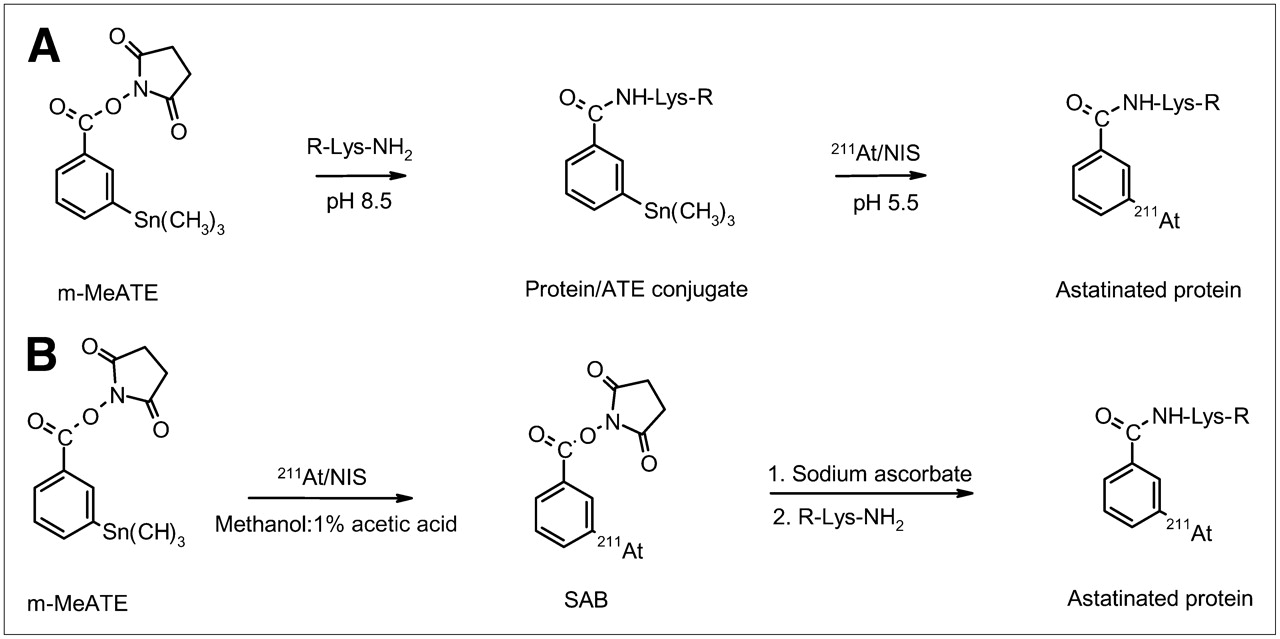
(A) Direct procedure. Conjugation of antibody (R-Lys-NH2) with m-MeATE and labeling of conjugated antibody. (B) Conventional procedure. Labeling of m-MeATE followed by conjugation of labeled reagent to antibody (R-Lys-NH2). SAB = N-succinimidyl 3-astatobenzoate.
Radioiodination of Antibody
Conjugate iodination of trastuzumab was conducted in essentially the same manner as conventional astatination, except that N-bromosuccinimide was used as the oxidizing agent.
In Vitro Quality Control
Radiochemical Purity.
The radiochemical purity of the astatinated and iodinated immunoconjugates was determined by methanol precipitation and size exclusion chromatography. Methanol precipitation was performed in triplicate. To 3-mL Ellerman tubes, 100 μL of 1% bovine serum albumin in PBS, 1- to 2-kBq samples of the radioactive products, and 500 μL of methanol were added. After 5 min, the total radioactive content of the tubes was measured with the γ-counter. The tubes were centrifuged, and protein-bound radioactivity in the pellet was measured. The radiochemical purity was calculated as the protein-bound activity divided by the total applied activity.
Size exclusion fast protein liquid chromatography (FPLC) was conducted with a Superdex-200 column and an Äkta-FPLC delivery system (GE Healthcare Bio-Sciences AB). Aliquots of the radioactive products containing ∼200 kBq were analyzed. The column was eluted with PBS at a flow rate of 0.5 mL/min. Fractions were collected, and radiochemical purity was determined as the eluted protein-bound activity divided by the activity injected into the system. Chromatograms are shown in Supplemental Figure 1 (supplemental materials are available online only at http://jnm.snmjournals.org).
Immunoreactivity and Affinity.
The biologic function of the antibodies after labeling was investigated by binding to SKOV-3 cells as described by Lindmo et al. (24). For determination of the immunoreactive fraction, a single-cell suspension of SKOV-3 cells was prepared at a concentration of 5 × 10⁶ cells per milliliter. The cells were serially diluted 1:2, and a constant amount (20 ng) of labeled antibodies was added to each dilution. The antibodies were allowed to react with the cells for 3 h at room temperature during gentle agitation. After incubation and repeated washing, the bound fraction in each dilution was determined by measuring the activity in the cells. Double inverse plots were derived from the data, and the immunoreactive fraction was calculated. Nonspecific binding of astatinated trastuzumab was examined by saturating the antigens on the SKOV-3 cells with an excess of trastuzumab.
For determination of the dissociation constant (Kd), the labeled antibodies (211At-trastuzumab [direct labeling or 2-step labeling] or 125I-trastuzumab [2-step labeling]) were serially diluted (1:2) from 4 to 0.0625 μg/mL in culture medium. From each dilution, 40 μL were taken and added to 5 × 10⁴ SKOV-3 cells (5 × 10⁵ cells per mL). The antibodies were allowed to react with the cells for 3 h at room temperature during gentle agitation. The total applied activity in the tubes was measured, the cells were washed, and bound radioactivity was counted. Saturation curves were derived from the data, and the apparent Kd values were calculated.
Serum Stability.
For determination of the in vitro stability of astatinated trastuzumab, triplicate samples of approximately 15 MBq of the astatinated antibody were incubated for 48 h at 37°C in freshly prepared human serum. At 1, 3, 6, 24, and 48 h, aliquots were taken from the serum mixture, and the antibody-bound astatine fraction was determined at each time by methanol precipitation. At 1 and 24 h, the bound fraction was also determined by size exclusion chromatography with the FPLC system.
Animal Study
The tissue distribution of astatinated trastuzumab was evaluated in non–tumor-bearing female BALB/C nu/nu mice (Charles River Laboratories International Inc.) at 4–6 wk of age. The mice were housed at 22°C with access to food and water ad libitum. Twenty mice were coinjected intravenously through the tail vein with 500 kBq of 211At-trastuzumab (labeled by the direct procedure) and 80 kBq of 125I-trastuzumab. Groups of 4 mice were sacrificed 1, 3, 6, 11, and 24 h after injection. At each time point, whole blood was collected by cardiac puncture, and tissues (neck, salivary glands, heart, lungs, liver, stomach, small intestine, large intestine, kidneys, spleen, and muscle) were excised. The tissues were weighed, and 125I and 211At activities were simultaneously measured in the γ-counter with a dual-window setting. Measurements were obtained from the tissue samples within 12 h after dissection to determine the 211At counts and then 4 d later (after the decay of 211At) to determine the 125I counts. The data were individually corrected for the spillover (<10%) of 125I into the 211At window and for radioactive decay. Results were expressed as the percentage of injected activity per gram of organ (%IA/g). Conventionally astatinated trastuzumab and iodinated trastuzumab were coinjected into another 20 mice, which were evaluated in the same manner.
RESULTS
m-MeATE Antibody Conjugation
The efficiency of binding of the reagent m-MeATE to the antibody was 70%−80%, as determined by tin quantification with ICP-OES analysis, corresponding to an average ratio of m-MeATE to antibody of ∼6:1. The stannyl residues remaining after astatination were efficiently quenched by iododestannylation with an excess of NIS after astatination. After labeling, approximately 80% of the stannyl residues remained on the antibody conjugate, and after NIS quenching, only ∼5% of the stannyl residues originally present on the antibody conjugate could be detected.
Radiolabeling
The labeling of the antibody conjugate was first studied by evaluation of the reaction kinetics, starting with reaction times from 5 to 30 min. The results showed no reaction time dependence in that interval, indicating that the reaction was completed within 5 min of reaction time. Next, shorter reaction times, that is, 10, 30, 60, and 120 s, were studied. Still no reaction time dependence was seen, indicating that the reaction was completed almost instantaneously. Radiochemical yields in the range of 85%−89% were obtained.
In the second study, the effect of the antibody conjugate concentration on the radiochemical labeling yield was investigated. The results showed little dependence on the immunoconjugate concentration. Even at a concentration of 0.125 mg/mL, the reaction was completed in less than 1 min, with a radiochemical labeling yield exceeding 70%.
Scaling of the chemistry to high activity levels, 100–500 MBq, showed that a high yield could be maintained. Activities of up to 500 MBq of 211At after irradiation, shipping, isolation, and workup were used for labeling, and products containing up to 400 MBq were obtained. The specific radioactivity of the high-activity astatinated trastuzumab was in the range of 0.34–1.0 GBq/mg. Because of the rapid reaction kinetics, the absorbed dose to the reaction volume was limited. When volumes in the range of 100–200 μL were used in the high-activity experiments, the absorbed doses were in the range of 130–400 Gy. Table 1 presents a summary of the synthesis and yield of 211At-trastuzumab by means of the direct procedure, including results of the studies of reaction time, concentration, and applied activity.
Synthesis and Yields of 211At-Trastuzumab
Results from the nonspecific labeling of unconjugated antibody indicated that approximately 30% of the initial 211At activity added to the reaction was apparently bound to the antibody.
Radiochemical Purity and Stability
The radiochemical purity of the directly labeled trastuzumab immunoconjugate was always above 97% after labeling and purification, as determined by methanol precipitation and FPLC analysis. However, after the labeling of unconjugated trastuzumab, only 10% of the activity was precipitated with the protein fraction.
Studies of in vitro stability in human serum showed that the directly astatinated trastuzumab immunoconjugate was stable, with a decrease in the radiochemical purity of only a few percentage points over the 48-h incubation time. More than 95% of the astatine was still associated with the antibody fraction after 24 h, as determined by size exclusion chromatography.
Immunoreactivity and Affinity
The immunoreactivity and the Kd values of directly astatinated trastuzumab were investigated along with those of conventionally astatinated and iodinated trastuzumab. The immunoreactive fractions (Kd values, mean ± SD) for trastuzumab astatinated by the direct procedure, trastuzumab astatinated by the 2-step procedure, and iodinated trastuzumab were 73%−87% (1.0 ± 0.06 nM), 75%−85% (0.44 ± 0.06 nM), and 75%−89% (0.29 ± 0.02 nM), respectively. Saturation curves are shown in Supplemental Figure 2. The nonspecific binding of the directly astatinated immunoconjugate was less than 1%, as determined by incubation with SKOV-3 cells saturated with an excess of unlabeled trastuzumab.
Biodistribution
A comparison of the tissue uptake in non–tumor-bearing nude mice of trastuzumab labeled by the direct procedure and trastuzumab labeled by the conventional 2-step procedure is shown in Figure 2. The results showed very similar distributions over the time period studied; however, small but significantly (P < 0.05) higher levels of uptake of conventionally produced 211At-trastuzumab were observed in several tissues. For example, at 6 h after injection, the uptake of 211At-trastuzumab produced by the 2-step procedure was higher than that of 211At-trastuzumab produced by the direct procedure in most of the organs studied. The tissue distributions of astatinated trastuzumab and iodinated trastuzumab (reference) are shown in Table 2 (direct astatination) and Table 3 (2-step astatination). The 211At/125I ratios in the tissues after the distribution of trastuzumab were well correlated, except for the uptake in the stomach. After 3 h, the uptake of astatine in the stomach was higher than that of iodine, reaching approximately twice the uptake of iodine at 6 h. The uptake of astatine in the thyroid was also higher than that of iodine, and there was an indication of higher uptake in the salivary glands. The higher uptake of astatine than of iodine was confirmed in both mouse studies, and a comparison of the 211At/125I tissue distribution ratios for trastuzumab indicated very similar distributions regardless of the astatination method used.

Tissue distribution of 211At after injection of 211At-trastuzumab labeled by direct procedure (□) and by conventional 2-step procedure (). Results are given as mean ± SD %IA/g. *Statistically significant difference (P < 0.05), as determined by Student t test.
Tissue Distribution Ratios After Coinjection in Mice of 211At-Trastuzumab Labeled by Direct Procedure and 125I-Trastuzumab Labeled by Conventional Procedure
Tissue Distribution Ratios After Coinjection in Mice of 211At-Trastuzumab and 125I-Trastuzumab Labeled by Conventional Procedure
DISCUSSION
One of the major challenges in the treatment of cancer is the elimination of occult disseminated cancer cells. Tumors recurring after primary curative treatment generally originate from invasive malignant cells (metastases), and adjuvant treatment is required to prevent or delay clinical tumor progress. For this purpose, α-particle emitters and carrier substances such as tumor-specific monoclonal antibodies have gained increasing interest for use in endoradiotherapy in patients with minimal residual disease (25,26). Among the α-particle emitters available, 211At appears to fit most of the requirements for such treatment (27). The main nuclear reaction used for 211At production is the 209Bi(α,2n)211At reaction, which requires a medium-energy cyclotron for α-particle acceleration (28,29). Unfortunately, few cyclotron facilities have the means to produce 211At, and the capacity at each facility is limited. Because of these limitations on production, high demands must be placed on distillation and workup yields after irradiation as well as on the efficiency of subsequent chemical procedures if the nuclide is to be used for clinical applications.
The main focus of the present study was improvement in labeling yields. For this purpose, a direct radiochemical reaction of antibody conjugates was studied. The antibodies were conjugated with the bifunctional labeling reagent m-MeATE before labeling, allowing direct astatination. The stannyl ester reacted with the primary amines, mainly the ε-lysine amines, on the antibody, resulting in an ε-lysyl-3-(trimethylstannyl)benzamide residue (Fig. 1A).
To our knowledge, this is the first study in which aryl-stannyl immunoconjugates have been used for astatine labeling. In all other reports concerning the astatination of antibodies with aryl-stannyl esters, the radiochemistry has been performed in 2 steps, that is, labeling of the reagent followed by conjugation to the antibody. The main reason for this strategy is that in addition to the reaction with the conjugate residue, astatine may bind with low stability directly to the protein, resulting in a product of poor quality. The latter result was confirmed in the present study by the direct labeling of unconjugated trastuzumab. The product was very labile, with less than 10% of the 211At stably attached to the antibody. A mechanism for the direct interaction of astatine with proteins has been proposed by Visser et al.; in this mechanism, astatine (unlike iodine, which mainly binds to tyrosyl residues) undergoes an electrophilic reaction with unsubstituted thiols on the protein (30). Another reason for not conjugating the antibody with the stannyl ester before labeling is that unreacted tin groups may be present on the protein after astatination, and these could contribute to toxicity in an in vivo application (31). However, contrary to these expected problems, the astatinated antibodies produced in the present study by direct astatination of preconjugated antibodies were of excellent quality. Before labeling of the antibody conjugate, NIS was used as the oxidizing agent to activate astatine. The resulting intermediate electrophilic species, after the oxidizing process in the reaction solvent, is probably the dihalide 211AtI or the polyhalide anion 211AtI2− (32). In addition to the electrophilic astatine, the triiodide anion (I3−) is also produced by NIS, as indicated by the yellow–brown color that gradually appeared in the NIS–methanol solvent. The triiodide anion has been shown to be a good electrophile (32) and, like activated astatine, will readily substitute for the stannyl groups on the protein (Supplemental Figure 3 and nuclear magnetic resonance data). This feature was taken advantage of in quenching most of the remaining unsubstituted tin groups on the antibody after the labeling reaction. However, it should be noted that the tyrosyl residues of the protein may also be subjected to iodination by the triiodide anion.
Compared with the conventional procedure, which generally gives overall radiochemical yields of approximately 30%−60% in a preparation time of 60 min, the direct astatination reaction proceeds almost instantaneously, with radiochemical yields in the range of 60%−80%. A reasonable explanation for the high yields and rapid reaction kinetics in the astatination of aryl-stannyl immunoconjugates is that the ε-lysyl-3-(trimethylstannyl)benzamide residues are hydrophobic and are therefore extremely exposed to the electrophilic substitution reaction, that is, astatination, in the protein buffer solvent. As long as these residues are in excess, they will probably be the main target for astatine. The use of NIS as an oxidant probably also contributes to the convenient chemistry in the sense that it rapidly forms the interhalogen 211AtI or 211AtI2− with liberated iodine in the NIS–methanol solvent; 211AtI and/or 211AtI2− can subsequently act as a good electrophile in the destannylation reaction.
The major obstacle in the production of high-activity levels of astatine-labeled antibodies by the conventional labeling technique is radiolysis in the reaction volume, which affects the astatine state in the labeling of the ATE reagent, subsequently affecting yields and antibody function after the conjugation step. Furthermore, conjugation of the astatinated reagent to the antibody is dependent on pH and on the antibody concentration. An antibody concentration above 2 mg/mL is required for reasonable labeling yields. For these reasons, it is virtually impossible to obtain both high yields and high specific activities of the final product by the 2-step procedure.
Unlike the conventional labeling procedure, the direct astatination reaction procedure shows little dependence on the concentration of the antibody conjugate. Even at concentrations as low as 0.125 mg/mL, the efficacy was high, with yields exceeding 70%. The rapid reaction kinetics in combination with only minor concentration dependence enables high-efficiency and high-specific-radioactivity labeling at a high level of astatine activity without leading to a detrimental absorbed dose to the reaction solvent. Indeed, the calculated absorbed doses did not exceed 400 Gy, even at initial labeling activities of up to 500 MBq.
All of the analytic investigations confirmed the quality of the product; these investigations included evaluation of the tin content before and after labeling, immunoreactivity, and stability in vitro and in vivo. Little sign of degradation was observed during the 48-h incubation of the astatinated product in freshly prepared human serum, demonstrating high in vitro stability. However, some aggregation of the astatinated trastuzumab was observed over time, probably because of the increased hydrophobicity of the conjugate. The immunoreactivity of directly labeled 211At-trastuzumab was compared with those of 125I-trastuzumab and 211At-trastuzumab produced by the 2-step procedure. The affinity data were in line with those of previous studies evaluating the characteristics of trastuzumab and labeled trastuzumab (33–35). However, a decrease in the affinity of directly astatinated trastuzumab by factors of 2 and 3 relative to the affinities of conventionally astatinated trastuzumab and iodinated trastuzumab, respectively, indicated an effect of the higher ratio of m-MeATE to antibody. This effect was previously addressed in studies with chelate-labeled antibodies, in which the chelate was attached to the antibody before labeling; in those studies, this effect was seen even at antibody-to-chelate ratios of 1:1–1:4 (36,37).
An animal study was performed for comparison of the biodistributions of this new procedure and the conventional 2-step 211At-labeling procedure. Iodinated trastuzumab was also included by coinjection with 211At-trastuzumab. As shown in Tables 2 and 3, the biodistribution of directly astatinated trastuzumab, expressed as the organ uptake ratio, correlated well with the uptake of both conventionally astatinated trastuzumab and iodinated trastuzumab (reference). The only organ uptake that deviated significantly was that of the stomach, in which the uptake of astatine was approximately twice that of iodine. The higher tissue uptake of astatine than of iodine in the stomach tissue is well documented in the literature, and the uptake increases when low-molecular-weight antibodies, such as F(ab′)2 and F(ab′) fragments, are used (19,38). However, a direct comparison of the distributions of 211At-trastuzumab produced by different procedures revealed only minor differences in tissue uptake over the 24-h period (Fig. 2). The data in Tables 2 and 3 and Figure 2 provide evidence that astatine was as stably attached to the antibody when the direct labeling method was used as when the 2-step labeling method was used.
Perhaps the most extraordinary feature of the direct labeling method is the specific radioactivity that can be obtained. As shown by Elgqvist et al., the specific activity of an astatinated antibody may be very important for the therapeutic outcome in a clinical application (39). In that study, calculations indicated that an increase in specific activity of 50% (from corresponding astatine-to-antibody molar ratios of 1:800 to 1:533) would increase the cumulated activity by ∼30%. With the direct method described here, a specific activity of astatinated trastuzumab of up to 1 GBq/mg could be produced, corresponding to an astatine-to-antibody molar ratio of ∼1:100.
CONCLUSION
A direct procedure for producing astatinated antibodies with a high level of activity and a high specific radioactivity was developed. Radiochemical labeling yields in the range of 67%−81% were achieved by conjugating the antibody before astatination. Because of the rapid reaction kinetics and low concentration dependence, high radiochemical yields could be obtained, even under high-activity reaction conditions. The maximum activities used in the present study were 100–500 MBq, yielding products of 67–400 MBq in a total preparation time of 15 min. The low-dose and low-concentration reaction conditions enabled the production of astatinated antibodies with very high specific radioactivity, and the procedure is expected to be applicable even at gigabecquerel levels. The quality of the directly labeled products was equal to that of the corresponding products obtained by the conventional 2-step procedure, as determined by in vitro analysis and biodistribution studies in mice. In conclusion, the direct astatination procedure provides a simple way to synthesize astatine-labeled proteins with radioactivity in the amounts required for clinical applications.
Acknowledgments
Thanks are due to Arvid Ödegard-Jensen at the Department of Nuclear Chemistry, Chalmers University of Technology, Gothenburg, Sweden, for help with the ICP-OES analysis and to Ingela Claesson and Helena Kahu at the Department of Oncology, Sahlgrenska Academy, University of Gothenburg, Gothenburg, Sweden, for help in culturing the tumor cells used and for help with the animal study. This work was supported by grants from the Swedish Cancer Society (grant 3548), the Swedish Research Council (grant K2007-53X-20321-01-3), and the King Gustaf V Jubilee Clinic Cancer Research Foundation in Gothenburg, Sweden.
Footnotes
-
COPYRIGHT © 2008 by the Society of Nuclear Medicine, Inc.
References
- Received for publication December 13, 2007.
- Accepted for publication June 2, 2008.


Jump to section
Related Articles
Cited By...
- Harnessing {alpha}-Emitting Radionuclides for Therapy: Radiolabeling Method Review
- Intraperitoneal {alpha}-Emitting Radioimmunotherapy with 211At in Relapsed Ovarian Cancer: Long-Term Follow-up with Individual Absorbed Dose Estimations
- Evidence of Local Concentration of {alpha}-Particles from 211At-Labeled Antibodies in Liver Metastasis Tissue
- Model of Intraperitoneal Targeted {alpha}-Particle Therapy Shows That Posttherapy Cold-Antibody Boost Enhances Microtumor Radiation Dose and Treatable Tumor Sizes
- Cure of Human Ovarian Carcinoma Solid Xenografts by Fractionated {alpha}-Radioimmunotherapy with 211At-MX35-F(ab')2: Influence of Absorbed Tumor Dose and Effect on Long-Term Survival
- The Advantage of Antibody Cocktails for Targeted Alpha Therapy Depends on Specific Activity
- Ex Vivo Activity Quantification in Micrometastases at the Cellular Scale Using the {alpha}-Camera Technique
- Intraperitoneal {alpha}-Particle Radioimmunotherapy of Ovarian Cancer Patients: Pharmacokinetics and Dosimetry of 211At-MX35 F(ab')2--A Phase I Study